 WhatsApp Us (24/7)
 +91 926 888 0303 (24/7)


Abstract: Young adults presenting with progressive neurological deficits and systemic features such as weight loss and night sweats pose a significant diagnostic challenge. Imaging that reveals widespread lytic skeletal lesions with a solitary pulmonary nodule often directs clinical suspicion towards an underlying malignancy. A previously healthy 24-year-old male presented with gradually progressive bilateral lower limb weakness, preceded by back pain, along with significant weight loss, loss of appetite, night sweats, and chronic dry cough. Neurological examination revealed paraparesis with areflexia in the lower limbs. Magnetic resonance imaging (MRI) of the spine showed multiple lytic lesions and a compression fracture at T10–T11 with significant nerve root compression. Fluorodeoxyglucose (FDG) positron emission tomography (PET)–computed tomography (CT) demonstrated a right middle lobe pulmonary nodule, mediastinal lymphadenopathy, and diffuse FDG-avid skeletal lesions. A malignancy workup, including bone marrow biopsy, was non-contributory. A CT-guided sternal biopsy revealed necrotising granulomatous inflammation, but no malignant cells or acid-fast bacilli. Pending culture results, the patient was started on empirical therapy based on the most probable aetiology. Gradual clinical improvement was observed, and definitive microbiological confirmation followed after 34 days. This case underscores the complexity of differentiating between infectious and malignant causes in patients presenting with systemic illness and multifocal bony lesions. High clinical suspicion, combined with histopathological and microbiological correlation, remains vital for accurate diagnosis.
Key words: Paraparesis, Multifocal Bony Lesions, Malignancy.
Introduction
Spinal involvement by systemic disease presents a broad differential diagnosis, ranging from infections to malignancies. When young adults present with progressive paraparesis, weight loss, and constitutional symptoms, metastatic malignancy is often high on the clinician’s radar — especially when imaging reveals lytic spinal lesions and a solitary pulmonary nodule. However, in tuberculosis (TB)-endemic regions such as Southeast Asia, certain infections can mimic malignancies both clinically and radiologically.
TB of the spine (Pott’s disease) accounts for approximately 50% of skeletal TB cases.1 Atypical presentations — such as multifocal non-contiguous vertebral involvement, absence of classical constitutional symptoms, or isolated pulmonary nodules — can obscure the diagnosis. Moreover, fluorodeoxyglucose (FDG) positron emission tomography (PET)–computed tomography (CT), though sensitive in detecting metabolically active lesions, does not reliably distinguish between infection and malignancy due to overlapping uptake patterns.2
We report a case of a young, immunocompetent male with rapidly progressive paraparesis, systemic features, and imaging highly suggestive of metastatic cancer, which ultimately revealed a very different aetiology. This case exemplifies the diagnostic dilemma posed by infections that masquerade as malignancies.
History
A 24-year-old male college student from Myanmar, belonging to a lower-middle socioeconomic background, presented to the outpatient department with the chief complaint of progressive bilateral lower limb weakness over the past two months.
He had been apparently well until six months prior, when he began experiencing lower back pain, initially dull and localised around the hip region, with no history of trauma, falls, lifting heavy objects, or exertion. The pain gradually progressed, radiating bilaterally to the lower limbs — from the hips to the feet — and worsened with movements and activities that increased intra-abdominal pressure, such as coughing or sneezing. Nonsteroidal anti-inflammatory drugs (NSAIDs) provided partial relief. Over time, the pain evolved into intermittent sharp, shooting pain, significantly impairing sleep.
Two months after the onset of back pain, he developed bilateral lower limb weakness, which was insidious in onset and gradually progressive. Initially, he could carry out daily activities independently, but over time required support and eventually became bedbound. The weakness involved both lower limbs equally, with no diurnal variation or fluctuation. He also reported tingling sensations in both lower limbs without numbness. There was noticeable muscle mass reduction, more prominent in the thighs than in the legs.
There was no involvement of the upper limbs and no history suggestive of bladder or bowel dysfunction. He denied symptoms of postural dizziness, gastrointestinal disturbances, autonomic dysfunction, palpitations, seizures, altered sensorium, speech disturbances, or cranial nerve involvement
Associated systemic symptoms included unintentional weight loss of approximately 15 kg over six months, with decreased appetite and night sweats, a chronic intermittent dry cough persisting for six months and persistent chest discomfort localised to the sternum for the same duration.
The patient had no history of fever, haemoptysis, chest pain, dyspnoea, orthopnoea, paroxysmal nocturnal dyspnoea, rash, joint pain, or constitutional features such as fatigue or malaise. There was no recent history of vaccination, foreign travel, or contact with known TB patients, nor any exposure to animals or pets. The patient also did not report any urinary, ear, nose, or throat (ENT), or gastrointestinal complaints. Past medical history was significant for treatment of pulmonary TB at 1.5 years of age; the strain was reportedly rifampicin-sensitive, and the patient completed a six-month course of treatment with both symptomatic and radiological resolution. There was no history of diabetes, hypertension, asthma, prior malignancy, or major surgeries. Family history revealed no known neurological or infectious disorders; however, it was notable that the patient’s elder brother was treated for adenocarcinoma of the lung at the age of 30. In terms of personal and social history, the patient was a nonsmoker, did not consume alcohol, and had no history of illicit drug use. The patient was non-vegetarian and had experienced sleep disturbances due to pain. This constellation of progressive neurological symptoms with systemic features prompted further evaluation.
Examination
General physical examination revealed no pallor, icterus, cyanosis, clubbing, lymphadenopathy, or oedema. Oral hygiene was normal, and the thyroid examination was unremarkable. Vital signs were stable. Systemic examinations of the respiratory, cardiovascular, and abdominal systems were normal. The central nervous system examination is as mentioned in Table 1.
| 1. | Higher mental function | Normal |
| 2. | Cranial nerve examination | Normal |
| 3. | Motor system | Upper limbs: Normal Lower limbs: 1. Power – 3/5 B/L 2. Tone – Decreased B/L 3. Attitude – Normal 4. Bulk – B/L wasting without any deformities 5. No involuntary movements 6. Straight leg raising test ++ |
| 4. | Reflexes | Upper limbs: Normal Lower limbs: 1. Superficial reflexes – Normal (flexor plantar response) 2. Deep tendon reflexes – Absent knee and ankle jerk B/L |
| 5. | Sensory system | Primary sensations – Normal Secondary sensations – Normal |
| 6. | Cerebellar function | Normal |
| 7. | Meningeal signs of irritation/ANS | Normal |
| 8. | Spine/skull deformity | Absent |
Table 1: Examination of central nervous system.
Abbreviations: ANS: Autonomic Nervous System; B/L: Bilateral.
Hospital Course and Investigations
The patient was admitted for further evaluation of bilateral lower limb weakness and systemic complaints. Initial baseline investigations, including complete blood count (CBC), liver function tests (LFT), renal function tests (KFT), and procalcitonin, were all within normal limits. Paired blood and urine cultures showed no growth, and there were no laboratory signs of ongoing infection.
Given the progressive paraparesis with back pain, a magnetic resonance imaging (MRI) of the spine was performed. It revealed the following:
- Multiple lytic lesions with abnormal patchy marrow signals involving the cervical, thoracic, and lumbosacral spine, raising suspicion of metastases versus infection
- A compression fracture at T10–T11, with significant nerve root compression, suggestive of radiculoneuropathy
In view of the extensive lytic lesions, constitutional symptoms (notably significant weight loss), and a family history of lung carcinoma, a whole-body18 FDG PET–CT scan was done which revealed the following:
- A 1.5 cm FDG-avid pleural-based nodule in the right middle lobe, with adjacent pleuro-pericardial thickening
- FDG-avid mediastinal lymphadenopathy
- Increased FDG uptake in the sternum
- Multiple FDG-avid lytic lesions throughout the spine (Figure 1), including the T10–T11 region with associated compression fracture (Figure 2).
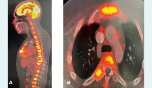
Figure 1: 18F-fluorodeoxyglucose (FDG) positron emission tomography (PET): A. Multiple FDG avid lytic lesions through spine, B. Sternal FDG enhancement.
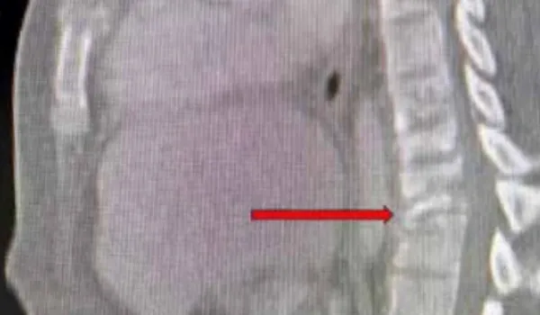
Figure 2: Compression fracture of vertebrae T10–T11 indicated by red arrow.
These findings were strongly suggestive of a primary lung malignancy with skeletal metastases. An oncology referral was made, and the provisional diagnosis was carcinoma of the lung with diffuse skeletal metastases and pathological vertebral fracture causing paraparesis.
To obtain histopathological confirmation, a CT/MRI-guided biopsy from one of the spinal lytic lesions and bone marrow aspiration and biopsy were planned.
Due to the patient’s severe osteoporosis and high fracture risk, spinal biopsy was deferred. Bone marrow examination was performed and returned normal, with no evidence of haematological malignancy or metastases.
Given the diagnostic uncertainty, further evaluation included:
- Cerebrospinal fluid (CSF) analysis via lumbar puncture, which showed:
- Normal glucose
- No cells or red blood cells (RBCs)
- Elevated protein (94 mg/dL) with albuminocytological dissociation, suggestive of possible arachnoiditis
- GeneXpert for Mycobacterium tuberculosis (MTB), adenosine deaminase (ADA), and acid-fast bacilli (AFB) stain: Negative
- CSF BioFire, bacterial/fungal cultures, and malignant cytology: All negative
- Contrast-enhanced MRI brain: Normal
- Workup for multiple myeloma (Serum protein electrophoresis [SPEP], serum free light chains, fluorescence in situ hybridisation [FISH] cytogenetics): Negative
- Serum calcium, vitamin D3, and parathyroid hormone (PTH) levels: Normal
- Extended infectious panel (sputum for AFB/GeneXpert, venereal disease research laboratory (VDRL), viral markers, urine Histoplasma antigen, serum galactomannan, and β-D-glucan): All negative
- Antinuclear antibody by immunofluorescence (ANA-IF): Negative
To obtain a histological diagnosis, a CT-guided sternal biopsy was pursued for its accessibility. Histopathology showed (Figure 3):
- Necrotising granulomatous inflammation
- No AFB
- No malignant ce
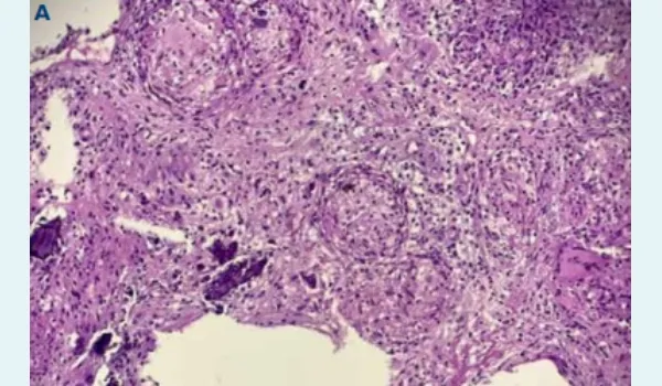
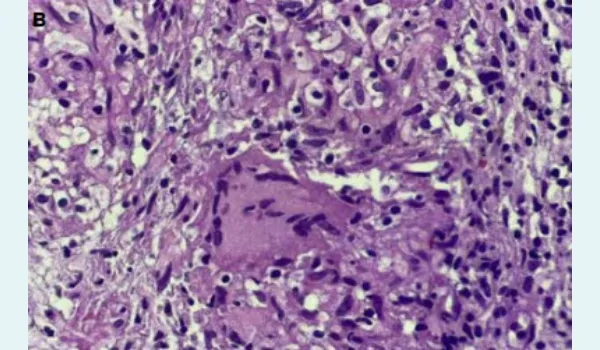
Figure 3: Necrotising granulomatous inflammation — haematoxylin and eosin stain: A. 10X, B. 40X.
Given the absence of malignant cells and the histological picture, empirical anti-TB therapy (ATT) was initiated at low doses while awaiting mycobacterial culture results. The patient was also managed conservatively for the vertebral compression fracture with spinal bracing and analgesics per neurosurgical advice.
After 34 days, culture of the sternal biopsy in BACTEC Mycobacteria growth indicator tube (MGIT) grew MTB complex, rifampicin-sensitive. By this time, the patient had already shown marked clinical improvement, including regained power in the lower limbs.
Final Diagnosis
- Pulmonary TB — Drug sensitive (presenting as a solitary pulmonary nodule)
- Pott’s spine — Multifocal skeletal TB (MSTB) with compression fracture at T10–T11 causing paraparesis
Treatment Initiated
The patient was started on standard first-line ATT comprising HRZE:
- H: Isoniazid 300 mg once daily (OD)
- R: Rifampicin 450 mg OD
- Z: Pyrazinamide 1000 mg OD
- E: Ethambutol 800 mg OD
Additionally, pyridoxine 20 mg OD was given for neuroprotection.
The planned treatment duration included an intensive phase of 2 months and a continuation phase of 10 months (with weight-based regimen modification).
Discussion
This case exemplifies the diagnostic complexity encountered when TB masquerades as metastatic malignancy, particularly in TB-endemic regions such as Southeast Asia.1 The constellation of findings — progressive neurological deficits, constitutional symptoms, and imaging showing widespread lytic skeletal lesions with a solitary pulmonary nodule — created a compelling picture strongly suggestive of malignancy, highlighting the challenges clinicians face in differentiating these conditions.
TB has long been recognised as a great mimicker of various diseases, particularly malignancy. In this case, several factors contributed to the initial diagnostic confusion. The patient's presentation with progressive bilateral lower limb weakness, significant weight loss (15 kg over 6 months), night sweats, and chronic cough, combined with imaging showing extensive lytic lesions, created a clinical picture highly suggestive of metastatic disease. The family history of lung adenocarcinoma in the patient's elder brother further reinforced clinical suspicion towards malignancy.
FDG PET–CT, while highly sensitive for detecting metabolically active lesions, has significant limitations in TB-endemic regions. Active TB granulomas demonstrate intense FDG uptake due to the high glucose metabolism of inflammatory cells, including macrophages, epithelioid cells, and lymphocytes surrounding the central necrotic nucleus. Studies have shown that TB causes false-positive results in 9 out of 11 staging cases and 12 out of 14 restaging cases when evaluated for malignancy.3 The maximum standardised uptake value (SUVmax) values often overlap significantly between TB and malignant lesions, with TB lesions commonly exceeding the traditional diagnostic threshold of 2.5.4 Some studies suggest that an SUVmax cut-off of 8.45 may be more appropriate for distinguishing TB from lung cancer in endemic areas.4
MSTB is an uncommon presentation, accounting for less than 5% of all bone TB cases.5 It typically occurs in immunocompromised individuals, making its occurrence in apparently immunocompetent patients, as in this case, particularly unusual.5 The condition can involve multiple non-contiguous vertebrae and other skeletal sites, making differentiation from skeletal metastases extremely challenging both clinically and radiologically. The rarity of this presentation contributes to diagnostic delays, as clinicians may not initially consider TB in the differential diagnosis of multifocal lytic lesions.
The histopathological finding of necrotising granulomatous inflammation without AFB initially presented a diagnostic challenge. However, studies have shown that the presence of necrotising granulomatous inflammation is highly suggestive of TB, with 85% of patients with necrotic granulomatous tissue having TB compared to only 14% with non-necrotic lesions.6 The absence of visible AFB does not exclude TB, as mycobacterial culture remains the gold standard for definitive diagnosis.6
The 34-day delay in culture confirmation underscores the challenges of rapid TB diagnosis. The use of BACTEC MGIT culture systems has improved detection rates and reduced diagnosis time compared to conventional methods. The eventual growth of rifampicin-sensitive MTB complex confirmed the diagnosis and guided appropriate therapy.
Myanmar ranks among the 30 countries with the highest TB burden globally and continues to face substantial barriers to effective TB control.7 High rates of untreated and drug-resistant TB present significant challenges to effective diagnosis. In such endemic settings, TB should always be considered in the differential diagnosis of destructive spinal processes, even when clinical and radiological features suggest malignancy
The patient's history of successfully treated pulmonary TB at 1.5 years of age raised questions about possible reactivation versus reinfection. While childhood TB treatment typically provides some immunity, reactivation or reinfection can occur, particularly in endemic areas with continued exposure risk.
Pott's disease accounts for approximately 50% of skeletal TB cases. The thoracic spine involvement, as seen in this patient with T10–T11 compression fracture, is particularly prone to neurological complications due to the narrow spinal canal and limited space for cord accommodation. The progressive paraparesis with areflexia in the lower limbs is consistent with the typical presentation of thoracic spine TB.
The decision to initiate empirical ATT based on necrotising granulomatous inflammation, despite negative microbiological studies, proved appropriate. The marked clinical improvement observed before microbiological confirmation supported the diagnosis. This approach is often necessary in TB-endemic regions where high clinical suspicion exists, particularly when tissue biopsy shows characteristic histopathological features.
The planned 12-month treatment duration (2 months intensive phase, 10 months continuation phase) follows standard recommendations for bone and joint TB, which typically requires longer treatment than pulmonary TB due to the avascular nature of bone tissue and slower drug penetration.
This case highlights that in regions where TB is endemic, the traditional clinical maxim 'when you hear hoofbeats, think horses, not zebras' may not always apply — TB often mimics other conditions. Continued clinical vigilance and comprehensive diagnostic evaluation are essential to ensure accurate diagnosis and optimal patient care.
Conclusion:
This case highlights the formidable challenge of distinguishing TB from metastatic malignancy, particularly in endemic regions where the disease often presents with atypical and deceptive features. MSTB with spinal involvement can closely mimic disseminated malignancy, both clinically and radiologically, leading to significant diagnostic uncertainty. Early recognition, careful integration of clinical, radiological and histopathological findings, and timely initiation of empirical therapy remain crucial for favourable outcomes. The patient’s clinical improvement prior to microbiological confirmation underscores the importance of maintaining a high index of suspicion for TB in similar presentations. Ultimately, this case reinforces the need for vigilance, multidisciplinary collaboration and context-specific diagnostic strategies to prevent misdiagnosis and ensure appropriate management.
Aryan Arora, Monica Mahajan, Pranjali Gupta, Tarini Madra. The Great Masquerader. MMJ. 2025,
September. Vol 2 (3).
References
- Tuli SM. Tuberculosis of the spine: a historical review. Clin Orthop. 2007;460:29–38.
- Gambhir S, Ravina M, Rangan K, et al. Imaging in extrapulmonary tuberculosis. Int J Infect Dis. 2017;56:237–47.
- Oh JK, Yoo IR, Oo JH, et al. F-18 FDG PET/CT findings of tuberculosis mimicking malignancy. J Nucl Med. 2011;52(supplement 1):1403–8.
- Lang S, Sun J, Wang X, et al. Asymptomatic pulmonary tuberculosis mimicking lung cancer on imaging: A retrospective study. Exp Ther Med. 2017;14(3):2180–8.
- Marudanayagam A, Gnanadoss JJ. Multifocal Skeletal Tuberculosis: A Report of Three Cases. Iowa Orthop J. 2006;26:151–3.
- Öztomurcuk D, Terzi Ö, Demirci C, et al. Investigation of Granulomatous Inflammations in Terms of Tuberculosis Diagnosis: A 5-Year Multi-center Laboratory Study. Turk Thorac J. 2022;23(1):11–6.
- Kaji A, Thi SS, Smith T, et al. Challenges in tackling tuberculosis on the Thai-Myanmar border: findings from a qualitative study with health professionals. BMC Health Serv Res. 2015;15:464.