 WhatsApp Us (24/7)
 +91 926 888 0303 (24/7)


Abstract: Small intestine transplantation remains a complex but definitive treatment for patients with irreversible intestinal failure, particularly in settings where parenteral nutrition (PN) services are limited. One of the critical challenges is achieving size compatibility between the donor organ and the recipient's abdominal domain. We describe a 50-year-old man, 160 cm in height and 65 kg in weight (body mass index [BMI] 25.3 kg/m2), who suffered from massive bowel gangrene due to thrombosis. He underwent resection of the entire small bowel 10 cm distal to the duodenojejunal (DJ) flexure. He had two episodes of central venous line-related sepsis, multiple hospital admissions, and lost 30 kg in 2 months. He underwent a novel technique of in-situ reduction of a deceased donor intestinal graft, based on the principles of living donor intestinal transplant (LDIT), wherein approximately 200 cm of the distal ileum was retrieved. The patient was extubated on the table and discharged with stable graft function. Regular follow-up for 3 months was uneventful. Recipients of other organs were stable at discharge. The success of this case highlights the technique’s feasibility, safety, and effectiveness. This innovative strategy could substantially expand the donor pool, particularly in resource-limited settings where paediatric or size-matched donors are scarce.
Key words: Deceased Donor Intestinal Transplant, Parenteral Nutrition, In-Situ Reduction, Small Bowel Gangrene, Donor–Recipient Size Mismatch.
Introduction
Patients suffering from progressive intestinal failure are at increased risk for significant complications associated with its management. These include intestinal failure or total parenteral nutrition (TPN)–associated liver disease, progressive loss of central vein access, and repeated life-threatening central venous catheter-associated infections requiring critical care.1 Overall, the number of patients undergoing small intestine transplantation has decreased in Western countries. This is multifactorial, including a better understanding of intestinal failure, post resection intestinal reconstruction procedures, newer intravenous lipid emulsions with improved tolerance to parenteral nutrition (PN), dedicated intestinal rehabilitation services, home TPN services, intestinal growth factors like teduglutide, and better central venous lines with antibacterial locks.2-4 In contrast, in South-East Asian countries, including India — where TPN services are not readily accessible — patients with intestinal failure have a dismal prognosis without intestinal transplantation. However, donor–recipient size discrepancies have limited the available pool of ideal donor organs, with the acceptable donor–recipient body weight ratio ranging from 0.76–1.1. This is especially true for smaller patients.5 Additional factors compounding this size discrepancy include the loss of abdominal domain, often resulting from multiple surgeries, meshes, or enterocutaneous fistulas that frequently occur in these patients.6 Attempts to use relatively larger grafts in recipients with a small abdominal domain can result in the development of compartment syndrome and vascular complications, including venous outflow obstruction. Strategies to overcome this issue include mesh or abdominal wall grafts to increase domain. The living donor intestinal transplant (LDIT) technique was described by Beneditti et al.7 However, in-situ reduction of the small intestine during deceased donor procurement and transplantation has not been described previously. We report the first case utilising this technique and describe the methodology of procurement and recipient outcomes herein.
Case Report
A 50-year-old patient, 160 cm in height and 65 kg in weight (body mass index [BMI] 25.3 kg/m2), suffered from massive bowel gangrene due to superior mesenteric artery (SMA) thrombosis. He underwent emergency exploratory laparotomy, where the entire small bowel 10 cm distal to the duodenojejunal (DJ) flexure up to the proximal ascending colon, was resected. The proximal jejunum was brought out as an end jejunostomy, and the ascending colon was brought out as a distal mucous fistula at another hospital. His hospital stay was prolonged, complicated by two episodes of central venous linerelated sepsis, after which a chemoport was inserted in the right internal jugular vein for administering TPN. He was discharged after 46 days of hospital admission, maintained on an alimental diet and PN. He required multiple hospital readmissions due to dehydration, and he lost 30 kg within 2 months. He was referred to our centre for small intestine transplantation. He was evaluated and listed for a deceased donor small intestine transplantation. Within 2 months of listing, he received 3 organ offers, but all of them had to be declined owing to his small abdominal size and weight. At this stage, his weight had decreased to 35 kg (BMI 13.67 kg/m2), and the abdomen appeared smaller compared to the initial admission. Finding an appropriately sized donor seemed challenging as most Indian adult donors on average weigh between 60–85 kgs. It was therefore decided to proceed with a reduced graft for the patient rather than waiting for an appropriate size-matched donor. Due to the unit's experience in performing living donor intestinal transplants, the retrieval was planned in the same manner as is done in case of a living donor, wherein approximately 200 cm of the distal ileum is retrieved. The patient received an offer from an 18-year-old female donor weighing 85 kg.
Donor surgery
The bowel was inspected and found to be appropriate for retrieval. A Cattell–Braasch manoeuvre was performed. The root of the mesentery was looped by creating a window on either side. The superior mesenteric vein (SMV) and the SMA were dissected and looped (Figure 1).
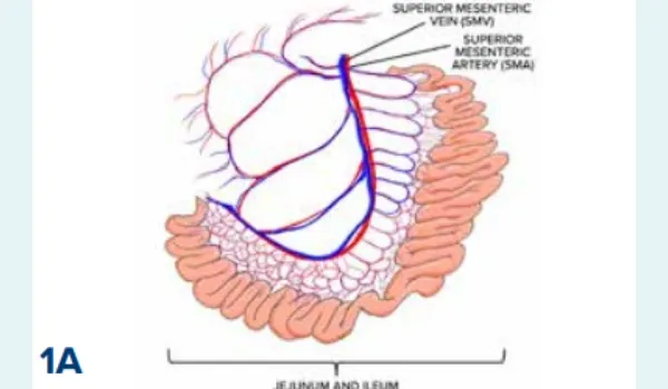
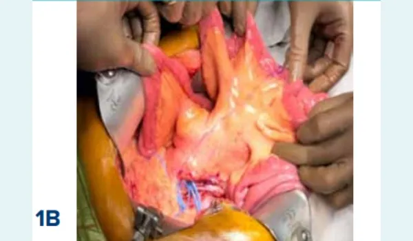
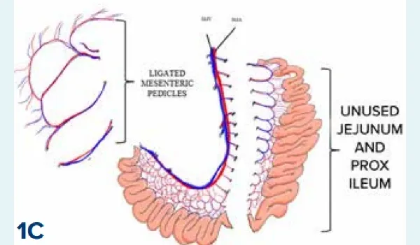
Figure 1 A–C: Deceased donor intestinal graft before reduction.
Abbreviations: SMA: Superior Mesenteric Artery; SMV: Superior Mesenteric Vein.
Two hundred centimetres of terminal ileum were measured, and the mesentery was divided carefully by ligating the branches of the vessels not supplying the graft on either side, with the apex at the looped mesenteric vessels (Figure 2).
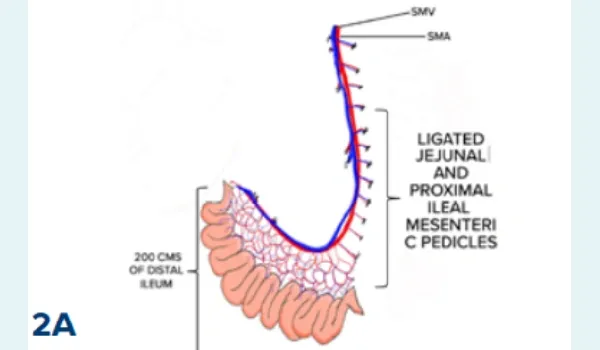
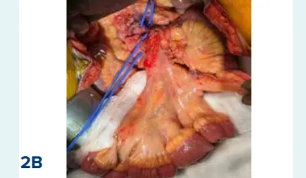
Figure 2 A and B: In-situ reduced deceased donor intestinal graft after ligating the mesenteric vessels.
Abbreviations: SMA: Superior Mesenteric Artery; SMV: Superior Mesenteric Vein.
The lymphatics of the ileal mesentery were preserved to ensure maintenance of the lymphatic drainage. The proximal end of the bowel was marked using a Prolene stitch for later identification at the bench. The distal SMA and SMV were clamped and divided prior to cross-clamp to retrieve the graft, which was perfused immediately with cold University of Wisconsin (UW) solution on the back table, as is done in the case of a living donor intestine graft. Retrieval of other organs, including heart, lungs, liver, and kidneys, proceeded as planned without affecting intestinal procurement. The operative duration for intestinal retrieval was 90 minutes. The donor tolerated the procedure well, with a pulse rate of 86/min, blood pressure of 130/80 mmHg, maintained on nor-adrenaline infusion at 0.08 mcg/kg/min, and arterial blood gas analysis showing lactate of 2 mmol/L. The graft was packed in a sterile container and shifted to the base hospital (Figure 3).
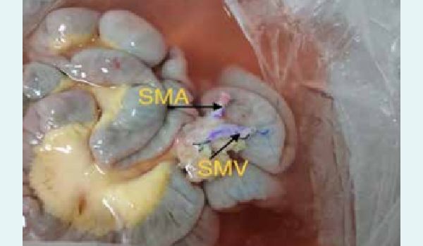
Figure 3: Benching: Superior mesenteric artery (SMA) and superior mesenteric vein (SMV) are marked for orientation.
Recipient surgery
In the recipient, the abdomen was explored. The infrarenal aorta and inferior vena cava (IVC) were exposed (Figure 4).
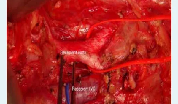
Figure 4: Recipient bed where the aorta and inferior vena cava (IVC) is looped Vascular conduits using the iliac artery and vein were used on the aorta and IVC. The graft was transplanted by anastomosing the SMA and SMV to the vascular conduits (Figure 5).
Figure 5: Implantation of graft superior mesenteric artery (SMA) and superior mesenteric vein (SMV) into recipient aorta and inferior vena cava, respectively.
The bowel was well perfused, and the proximal and distal bowel anastomoses were performed, and a loop ileostomy was created proximal to the distal anastomosis. The mesenteric edges were sutured retroperitoneally to secure the graft and prevent rotation of the vascular pedicle. As the lymphatic drainage of the graft was kept intact up to the edge of the graft mesentery, spontaneous collateral formation with native retroperitoneal lymphatics was expected post-transplantation. Abdominal closure was completed immediately, with peak ventilator pressures unchanged following closure. The patient was extubated and shifted to the intensive care unit (ICU).
Postoperative course
Standard triple immunosuppression comprising tacrolimus, mycophenolate, and steroids was administered. The stoma began functioning on postoperative day (POD) 3, after which oral feeds were introduced progressively as per tolerance. The post-operative course was complicated by an anastomotic bleed on POD 7, due to dual anticoagulation (low molecular weight heparin [LMWH] and Ecosprin), which resolved by conservative management. Weekly protocol biopsies showed no rejection. Complete enteral autonomy was achieved on POD 18, after which the patient was discharged with stable graft function. Regular follow-up for 3 months was uneventful. The patient continues to be at home with stable graft function. Recipients of other organs were also stable at discharge.
Discussion
Small intestine transplantation remains a complex but definitive treatment for patients with irreversible intestinal failure, particularly in settings where PN services are limited or ineffective. One of the most critical technical challenges in this procedure is achieving size compatibility between the donor organ and the recipient's abdominal domain. This becomes particularly relevant in smaller adult or paediatric recipients, where transplantation of a full-sized intestinal graft from a standard adult donor can result in abdominal compartment syndrome, compromised graft perfusion, and even loss of the graft.
Traditionally, to address size mismatch, options include the use of prosthetic meshes for abdominal wall closure or vascularised composite grafts such as the rectus abdominis muscle. While these techniques can enable closure, they carry significant risks, including mesh infection, hernia formation, increased operative time, and potential for additional graft-related complications such as rejection of the vascularised flap.8,9
An alternative and increasingly attractive approach is graft size reduction. Prior studies, including those by Heaton et al., have described ex-situ reduction methods, where the small bowel is tailored during the back-table preparation by resecting a jejunal segment.10 However, ex-situ techniques may carry risks of inadvertent vascular or lymphatic injury, increased cold ischaemia time, and unanticipated bleeding during reperfusion.
In this case, we describe a different in-situ reduction technique, modeled on principles of living donor intestinal transplantation, as described by Beneditti et al.7 The familiarity of the transplant team with living donor procedures allowed for precise vascular ligation under direct vision in a warm setting, ensuring preservation of critical vasculature and lymphatic drainage. Key advantages of this method include:
- Controlled reduction of the graft tailored to the recipient’s abdominal capacity
- Early procurement of the intestinal graft without interfering with the retrieval of other abdominal organs
- Reduction of back-table benching time, thereby shortening cold ischaemia and improving graft perfusion post-revascularisation
- Preservation of mesenteric lymphatics, facilitating spontaneous lymphatic drainage and reducing the risk of postoperative chyle leak
The patient had already lost substantial body mass and experienced repeated sepsis and central venous access complications. The in-situ reduction approach provided a critical solution in an otherwise high-risk scenario, permitting primary abdominal closure and excellent graft function. The patient achieved full enteral autonomy by POD 18 and has maintained stable graft function without signs of rejection.
Conclusion:
To the best of our knowledge, this is the first reported case in the literature of an in-situ reduction of a deceased donor small intestinal graft. The success of this case highlights the technique’s feasibility, safety, and effectiveness. This innovative strategy could substantially expand the donor pool by making larger adult donor grafts usable for smaller recipients, particularly in resource-limited settings like India, where paediatric or size-matched donors are scarce. Additionally, this case demonstrates that the technique can be done without a significant impact on the procurement of other organs.
Gaurav Chaubal, Hunaid Hatimi, Aditya Nanavati, Amith Pakkala, Deepti Ramachandra, Amith Sreekanth
Harshit Chakshota, Premal Narkhede, Blossom D’souza, Pallavi R Kumar, Samir Shah, Uday Sanglodkar,
Alisha Chaubal, Hardik Shah, Tejas Joshi, Abdul S Ansari, Tejaswini Ranade, Radhika Ruhatiya, Prateek
Gupta, Saranya Narayanan, Uttkarsha Nikale, Wasim Khot. Novel Technique of In-Situ Reduction of a
Deceased Donor Small Intestine Graft to Improve Access in Donor–Recipient Mismatch. MMJ. 2025,
September. Vol 2 (3).
References
- Kaufman SS, Atkinson JB, Bianchi A, et al. Indications for pediatric intestinal transplantation: a position paper of the American Society of Transplantation. Pediatr Transplant. 2001;5(2):80–7.
- Horslen SP, Ahn YS, Wood NL, et al. OPTN/SRTR 2022 Annual Data Report: Intestine. Am J Transplant. 2024;24(S2S1):S266–S304.
- Pironi L, Agostini F, Guidetti M. Intravenous lipids in home parenteral nutrition. World Rev Nutr Diet. 2015;112:141–9.
- Pironi L, Goulet O, Buchman A, et al. Outcome on home parenteral nutrition for benign intestinal failure: a review of the literature and benchmarking with the European prospective survey of ESPEN. Clin Nutr. 2012;31(6):831–45.
- Fishbein TM, Bodian CA, Miller CM. National sharing of cadaveric isolated intestinal allografts for human transplantation: a feasibility study. Transplantation. 2000;69(5):859–63.
- Carlsen BT, Farmer DG, Busuttil RW, et al. Incidence and management of abdominal wall defects after intestinal and multivisceral transplantation. Plast Reconstr Surg. 2007;119(4):1247–55.
- Testa G, Panaro F, Schena S, et al. Living related small bowel transplantation: donor surgical technique. Ann Surg. 2004;240(5):779–84.
- Cipriani R, Contedini F, Santoli M, et al. Abdominal wall transplantation with microsurgical technique. Am J Transplant. 2007;7(5):1304–7.
- Grevious MA, Iqbal R, Raofi V, et al. Staged approach for abdominal wound closure following combined liver and intestinal transplantation from living donors in pediatric patients. Pediatr Transplant. 2009;13(2):177–81.
- Delrivière L, Muisan P, Marshall M, et al. Size reduction of small bowels from adult cadaveric donors to alleviate the scarcity of pediatric size-matched organs: an anatomical and feasibility study. Transplantation. 2000;69(7):1392–6.