 WhatsApp Us (24/7)
 +91 926 888 0303 (24/7)


Abstract: We present a case of a 56-year-old man with progressive dyspnoea and dry cough for over one month, initially treated as community-acquired pneumonia without improvement. Despite antibiotics and steroids, his respiratory function worsened, accompanied by haemoptysis and escalating oxygen needs, necessitating intubation. Laboratory evaluation revealed elevated C-reactive protein (CRP), negative infectious disease panel, a positive myositis panel (anti-PL-12), and SSA/Ro52 antibodies. Chest computed tomography (CT) showed extensive subpleural ground-glass opacities consistent with rapidly progressive interstitial lung disease (ILD). Given the diagnosis of anti-synthetase syndrome (ASS), high-dose pulse methylprednisolone (500 mg intravenous [IV] for 3 days) and rituximab (1 g IV, two doses) were instituted. The patient exhibited marked clinical and radiologic improvement, achieving extubation on Day 10. Post-therapy imaging demonstrated near resolution of ground-glass opacities, leaving reticulations indicative of chronic fibrosis.
This case underscores the significance of early recognition and aggressive immunomodulation in ASSassociated ILD. High-dose steroids and rituximab may offer critical benefits in refractory, rapidly progressive lung involvement. A multidisciplinary approach remains essential, and further research is needed to establish standardised treatment protocols.
Key words: Anti-Synthetase Syndrome, Interstitial Lung Disease (ILD), Rapidly Progressive ILD, Anti-PL-12 Antibody, Rituximab Therapy
Introduction
Diffuse parenchymal lung disease (DPLD), also known as interstitial lung disease (ILD), is classified on the basis of shared clinical, radiographic, physiological, or pathological features. The complexity in comprehending this category of diseases arises from the intricate terminology used. In reality, the pathogenic progression entails a sequence of inflammation and fibrosis that extends beyond merely affecting the interstitial bed, as suggested by the name ILD. In DPLD, the pathology alters the lung parenchyma, encompassing the alveoli, alveolar ducts, and bronchioles.1
One condition in which ILD occurs is anti-synthetase syndrome (ASS). It is an autoimmune disease characterised by antibodies directed against an aminoacyl transfer t-RNA synthetase, along with clinical features that can include ILD, myositis, Raynaud’s phenomenon, and arthritis.2 ASS is categorised as an idiopathic inflammatory myopathy (IIM), and is more likely to be associated with ILD than other IIMs, such as dermatomyositis (DM) and polymyositis (PM), with which it shares similar features. The ILD observed in individuals with ASS is frequently intense and progresses rapidly, thus contributing significantly to the elevated morbidity and mortality linked to this syndrome.3
Case Report
A 56-year-old male presented to the emergency department with complaints of shortness of breath and dry cough for one month. The patient had no history of fever, weight loss, or haemoptysis, and was admitted to the ward. He denied any other symptoms, and reported no relevant family history. The patient had previously been treated elsewhere as a case of community-acquired pneumonia based on clinical and radiological findings (Figure 1A).
On examination, the patient was dyspnoeic; the heart rate was 92/min, blood pressure was 110/70 mmHg, and room-air oxygen saturation was 90%, with bilateral crepitations on auscultation. He was initially managed with intravenous (IV) antibiotics, oxygen supplementation, and other supportive measures.
IDespite treatment, his respiratory distress worsened, with new onset haemoptysis and increasing oxygen requirements. The patient was intubated and started on invasive mechanical ventilation. Investigations were sent (enlisted in Tables 1 and 2), and a repeat computed tomography (CT) scan of the chest was performed (Figure 1B) on Day 5 that showed increase in ground-glass opacities indicating worsening of condition.
Upon investigation, the patient tested positive for elevated C-reative protein (CRP), myositis panel (anti-PL-12), and SSA/Ro52 antibodies, and therefore met the diagnostic criteria (Table 3) for ASS. As the patient was not responding to the initial line of treatment involving antibiotics and steroids, on Day 5 he was subsequently treated with IV methylprednisolone 500 mg/day for three doses and 1 gm rituximab for two doses, two weeks apart. Following this, the patient showed significant improvement and was extubated on Day 10. A CT scan was repeated post extubation to assess for sequelae (Figure 1C).
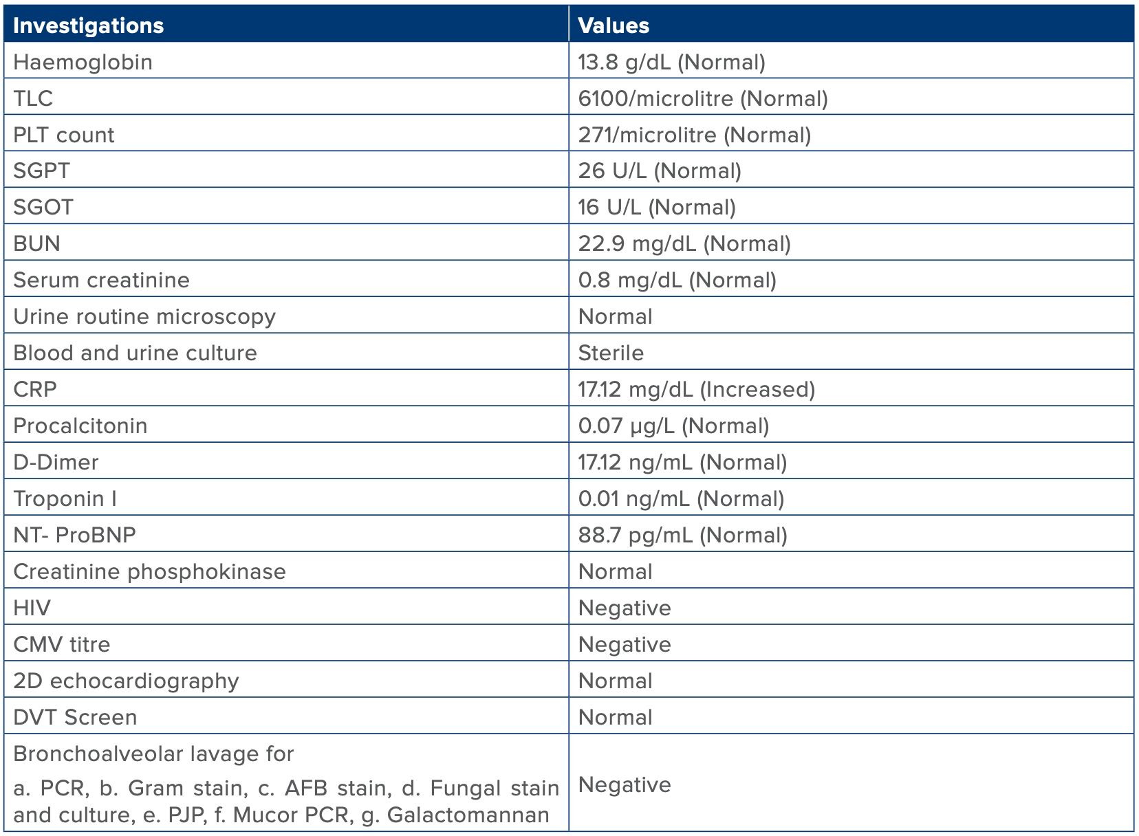
Table 1: Blood and basic investigation.
Abbreviations: AFB Stain: Acid-Fast Bacilli Stain; BUN: Blood Urea Nitrogen; CMV Titre: Cytomegalovirus Antibody Titre; CRP: C-Reactive Protein; DVT: Deep Vein Thrombosis; HIV: Human Immunodeficiency Virus; NT-proBNP: N-terminal pro–B-type natriuretic peptide; PCR: Polymerase Chain Reaction; PJP: Pneumocystis jirovecii pneumonia. PLT: Platelet; SGOT: Serum Glutamic-Oxaloacetic Transaminase; SGPT: Serum Glutamic Pyruvic Transaminase; TLC: Total Leucocyte Count.
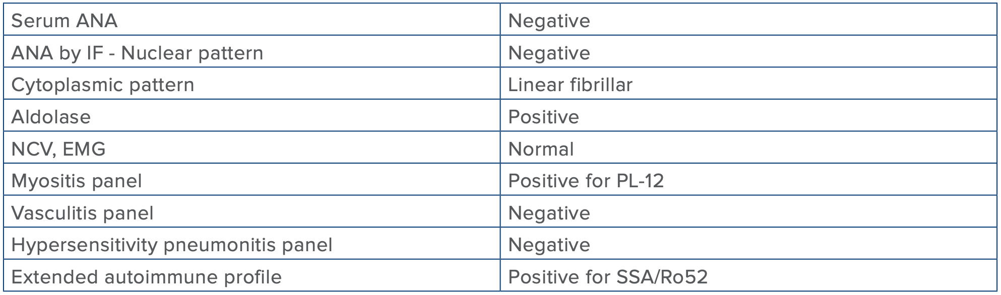
Table 2: Specific investigations done for the patient.
Abbreviations: ANA: Anti-Nuclear Antibody; EMG: Electromyography; IF: Indirect Immunofluorescence; NCV: Nerve Conduction Velocity.
Solomon et al. Diagnostic Criteria4
Definitive: the presence of ARS autoantibody
Plus, two major or one major and two minor criteria
- Major:
- Interstitial lung disease
- Dermatomyositis or polymyositis by Bohan and Peter criteria
- Minor:
- Arthritis
- Fever
- Mechanic’s hands
Connors et al. Diagnostic Criteria2
Definitive: the presence of ARS autoantibody
Plus, one or more of the following:
- Interstitial lung disease
- Arthritis
- Raynaud’s phenomenon
- Fever
- Mechanic’s hands
Abbreviations: ARS: Aminoacyl t-RNA Synthetase. Conduction Velocity.
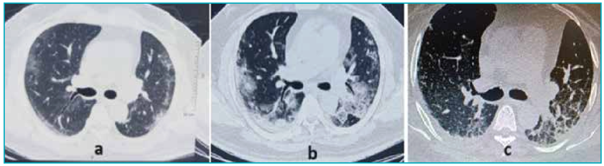
Figure 1: Computed tomography (CT) of the chest: A. A small focal area of patchy ground-glass opacity in the subpleural area of the lateral segment of the right middle lobe. Curvilinear ground-glass opacities with stranding in subpleural areas of the superior segment of bilateral lower lobes, B. Day 5: In comparison to prior high-resolution computed tomography (HRCT), an increase in size and attenuation of the ground-glass opacities in the subpleural areas of the superior segments of bilateral lower lobes. Focal ground-glass opacity in the lateral segment of the right middle lobe also shows an increase in attenuation, C. Day 10: Scan shows near complete resolution of the ground-glass opacities in both lower lobes with increased reticulations and strandings representing chronic fibrotic changes post pulse therapy
Discussion
ASS is a rare, multisystemic autoimmune disease with variable symptoms ranging from myositis, ILD, and non-erosive arthritis. The diagnosis of ASS can be facilitated by employing the precise criteria outlined by Solomon et al., and Connors et al. (Table 3).2,4
Six anti-aminoacyl transfer RNA (t-RNA) synthetase autoantibodies have been associated with ASS: anti-histidyl-tRNA synthetase (anti-Jo-1), anti-isoleucyl-tRNA (anti-OJ), anti-threonyl-tRNA (anti-PL7), anti-alanyl-tRNA (anti-PL12), anti-glycyl-tRNA (anti-EJ), and anti-asparaginyl-tRNA (anti-KS). Amongst these, the most common and well-recognised antibody is anti-Jo-1, detected in approximately 20%–30% of such patients.5
Essential components of the diagnostic process include high-resolution computed tomography (HRCT) imaging, which may show nodules, micronodules, linear opacities, irregular interfaces, ground-glass opacities, fibrosis with or without honeycombing, consolidation, traction bronchiectasis, and bronchiectasis.6 Among the observed patterns, nonspecific interstitial pneumonitis (NSIP) is the most common, followed by organising pneumonia.7 Bronchoscopic or muscle biopsy may seldom be required; however, if the patient’s condition permits, a bronchoscopic lavage prior to initiating immunosuppression may be performed to rule out secondary infection.
Currently, there are no established treatment protocols or guidelines for individuals affected by this condition. The first-line of treatment is corticosteroids. Other immunosuppressants such as azathioprine, mycophenolate mofetil, tacrolimus, rituximab, and cyclophosphamide may be used in conjunction with steroids as augmentative therapy for pulmonary and muscular movement. Due to the rarity of the disease, no large-scale treatment trials have been conducted. The available medical literature primarily consists of individual case reports and small case series, with no head-to-head trials. Some reports comparing cyclophosphamide with rituximab have found rituximab to be associated with better progression-free survival at two years, leading some authors to consider it a firstline option.8,9 The use of pulse steroids as a last resort in refractory autoimmune conditions is well-established and may be a viable option in this setting, even though the evidence for its use in ASS remains scarce.10
Contribution
All the authors were involved in the literature search, planning, conducting, writing the original manuscript draft, and editing. All the authors have agreed with the submitted manuscript. Dr. Nikhil Raj is the corresponding author and guarantor for all.
Acknowledgments
The authors would like to thank the residents and interventional pulmonology staff for their support in patient care and data collection.
Conflicts of interest
The authors declare no conflicts of interest regarding the current study.
Funding
The current research received no external funding.
Availability of data and material
The clinical data and the study materials are available from the corresponding author upon reasonable request.
Patient consent
Written informed consent has been obtained from the patient or their legally authorised representative.
CONCLUSION:
High doses of immune suppression remain the cornerstone therapy for refractory ASS, and insufficient recognition of ASS among primary care physicians leads to delayed referrals to specialists, contributing to the advancement of the illness. Larger studies are needed to standardise therapy for ASS.
Vivek Nangia, Nikhil Raj, Navin Dalal, Anshul Mittal, Amina Mobashir, Manu Madan, Parth Rajdev,
Swapnil Pathak. A Rare Case of Rapidly Progressive Diffuse Parenchymal Lung Disease. MMJ. 2025, September. Vol 2 (3).
References
- Antoine MH, Mlika M. Interstitial Lung Disease. [2023]. In: StatPearls [Internet]. Treasure Island (FL): StatPearls Publishing; 2024. Available from: https://www.ncbi.nlm.nih.gov/books/ NBK541084/.
- Connors GR, Christopher-Stine L, Oddis CV, et al. Interstitial lung disease associated with the idiopathic inflammatory myopathies: what progress has been made in the past 35 years? Chest. 2010;138(6):1464–74.
- Kalluri M, Oddis CV. Pulmonary manifestations of the idiopathic inflammatory myopathies. Clin Chest Med. 2010;31(3):501–12.
- Solomon J, Swigris JJ, Brown KK. Myositis-related interstitial lung disease and antisynthetase syndrome. J Bras Pneumol. 2011;37:100–9.
- Rasendrakumar A, Khanna A, Bakhai S. Early Recognition and Successful Treatment of Anti-synthetase Syndrome. Cureus. 2022;14(1):e21786.
- Wells M, Alawi S, Thin KYM, et al. A multidisciplinary approach to the diagnosis of antisynthetase syndrome. Front. Med. 2022;9:959653.
- Huang K, Aggarwal R. Antisynthetase syndrome: A distinct disease spectrum. J Scleroderma Relat Disord. 2020;5(3):178– 91.
- Langlois V, Gillibert A, Uzunhan Y, et al. Rituximab and Cyclophosphamide in Antisynthetase Syndrome-related Interstitial Lung Disease: An Observational Retrospective Study. JRheum Supplements. 2020;47(11):1678–86.
- Zekić T. Rituximab as the first-line therapy in anti-synthetase syndrome-related interstitial lung disease. Rheumatol Int. 2023;43(6):1015–21.
- Mortel MR, Ochieng P, Salvatore M, et al. Pulse steroids for refractory Antisynthetase Syndrome: A booster therapy? Chest. 2015;148(4):408A.