 WhatsApp Us (24/7)
 +91 926 888 0303 (24/7)


Introduction
Foetus papyraceus is a rare complication of twin pregnancy where one twin dies in the early second trimester, and the dead foetus is retained for at least 10 weeks. During this time, the deceased foetus becomes compressed between the uterine wall and the membrane of the surviving twin. The absorption of amniotic fluid leads to a mummified, parchment-like appearance of the retained foetus.1 An intrauterine diagnosis of foetus papyraceus is warranted because it can lead to morbidity and mortality of both the surviving foetus and the mother.
Case report
A 29-year-old homemaker was booked and supervised in our hospital from the beginning of her in vitro fertilizationconceived dichorionic diamniotic (DCDA) twin pregnancy. A single foetus demise occurred at 22 weeks, but the healthy twin continued to grow normally. Gestational diabetes was detected during antenatal screening, and was managed with insulin, lifestyle measures, and close foeto-maternal surveillance. She was admitted at 36 weeks and 2 days for lower segment caesarean section (LSCS), and a live baby was born. Foetus papyraceous was also delivered and shown to the family (Figure 1). The postoperative period was uneventful. The patient was discharged in good condition with appropriate advice.
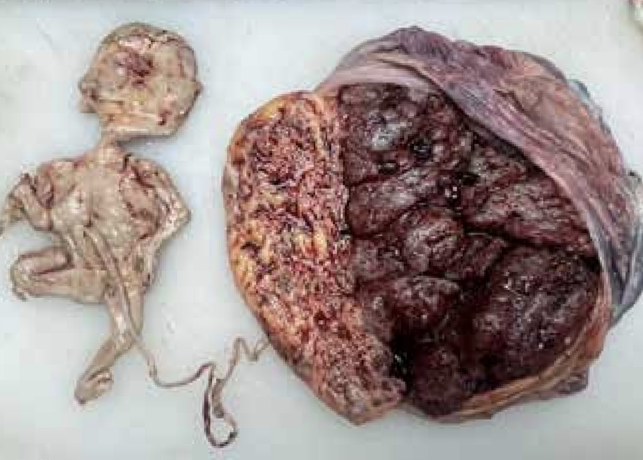
Figure 1: Foetus papyraceous with thin umbilical cord, and placenta. Healthy placenta and a healthy umbilical cord (left side)
Foetus papyraceous, also known as mummified foetus or foetus compresses, or vanishing twin refers to a deceased foetus that becomes mummified, flattened, and compressed within the uterus after death. This condition results in the dry, parchment-like remains of the dead foetus without signs of maceration. The foetus is compressed between membranes and the uterine wall, leading to the absorption of the amniotic fluid and a desiccated, parchment-like appearance.2 It is a rare condition, with an incidence of 1 in 12,000 live births, mainly observed in twin pregnancies. The death of a twin generally occurs in the second trimester of pregnancy, with retention of the foetus for a minimum of 10 weeks.3 It can manifest in both monochorionic and dichorionic twin pregnancies, although, it is more common in monochorionic twin pregnancies and can be diagnosed early through imaging studies like ultrasonography during antenatal care visits.4 Its aetiology remains unknown, although it is associated with twin-to-twin transfusion syndrome, genetic abnormalities, and improper cord implantation, such as velamentous cord insertion.
Foetus papyraceous is associated with an increased risk of morbidity and mortality for both the surviving twin and the mother. Perinatal mortality in monochorionic twin pregnancies is three times more common than in dichorionic twin pregnancies due to unequal sharing of the placenta and the presence of vascular connections. No association with parity or maternal age has been identified. The surviving infant may develop complications due to thrombi and other factors released from the dead foetus, leading to intrauterine growth retardation, congenital malformations, and prematurity. Severe hypoxia and hypotension in the surviving foetus can lead to hypoxic-ischaemic encephalopathy, also known as multicystic encephalomalacia. This condition can occur in 20% of surviving twins. Similarly, there is an increased incidence of cerebral impairment. Twin embolization syndrome is another serious complication that occurs in 25% of surviving twins after the demise of one twin.
Maternal complications may include preterm labour, consumptive coagulopathy, labour dystocia, and sepsis as a result of the retention of a dead foetus.
Our patient’s diagnosis of dichorionic twin pregnancy with foetus papyraceous during the second trimester was facilitated by excellent diagnostic facilities. The mother was closely monitored for consumption coagulopathy and maternal infections, with haematological, biochemical, and coagulation parameters, along with serial ultrasound examinations during the entire antenatal period as well as after delivery Both the mother and the surviving twin were healthy at six months of follow-up.
Source(s) of support: Nil
References
- Awoyesuke PA. Fetal Papyraceus Discovered at Second stage of Labour in an Unbooked patient: A Case Report. As J Case Rep Med Heal 2019;2(2):1-5.
- Singh A. Foetus Papyraceus. In Konar H, editor. D.C. Dutta’s Textbook of Gynecology. 10th ed. New Delhi: Jaypee Brothers Medical Publishers; 2023. P123-25.
- Wijeratne D, Fiander A. Gynaecological disease in the developing world: a silent pandemic. Obstet Gynecol. 2018;20(4):237-44.
- Álvarez-Acuña AM, López-Álvarez LM, Lozada-Martínez ID, et al. Intraoperative finding of papyraceous twin etus in pregnant woman without specialized follow-up: A summary of difficulties in accessing prenatal services in low-and middle-income countries. International Journal of Surgery Case Reports. 2022;95:107234.
- Dickey RP, Taylor S, Lu PY, et al. Spontaneous reduction of multiple pregnancy: incidence and effect on outcome. Am J Obstet Gynecol. 2002;186(1):77-83.
- Vashistha P, Kashyap P, Kumar A, et al. Fetus papyraceous, a rare complication of twin pregnancy: case report. Int J Reprod Contracept Obstet Gynecol. 2021;10:361-3.
- Upreti P, Malik N, Kumar R, et al. Fetus Papyraceous in Diamniotic Dichorionic Twins: A Rare Entity. J South Asian Feder Obst Gynae. 2023;15(3):347-48.