 WhatsApp Us (24/7)
 +91 926 888 0303 (24/7)


Abstract: Radiotherapy, with its colloquial use, is widely known to be painful and to cause damage to the skin by burning or scarring. This has led to fear, anxiety and aversion among many patients to proceed with radiation many times.
Even doctors and oncologists from other specialities sometimes fear the side effects of radiation and do not want to subject their patients to the perils of the treatment and hence, put the patients on some other form of treatment that might be less effective or more expensive. Radiation, however, is a very effective treatment for various cancers with a very tolerable toxicity profile when done at an experienced centre under the supervision of an experienced and properly trained radiation oncologist. In this modern era when everything is technologydriven, radiotherapy also has improved by leaps and bounds with modern linear accelerators having utmost precision being widely available for treatment. Radiotherapy plays a crucial role in the treatment of various head and neck malignancies, brain tumours, gynaecological malignancies, lung cancer, breast cancer, palliation of skeletal and brain metastases and many more. Apart from achieving palliation, radiation can also decrease the disease burden or at least stabilize it. Achieving adequate local control is also important on many occasions to provide a better quality of life. It is important to understand that even for widespread metastatic disease, the end of life should be respectful, and the way of dying should also be socially and emotionally acceptable both to patients and caregivers. For example, controlling raised intracranial pressure or symptoms arising out of brain metastases should be important in terms of providing a better quality of life.
Here, we discuss one such instance where radiation was unnecessarily denied on the pretext of side effects
Key words: Brain metastases, Brain radiation, TKIs, Local control, Quality of life
Introduction
Yes, radiation can be a double-edged sword if used inappropriately and at a less trained centre. Diligent planning and adopting effective methods for reproducibility in the presence of the latest techniques of delivering radiation like Intensity Modulated Radiation Therapy (IMRT), Image Guided Radiation Therapy (IGRT), Volumetric Modulated Arc Therapy (VMAT), and helical tomotherapy has made it possible to implement radiation treatment with highest accuracy in targeting the tumour and simultaneously protecting the critical structures therefore, tightly controlling the toxicities.
Case discussion
A 38-year-old lady, an engineer by profession, and a known case of carcinoma lung with distant metastases including multiple brain metastases, presented with significant stuttering causing interference in her social life and causing great mental agony. She also had severe headache, heaviness in head, vertigo and ataxic gait. The magnetic resonance imaging (MRI) scan showed multiple lesions scattered throughout the brain in bilateral cerebral hemispheres, right cerebellar hemisphere, midbrain, and right basal ganglia. The largest lesion was of 3.1cm in its greatest dimension in the left temporal region with significant perilesional oedema (Figures 1 & 2).
Her symptoms had been persistent for several months and other than offering a surgical removal of one of the cerebellar lesions, no other local treatment was offered to her. She was put on a new generation tyrosine kinase inhibitor (TKI) to avoid radiation-associated side effects. However, there was no improvement in her central nervous system (CNS) symptoms.
She was subsequently planned for whole brain radiation to a dose of 30Gy in 10 fractions along with simultaneous integrated boost (SIB) to larger lesions to a dose of 40Gy in 10 fractions. Radiation was planned with hippocampus sparing using helical tomotherapy under daily image guidance with the necessary protocols being followed.
Response evaluation contrast MRI of brain was done after one month which showed significant reduction in all the brain lesions, complete resolution of the lesion in right basal ganglia and the largest lesion in left temporal area decreased to 1.8cm in its largest dimension. Patient experienced dramatic resolution of CNS symptoms after radiation (Figure 3) including significant improvement of stuttering.
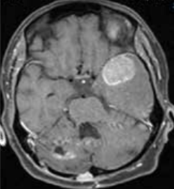
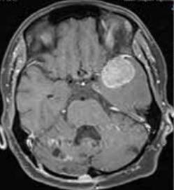
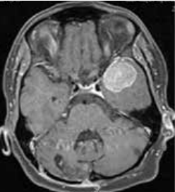
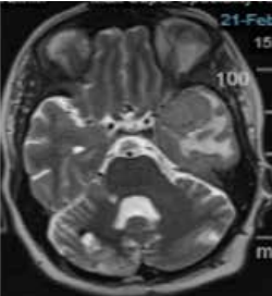
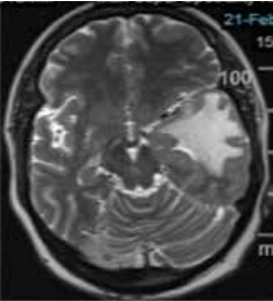
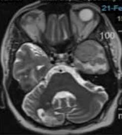
Figure 1: Metastatic lesions at presentation
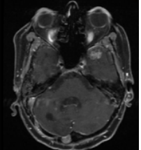
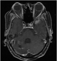
Figure 2: Left temporal lesion and perilesional oedema
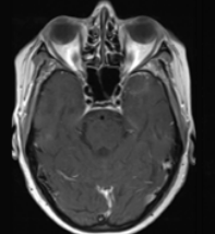
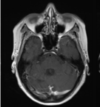
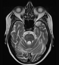
Figure 3: Contrast-enhanced Magnetic Resonance Imaging (CE MRI) brain 1-month post-radiation
Patient continued her follow up and a scan at 4 months showed further reduction of all brain lesions without any evidence of intracranial progression. Unfortunately, she had intrapulmonary progression and was started on next line of systemic therapy. Patient did not complain of any loss of memory or neurocognition until 10-months post-radiation, the biggest fear of whole-brain radiation.
She continued systemic therapy for disease progression at the primary site and subsequent MRI more than 1 year after treatment showed further decrease in size of the temporal lesion with excellent intracranial control (Figure 4).
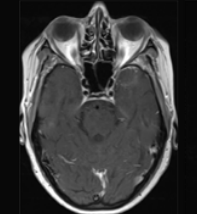
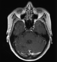
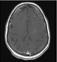
Figure 4: CE MRI Brain 1-year post-radiation
Discussion
Such tremendous response cannot be undermined, and radiation should be considered as an important mode of treatment in acquiring disease and symptom control in case of brain metastases despite having various new targeted drugs and immunotherapy in the management of metastatic stage IV lung cancer, breast cancer or melanoma to name a few among many other malignancies.1
Many of the systemic agents have very poor CNS penetration and yet are offered as the first line treatment in patients with symptomatic brain metastases where, treating with radiation could benefit the patients to a larger extent. As in this current patient, she was not even offered radiation for a long time and was put on a TKI which failed to show any benefit1. Whole brain radiation with hippocampus sparing can be effective in such scenarios.
Disclosure
None
Conflict of interest
None
CONCLUSION:
This case highlights the benefit of the hippocampus sparing whole brain radiation. In recent years, technology has made it possible to reduce the dose to the hippocampus to a great extent which helps in preserving neurocognition. It becomes important to select proper cases as hippocampus-sparing radiation is not recommended for brain metastases present within 5mm of the hippocampus.2 Adding memantine during and after whole brain radiation can also help in preserving neurocognition.3 The decision of avoiding brain radiation must be discussed with the radiation oncologists before denying it in pursuit of preserving neurocognition
References
- Mantovani C, Gastino A, Cerrato M, et al. Modern radiotherapy for management of brain metastases from non-small cell lung cancer: Current Approaches and future directions; Front Oncol. 2021;11:772789.
- Gondi V, Tolakanahalli R, Mehta MP, et al. Hippocampal-sparing whole-brain radiotherapy: a "how-to" technique using helical tomotherapy and linear accelerator-based intensity-modulated radiotherapy. Int J Radiat Oncol Biol Phys. 2010;78(4):1244-52.
- Brown PD, Pugh S, Laack NN, et al; Radiation Therapy Oncology Group (RTOG). Memantine for the prevention of cognitive dysfunction in patients receiving whole-brain radiotherapy: a randomized, double-blind, placebo-controlled trial. Neuro Oncol. 2013;15(10):1429-37