 WhatsApp Us (24/7)
 +91 926 888 0303 (24/7)


Abstract: Here, we present an interesting case of a germ-cell tumour arising from the optic chiasma, a location typically associated with gliomas or nerve sheath tumours. This report highlights the rarity of germ-cell tumours in this location and underscores the challenges in diagnosis due to the overlap of symptoms with more common chiasmatic lesions. The case represents the importance of surgical biopsy despite being located at clinically important locations. The crucial change in the adjuvant therapy based on histopathological diagnosis can potentially give patients a good prognosis. Moreover, the case emphasizes the role of timely intervention in improving visual outcomes in paediatric patients.
Key words: Optic Chiasma Germinoma, Intracranial Germ Cell Tumours, Chiasmatic Tumour Biopsy, Paediatric Brain Tumours, Tumour Decompression Surgery
Introduction
Intracranial germ-cell tumours (GCTs) comprise <% of all central nervous system (CNS) tumours and 3.8% of paediatric brain tumours.1 Germinoma is a more frequent tumour type of intracranial GCTs that occurs in the paediatric age group.2 The pineal, suprasellar, basal ganglia regions and thalamus are the commonly reported locations of germinoma. Primary germinoma of the optic nerve or chiasm is extremely rare, with limited reported cases in the literature.3 Such rare presentations pose significant diagnostic challenges, particularly in distinguishing them from more prevalent conditions like optic pathway gliomas. The unique presentation and management of this case contribute to the limited existing literature on optic chiasma germinomas
Case report
A 16-year-old female presented to BLK- Max with diminished vision predominantly affecting the left eye more than the right, over 15 days. She initially sought medical attention in her home country due to delayed menarche. During evaluation, she was diagnosed with central diabetes insipidus 6 months ago and was subsequently placed on desmopressin supplementation. The magnetic resonance imaging (MRI) scan showed a chiasmaticsupra chiasmatic lesion extending into the hypothalamus (Figure 1).
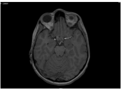
Figure 1(A): T1 axial images showing optic nerve and chiasma with tumour arising from it.
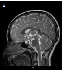
Figure 2: T2 images showing the tumour arising from optic chiasm. A. Sagittal; B. Coronal; C. Axial
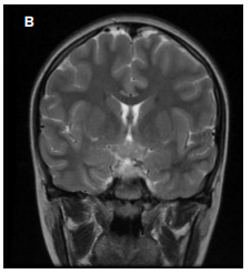
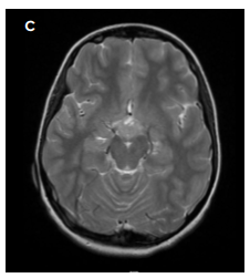
The patient was examined for features of neurofibromatosistype 1 (NF1), but no markers were identified. Recently, she started having visual deficits in the form of temporal field cuts, prompting her to seek further evaluation at our centre. On evaluation a 23x20.7x15mm contrast-enhancing lesion arising from the optic chiasma was identified (Figures 2,3).
The lesion involved the optic chiasm, hypothalamic-pituitary axis with involvement of pituitary stalk, which explained the symptomatology. The absence of neurocutaneous markers of NF-1, created a diagnostic dilemma. The patient was subsequently planned for craniotomy and tumour decompression with biopsy, (Figure 4), which was successfully completed without causing any additional visual deficits.
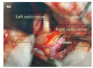
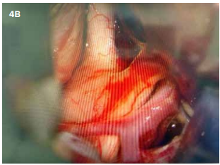
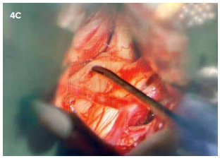
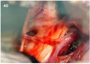
Figure 4: A & B. Tumour location as seen on intra operative microscope. The brownish mass arising from chiasm is identified as tumour. C. Tumour capsule opened, and decompression being done. D. Final image showing tumour decompressed tumour.
Rather, the patient’s temporal field cuts resolved significantly following the procedure. The biopsy revealed the lesion to be a germinoma, an extremely rare finding for a tumour originating from the chiasm (Figure 5).
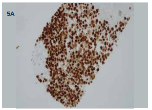
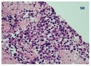
Figure 5: Microscopic images of the tumour; A. Immunohistochemistry (IHC) image; B. Haematoxylin and Eosin (H&E)
Discussion
The usual tumours of chiasmal origin (glioma) have poor prognosis. However, germinomas, being radio- and chemosensitive offer a more favourable outcome for the patient. These cases are a testament to the surgical acumen needed to address the tumours located at such critical areas without inducing postoperative deficits. The role of histopathological confirmation cannot be overstated, as it directs the course of further management. Timely surgical intervention followed by adjuvant therapy has been shown to significantly improve both survival rates and quality of life in patients with intracranial germinomas. Moreover, early diagnosis and treatment are crucial in preventing irreversible visual damage, especially in paediatric patients where the risk of long-term deficits is higher. This case underscores the importance of a multidisciplinary approach, involving neurosurgeons, oncologists, and radiologists, to optimize patient outcomes in such complex and rare presentations. Diagnosing a germinoma provides these young lives a chance to live a longer, productive life supported by available adjuncts in the postoperative period
References
- McCarthy BJ, Shibui S, Kayama T, et al. Primary CNS germ cell tumours in Japan and the United States: an analysis of 4 tumour registries. Neuro-oncology. 2012;14(9):1194-1200.
- Yeo KK, Nagabushan S, Dhall G, Abdelbaki MS. Primary central nervous system germ cell tumours in children and young adults: A review of controversies in diagnostic and treatment approach. Neoplasia. 2023;36:100860.
- Abbas M, Enani MZ, Alsabban Z, Meliti A, Homoud M. Primary anterior visual pathway germinoma in a 13-year-old boy: A case report. Surg Neurol Int. 2024;15:48.