 WhatsApp Us (24/7)
 +91 926 888 0303 (24/7)


Abstract:
Although presacral masses are uncommon, surgical resection is advised if they are identified, particularly in symptomatic patients. Additionally, there is a chance that the tumour could be malignant. Numerous surgical techniques, such as posterior, anterior, and combined abdominosacral approaches, have been documented for the resection of presacral tumours. Laparoscopic or robotic methods for resecting presacral masses are becoming increasingly common with the advent of minimally invasive techniques. Here, we present a unique case of a large presacral mass presenting with urinary retention and constipation. The mass was excised with robotic assistance. It was removed successfully without any concomitant injury to urinary bladder or rectum. The patient’s urinary and bowel obstructive symptoms resolved immediately following surgery. Roboticassisted excision of a presacral mass offers advantages, including enhanced magnified vision and improved instrument articulation.
Key words: Presacral Mass, Mature Teratoma, Urinary Retention.
Introduction
The majority of documented series on presacral masses and tumours are case reports, as these lesions are uncommon. Owing to the presence of various embryological remnants and distinct tissue types in the presacral region, a wide spectrum of tumour forms can develop. Presacral tumours are categorised into five groups based on their origin: congenital, inflammatory, neurogenic, osseous, and miscellaneous.1 These tumours can be benign or malignant.2 On magnetic resonance imaging (MRI), solid lesions indicate malignant characteristics.3 Most presacral tumours are discovered incidentally and do not exhibit any specific symptoms.4 Due to the possibility of growth or malignant change, surgical excision is advised upon diagnosis, in both symptomatic and asymptomatic patients.5
Numerous surgical techniques have been documented for the resection of presacral tumours. While posterior, anterior, and combined abdominosacral approaches are frequently used, transvaginal and transrectal techniques have also been used to resect presacral malignancies.
With the advent of minimally invasive surgery, laparoscopic and robotic methods for resecting presacral tumours are increasingly being adopted for presacral tumour resection. Here, we describe a case of a presacral mass that was successfully excised using a robotic anterior approach.
Case report
A 55-year-old-woman presented to her physician with acute urinary retention. She was catheterised and underwent imaging. Ultrasound (USG) followed by computed tomography (CT) showed a 285-cc sized hypodense collection with enhancing wall in the presacral region, along with air foci within the collection (Figure 1A). The patient was planned for robotic excision of the pelvic mass via an anterior approach.
Initially, during dissection posterior to the rectum, the cyst could not be localised. A Romson 24-French abdominal drain tube was therefore inserted rectally to help demarcate the rectum and cyst boundaries. Once localised, the dissection planes became much clearer. The rectum and mesorectum were preserved during medial dissection (Figure 1B), and the presacral venous plexus was preserved posteriorly. Laterally, care was taken to avoid injury to the ureter and autonomic nerves. The distal dissection proved most challenging, as the uterus and other pelvic viscera, including the rectal wall, limited visualisation of the dissection plane. To facilitate the procedure, the cyst was decompressed and its contents aspirated. Careful traction and counter-traction of the surgical plane was essential for achieving a successful bloodless dissection. The specimen was extracted through the right lower quadrant trocar site after extending the incision by about 3 cm (Figure 1C). The robotic resection was completed without significant intraoperative events. Histopathology confirmed the diagnosis of a mature cystic teratoma (Figure 1D). The patient was discharged home on postoperative day 3 without any complications.
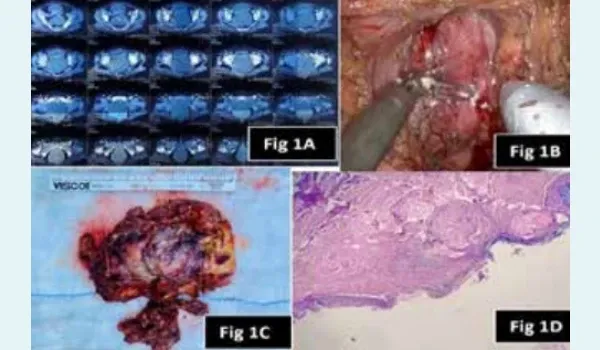
Figure 1A: Computed tomography (CT) images showing solid cystic mass in presacral region pushing the urinary bladder and rectum anteriorly.
Figure 1B: Intraoperative picture showing the glistening anterior surface of the cyst wall. Rectum is seen pushed on left side.
Figure 1C: Gross specimen showing the collapsed cyst with variegated consistency.
Figure 1D: Histopathology with haematoxylin and eosin-stained sections, showing a cyst wall lined by stratified squamous and ciliated columnar epithelium, with the presence of skin adnexal structures, mature adipose tissue, and smooth muscle.
Discussion
A traditional technique that offered a short path of entry into the presacral area and adequate surgical access was the posterior approach, also known as the Kraske trans-sacral approach. However, division of the pelvic floor muscles and coccygectomy or sacrectomy could result in postoperative discomfort and a potential risk of faecal incontinence.6 For larger tumours extending downward to as low as S4 and upward as far as the sacral promontory, an abdominosacral approach is advised. When there is no nerve involvement and the tumour’s inferior margin lies above S4, the anterior or abdominal approach is recommended.7 This approach allows excellent exposure of the ureters, pelvic autonomic nerves, and iliac arteries.8
Although the anterior approach often necessitates a long midline incision, the advent of minimally invasive techniques has enabled resection of presacral tumours using anterior robotic or laparoscopic techniques.9 Given the potential for malignant transformation, it is crucial to achieve complete resection of presacral masses.10 In summary, a laparoscopic anterior approach is both feasible and safe for the resection of presacral tumours. Care should be taken to avoid injury to vascular and neurological structures, visceral organs, or inadvertent perforation of the cystic lesion.11 In this case, we employed the anterior robotic technique to successfully excise the presacral tumour.
Conclusion
The robotic anterior approach is a safe and viable option for the resection of presacral masses. It is important to avoid injury to vascular, neurological, and visceral organs. Presacral masses rarely present with urinary retention and constipation. This case was unique in that the patient presented with acute urinary retention. The robotic approach enabled superior delineation of the surrounding pelvic anatomy, thereby facilitating a precise and effective surgical resection.
Shailesh Chandra Sahay, Pawan Kesarwani, Pramod K Karan, Abhijeet Jha, Shubhankar Powar. Robotic
Assisted Excision of a Giant Presacral Mass Presenting as Urinary Retention. MMJ. 2025, June. Vol 2 (2).
References
- Aranda-Narvaez JM, Gonzalez-Sanchez AJ, Montiel-Casado C, et al. Posterior approach (Kraske procedure) for surgical treatment of presacral tumors. World J Gastrointest Surg. 2012;4:126-130.
- Hobson KG, Ghaemmaghami V, Roe JP, et al. Tumors of the retrorectal space. Dis Colon Rectum. 2005;48:1964-1974.
- Yang DM, Kim HC, Lee HL, et al. Squamous cell carcinoma arising from a presacral epidermoid cyst: CT and MR findings. Abdom Imaging. 2008;33:498-500.
- Simpson PJ, Wise KB, Merchea A, et al. Surgical outcomes in adults with benign and malignant sacrococcygeal teratoma: a single-institution experience of 26 cases. Dis Colon Rectum. 2014;57:851-857.
- Mathis KL, Dozois EJ, Grewal MS, et al. Malignant risk and surgical outcomes of presacral tailgut cysts. Br J Surg. 2010; 97:575-579.
- Criss CN, Grant C, Ralls MW, et al. Robotic resection of recurrent pediatric lipoblastoma. Asian J Endosc Surg. 2019; 12:128-131.
- Nieuwenhuis DH, Gagner M, Consten EC. The endoscopic perineal approach to the presacral space: an excision biopsy. J Laparoendosc Adv Surg Tech A. 2009;19:799-801.
- Lim SW, Huh JW, Kim YJ, et al. Laparoscopy-assisted resection of tailgut cysts: report of a case. Case Rep Gastroenterol. 2011;5:22-27.
- Tobias-Machado M, Hidaka AK, Sato LLK, et al. Laparoscopic resection of prescral and obturator fossa schwannoma. Int Braz J Urol. 2017;43:566.
- Poskus E, Makunaite G, Kubiliute I, et al. Case report: Laparoscopic approach in the treatment of presacral lipoma. Ann Med Surg (Lond). 2018;35:64-66.
- Kim CW, Lee SH. Laparoscopic Resection of Presacral Tumor: A New Approach in the Era of the Minimally Invasive Surgery. J Minim Invasive Surg. 2019;22(3):131-133.