 WhatsApp Us (24/7)
 +91 926 888 0303 (24/7)


Abstract:
Kikuchi-Fujimoto Disease (KFD) is a rare, benign, self-limiting cervical lymphadenitis of unknown aetiology, with a worldwide distribution and a higher prevalence among Japanese and other Asiatic individuals. It predominantly affects young adults under the age of 30, predominantly women. It can closely mimic infective and immunological disorders and is often reported as a cause of prolonged fever of unknown origin. Although the clinical course is typically benign, KFD has been often misdiagnosed as malignancy or infection. In this case report, we present a case of a 20-year-old male, a known case of hypothyroidism, who presented with highgrade fever with chills for one month and multiple swellings over the neck. He was evaluated and diagnosed as a case of KFD on an excisional biopsy of a left cervical lymph node and was treated with steroids and other symptomatic measures.
Key words: Kikuchi-Fujimoto Disease (KFD), Histiocytic Necrotising Lymphadenitis, Excision Biopsy
Introduction
Kikuchi-Fujimoto Disease (KFD) is a rare, benign, self-limiting cervical lymphadenitis of unknown aetiology, with a worldwide distribution and a higher prevalence among Japanese and other Asiatic individuals. It predominantly affects young adults under the age of 30, predominantly women. It can closely mimic infective and immunological disorders and is often reported as a cause of prolonged fever of unknown origin. Although the clinical course is typically benign, KFD has been often misdiagnosed as malignancy or infection. In this case report, we present a case of a 20-year-old male, a known case of hypothyroidism, who presented with highgrade fever with chills for one month and multiple swellings over the neck. He was evaluated and diagnosed as a case of KFD on an excisional biopsy of a left cervical lymph node and was treated with steroids and other symptomatic measures.
Although benign, KFD is often mislabelled as an infection or malignancy.3 It was originally described in 1972 in Japan by Dr. Masahiro Kikuchi and Y. Fujimoto.4,5 The exact pathophysiology remains unclear, but the disease is thought to be triggered by an autoimmune condition, infection, or a physical injury, leading to an inflammatory cascade.4,5 The disease onset is generally acute or subacute, evolving over a period of 2-3 weeks. Generalised lymphadenopathy is rare, although cervical lymphadenopathy is almost always present. The lymph nodes are typically mobile, solitary, and painful, with sizes ranging from 0.5 cm to 4 cm, and rarely exceeding 6 cm. The most commonly involved lymph nodes are located in the posterior and anterior cervical triangles, but may also occur in the axillary, supraclavicular, mediastinal, inguinal, peritoneal, or retroperitoneal regions.
Kikuchi’s disease is extremely rare, and its exact incidence remains uncertain. It can affect individuals of all age groups but is most commonly seen in individuals under 30 years of age.3 Kikuchi’s disease, is most frequently observed in Asian populations, with rare sporadic cases reported in the United States and Europe.
Case report
A 20-year-old male, resident of North India, a known case of hypothyroidism, presented with multiple neck swellings and high-grade fever with chills, persisting for one month. No other localising symptoms or signs were elicited. There was no prior history of tuberculosis.
On clinical examination, multiple enlarged lymph nodes were noted in the left axilla and cervical region. They were soft in consistency, mobile, painless, and measured approximately 3 x 2.5 cm. Lymph nodes were not palpable in other parts of the body. Systemic examination findings were within normal limits
Laboratory investigations including complete blood count (CBC), liver function tests (LFT), kidney function tests (KFT), peripheral smear, procalcitonin, and urine routine analysis were within normal limits. Peripheral smear for malaria parasites (performed in another hospital) was negative. Tests for Widal, scrub typhus, and viral markers-Human Immunodeficiency Virus types 1 and 2 (HIV-1 and HIV-2), Hepatitis C Virus (HCV) and Hepatitis B surface Antigen (HBsAg)—were negative. Paired urine and blood cultures were sterile. Transthoracic echocardiography (TTE) showed no evidence of vegetations, clots, or pulmonary embolism.
The erythrocyte sedimentation rate (ESR) was mildly raised at 41 mm in the first hour, and C- reactive protein (CRP) was 1.7. Serum ferritin (430 ng/mL) and lactate dehydrogenase (LDH) (384 U/L) were also mildly raised.
Tests for Mantoux, Monospot, serum angiotensin-converting enzyme (ACE) Levels, antinuclear antibodies (ANA) by indirect immunofluorescence assay (IFA) and screening profile, antistreptolysin O (ASO) titre, coronavirus disease 2019 reverse transcription polymerase chain reaction (COVID 19 RT-PCR), influenza RT-PCR, Brucella immunoglobulin M (IgM) and immunoglobulin G (IgG), cytomegalovirus (CMV) IgM, EpsteinBarr Virus viral capsid antigen (EBV VCA) IgM, and Toxoplasma serology were all negative.
Contrast-enhanced computed tomography (CECT) chest showed multiple enlarged, homogenous axillary lymph nodes, as shown in Figure 1
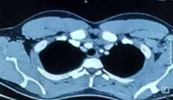
Figure 1: Contrast-enhanced computed tomography (CECT) chest (performed externally) showed at least three enlarged homogenous axillary lymph nodes, largest measuring 3.8 X 2.7 cm.
Ultrasound (USG) of the axilla performed in another hospital, showed multiple enlarged left axillary lymph nodes (2.1 x 1.4 cm) with necrosis. A USG-guided fine needle aspiration cytology (FNAC) of the cervical lymph node was performed, along with immunohistochemistry (IHC) studies, followed by an excision biopsy of the lymph node.
FNAC of the cervical lymph node showed moderate cellularity comprising a polymorphous population of lymphoid cells, with the presence of macrophages and occasional immunoblasts. The background showed necrotic areas; however, no wellformed granulomas were identified.
Subsequent excision biopsy of the lymph node showed a distorted nodal architecture (Figure 2). Pale areas were noted within the lymph node, composed of eosinophilic, granular material, and karyorrhectic debris (Figure 3), interspersed with lymphocytes and histiocytes. These areas notably lacked neutrophils and giant cells. Other areas showed primary and secondary lymphoid follicles. No granulomas were identified, and no acid-fast bacilli (AFB) were seen on Ziehl-Neelsen stain.
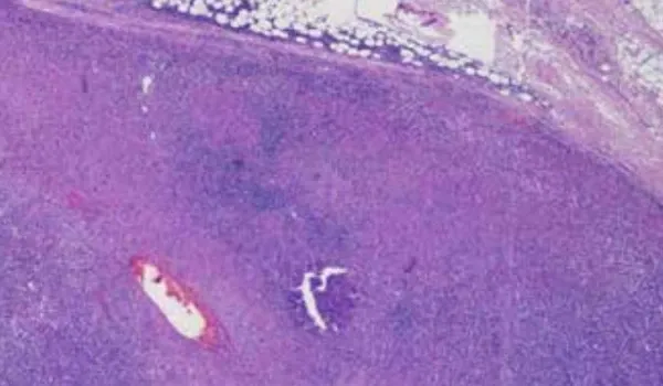
Figure 2: Lymph node with distorted architecture and pale necrotic areas.
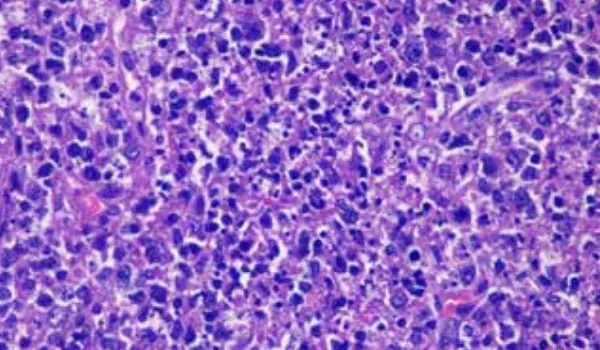
Figure 3: Lymph node exhibiting karyorrhectic debris devoid of neutrophils.
Immunohistochemical stains, including Cluster of Differentiation (CD)3, CD20, CD15, CD30, Paired Box Protein 5 (PAX5), Epstein-Barr Virus Latent Membrane Protein (EBV-LMP), B-cell lymphoma 2 (Bcl2), terminal deoxynucleotidyl transferase (Tdt), and Ki67 proliferation index were done, and the possibility of a lymphoproliferative disorder was completely excluded. Based on the morphological findings and IHC results, a diagnosis of necrotising lymphadenitis suggestive of KFD was rendered.
The patient was started on oral steroids, showed signs of improvement, and was discharged from hospital on oral steroids with advice for follow-up through the outpatient department (OPD).
Discussion
When evaluating a patient for KFD, it is essential to initiate an infectious work-up guided by the patient’s medical history. This should include recent infectious illness, travel history, exposure to animals or tuberculosis, and current or recent antibiotic use.
The most common presentation of KFD includes localised or generalised lymphadenopathy, fever, fatigue, weight loss, hepatosplenomegaly, and rash. A definitive diagnosis requires excisional lymph node biopsy, as IHC analysis is necessary.5,6
The exact aetiology of KFD is not known, although infectious agents such as EBV, HIV, Herpes Simplex Virus (HSV), dengue virus, Human T-lymphotropic Virus type 1 (HTLV 1), parvovirus B19, Toxoplasma gondii, Yersinia enterocolitica, Bartonella spp., and Brucella spp. may have a role.
Serological testing for various viral and bacterial causes of lymphadenitis, along with an autoimmune work-up including family history, should be considered based on the exposure history and clinical presentation. For suspected tuberculous lymphadenitis, Mantoux test, or interferon-gamma release assay (IGRA) should be performed.
Peripheral flow cytometry may be contemplated in cases with a suspicion of malignancy. Histological and immunohistochemical analysis are important for excluding malignancy.
Diagnosing KFD necessitates an excisional biopsy of an enlarged lymph node, with IHC playing a crucial role in excluding other differential diagnoses.7 KFD is characterised by three histological stages—proliferative, necrotising, and xanthomatous.
A notable absence of neutrophils or eosinophils is evident across all these stages, and serves as a crucial distinguishing feature between KFD and infectious aetiologies.6-8 Furthermore, microscopic examination and culture studies typically yield negative results.
Laboratory investigations may reveal raised CRP, ESR, ferritin, aminotransferases, and LDH. Leukopenia occurs in 20%-58% of cases.
Characteristic histopathologic findings of KFD include irregular paracortical areas of coagulative necrosis with abundant karyorrhectic debris, distortion of nodal architecture, and large number of different types of histiocytes at the margin of necrotic areas.
Neutrophils are characteristically absent, and plasma cells are either absent or scarce. Atypia in the reactive immunoblastic component may mimic lymphoma. The immunophenotype shows a predominance of T cells with few B cells.9
Differential Diagnosis
Distinguishing KFD from lymphomas and infectious aetiologies is critical, and additional support for the histologic diagnosis can be obtained through cultures and serological testing. Although systemic lupus erythematosus (SLE) lymphadenitis may resemble KFD histologically, the presence of haematoxylin bodies in SLE lymphadenitis aids differentiate it from KFD.10
Infectious diseases
- Viral: EBV, CMV, Infectious hepatitis, HSV, human herpesvirus-6 (HHV-6), varicella-zoster virus (VZV), rubella, measles, HIV, and human herpesvirus-8 (HHV- 8)
- Bacterial: Streptococci, staphylococci, tuberculosis, brucellosis, plague, and chancroid
- Fungal: Histoplasmosis, coccidioidomycosis, and paracoccidioidomycosis
- Chlamydial: Lymphogranuloma venereum (LGV), and trachoma
- Parasitic: Toxoplasmosis, leishmaniasis, trypanosomiasis, and filariasis
- Rickettsial: Scrub typhus, rickettsial pox, and Q fever
Immunologic diseases
Rheumatoid arthritis (RA), juvenile RA, mixed connective tissue disease (MCTD), SLE, dermatomyositis, and Sjögren’s syndrome.
Malignant diseases
Hodgkin’s lymphoma, non-Hodgkin lymphoma (NHL), hairy cell leukaemia (HCL), malignant histiocytosis, and amyloidosis.
Lipid storage diseases
Gaucher’s disease, Niemann-Pick disease, Fabry disease, and Tangier disease.
Endocrine diseases
Hyperthyroidism.
Other disorders
Castleman’s disease (a rare disorder that affects lymph nodes and organs), sarcoidosis, severe hypertriglyceridaemia, Kawasaki disease, histiocytosis X, and lymphomatoid granulomatosis.8
Management
No specific treatment exists for KFD. It follows a self-limiting course, typically resolving spontaneously within 1-6 months, and has an excellent prognosis.
Supportive management is the mainstay of treatment. Corticosteroids and immunosuppressants may be considered for severe extranodal or generalised KFD or recurrent cases.
Acknowledgements
We sincerely thank Dr. Sandeep Budhiraja, Dr. Vinay Labroo, and Dr. Sharique Ahmed from Max Super Speciality Hospital, Noida, for their invaluable support in the preparation of this case report. Their guidance in data analysis, manuscript preparation, and literature review was instrumental in shaping the final publication.
Supportive management is the mainstay of treatment. Corticosteroids and immunosuppressants may be considered for severe extranodal or generalised KFD or recurrent cases.
Conclusion
Although KFD is rare, it should be considered in the differential diagnosis of a young patient presenting with fever and cervical lymphadenopathy. While it is historically more common in females, recent studies suggest a nearly equal incidence among males and females. Due to its characteristic overlap with conditions like tuberculous lymphadenitis and lymphoma, KFD remains an arduous diagnosis for physicians. Therefore, clinicians must remain vigilant and consider KFD as a potential diagnosis to prevent mismanagement. Histological studies are essential to exclude other possible causes of lymphadenopathy.8 Early recognition of disease is important to prevent complications.
Vinay Labroo, Sharique Ahmed, Sayani Bera, Madhura More. Kikuchi Disease in a Male: A Rare Case
Report. MMJ. 2025, June. Vol 2 (2).
References
- Sudhakar MK, Sathyamurthy P, Rajendran IE, et al. Kikuchi's disease: A case report from south India. International Journal of Case Reports and Images. Int J Case Rep Images. 2011;2(2):15-8.
- Ganpule AP, Chabra JS, Singh AG, et al. Case Report: Kikuchi-Fujimoto disease: a diagnostic and therapeutic dilemma following pretransplant nephrectomy for a 2.35 Kg kidney. F1000Res. 2016;5:1407.
- Masab M, Surmachevska N, Farooq H. Kikuchi-Fujimoto Disease. Updated 2023 Oct 29. In: StatPearls [Internet]. Treasure Island (FL): StatPearls Publishing; 2025 Jan–. Available from: https://www.ncbi.nlm.nih.gov/books/ NBK430830/.
- Bosch X, Guilabert A. Kikuchi-Fujimoto disease. Orphanet J Rare Dis. 2006;1:18.
- Dumas G, Prendki V, Haroche J, et al. Kikuchi-Fujimoto disease: retrospective study of 91 cases and review of the literature. Medicine (Baltimore). 2014 Nov;93(24):372-382. Erratum in: Medicine (Baltimore). 2014 Nov;93(24):414.
- Harrison J, Sukumaran S, Vijayan V. Systemic Lupus Erythematosus Lymphadenopathy Presenting as Kikuchi-Fujimoto Disease in an Adolescent. Cureus. 2023;15(2):e35304.
- Mallick A, Mohapatra MM, Babu VM, et al. All necrotizing nodes are not tuberculosis - A report of two cases. Indian J Tuberc. 2022;69(4):695-698.
- Joean O, Thiele T, Raap M, et al. Take a second look: it's Kikuchi's disease! A case report and review of literature. Clin Pract. 2018;8(4):1095.
- Aminiafshar S, Namazi N, Abbasi F. Kikuchi-fujimoto disease in 21-year-old man. Int J Prev Med. 2013;4(8):964-6.
- Tsang WY, Chan JK, Ng CS. Kikuchi's lymphadenitis. A morphologic analysis of 75 cases with special reference to unusual features. Am J Surg Pathol. 1994;18(3):219-31.