 WhatsApp Us (24/7)
 +91 926 888 0303 (24/7)


Abstract:
Gonadal vein thrombosis (GVT) is an uncommon clinical entity typically associated with postpartum or postoperative states. Its occurrence in the context of systemic autoimmune diseases is rare and diagnostically challenging, particularly when presenting as fever of unknown origin (FUO).
We report the case of a 39-year-old woman presenting with a six-month history of painful red discoloration of the fingertips, right lower quadrant abdominal pain, and five months’ history of persistent low-grade fever. Initial evaluation revealed systemic symptoms including significant fatigue, weight loss, dry cough, and alopecia. Physical examination showed cervical lymphadenopathy, painful oral ulcers, malar rash, and bilateral lower limb oedema. Laboratory investigations revealed leukopenia, elevated inflammatory markers, hypoalbuminemia with nephrotic-range proteinuria, positive antinuclear antibody (ANA) with high titres, and hypocomplementemia. Imaging identified a right gonadal vein thrombosis and multiple fluorodeoxyglucose (FDG)-avid lymph nodes on positron emission tomography - computed tomography (PET-CT). The clinical picture, combined with serological findings, was consistent with a diagnosis of mixed connective tissue disease with features overlapping systemic lupus erythematosus (SLE) and polymyositis. A secondary antiphospholipid syndrome (APS) was considered likely in the context of thrombosis and positive beta-2 glycoprotein antibodies and anti-cardiolipin antibodies.
This case underscores the importance of considering systemic autoimmune diseases in the differential diagnosis of FUO and thrombosis at unusual sites. GVT, though rare, may be the first manifestation of an underlying connective tissue disease or APS, and warrants comprehensive immunological and haematological evaluation.
Key words: FUO, Gonadal Venous thrombosis, Mixed Connective Tissue Disease, APS.
Introduction
Gonadal vein thrombosis (GVT) is a rare vascular condition most commonly observed in the postpartum period or following pelvic surgery. It is infrequently encountered in non-obstetric settings and seldom reported as a primary manifestation of systemic autoimmune or connective tissue diseases. The clinical presentation of GVT can be subtle and often mimics other causes of abdominal pain, making diagnosis challenging.
When accompanied by systemic features such as fever of unknown origin (FUO), generalised lymphadenopathy, and mucocutaneous involvement, the differential diagnosis broadens significantly, requiring consideration of inflammatory, infectious, and malignant aetiologies.1
Systemic autoimmune diseases such as systemic lupus erythematosus (SLE), Sjögren’s syndrome, systemic sclerosis, and polymyositis are known to present with protean manifestations that may mimic infection or malignancy.2 Vascular complications, particularly thrombosis in unusual locations, may be the initial or sole manifestation of underlying antiphospholipid antibody syndrome (APS), which can be either primary or secondary to a systemic autoimmune condition.
We present an unusual case of GVT in a previously healthy woman, presenting with FUO and multisystem involvement, ultimately diagnosed as mixed connective tissue disease (MCTD) with overlapping features of SLE and polymyositis. This case highlights the diagnostic complexity of GVT in the autoimmune context and underscores the need for a high index of suspicion for systemic diseases in patients presenting with FUO and atypical thrombotic events.
Case Report
A 39-year-old woman from Rajasthan, India, presented to the internal medicine outpatient department with a constellation of symptoms that had evolved over the previous six months. Her primary complaints included episodic painful red discoloration of the fingers, persistent right lower quadrant abdominal pain, and low-grade fever.
The digital symptoms began six months prior and were characterised by sudden-onset, intermittent, painful erythema localised to the distal palmar aspect of all fingers. The episodes were non-progressive and relieved transiently by immersion in cold water. There were no features suggestive of gangrene, colour changes extending to the hands, or associated temperature fluctuations. The pain was severe enough to impair fine motor function.
Simultaneously, she developed right lower quadrant abdominal pain, initially dull and intermittent but progressively intensifying over time to a continuous, stabbing quality, rated as 10/10 in severity. The pain was non-radiating, unresponsive to oral paracetamol or NSAIDs, and eventually required intravenous analgesics during hospitalisation. There was no diurnal variation or known precipitating factors.
Five months prior to presentation, she began experiencing persistent low-grade fever (100–101°F) without chills or rigors. The fever was insidious in onset and recurred after the effect of antipyretics waned. It was associated with severe fatigue and myalgia, significantly limiting her activities of daily living.
Systemic review revealed additional symptoms, including a dry, non-productive cough of four months' duration without diurnal or positional variation; approximately 10 kg of unintentional weight loss over six months; progressive fatigue, diffuse scalp hair thinning with visible patchy hair loss; and proximal muscle weakness, manifesting as difficulty rising from a chair or lifting her arms above her head.
There was no history of photosensitivity, oral or genital ulcers prior to this episode, Raynaud’s phenomenon, joint pain, sicca symptoms, dysphagia, chest pain, dyspnoea, palpitations, urinary symptoms, or mood disturbances. She denied any recent infections, travel, allergen exposures, or occupational risk factors. There was no personal or family history of autoimmune, thrombotic, or malignant conditions.
Her menstrual history was unremarkable, with regular cycles and no abnormal bleeding or dysmenorrhoea. Obstetric history included two full-term deliveries without complications. She was not on any medications prior to the onset of symptoms and had no known drug or food allergies.
Examination
On presentation, the patient was lying comfortably in bed, alert, and well-oriented to time, place, and person. Her gait was normal.
General examination
The pathophysiology of CTS involves a cascade of events initiated by increased pressure within the carpal tunnel. Normal pressure ranges from 2-10 mmHg, but can exceed 30 mmHg in CTS patients, particularly during wrist flexion or extension. 11 This elevated pressure leads to several pathophysiological changes:
-
Pallor: Present
-
Icterus, cyanosis, clubbing:Absent
-
Lymphadenopathy: A single, non-matted, non-tender cervical lymph node measuring approximately 8 × 6 cm was palpable in the right posterior triangle, supraclavicular in location (2 cm above the clavicle). It was soft to firm in consistency, not fixed to overlying skin or underlying structures. There was no erythema, fistula, sinus formation, or discharge associated with it. No other lymph nodes were appreciable.
-
Oedema: Bilateral grade 3 pitting oedema was noted in the lower limbs, extending from both feet up to the knees. There was no skin induration.
Oral examination
Multiple small, painful, round-to-oval ulcers measuring 1–2 cm were noted over the oral mucosa. These ulcers had a red base with a pale border and were diffusely distributed without evidence of discharge or induration at the base. The patient exhibited difficulty opening the mouth, with opening limited to two finger breadths.
Neck and thyroid examination
The thyroid gland was not enlarged and was non-tender. It moved with deglutition and protrusion of the tongue and was freely mobile. However, diffuse hardening of the right thyroid cartilage was appreciated. No overlying erythema was present.
Systemic examination
-
Cardiovascular system (CVS): S1 and S2 heart sounds were normal. No murmurs were heard.
-
Central nervous system (CNS): Higher mental functions, cranial nerves, motor and sensory systems, reflexes, gait, and autonomic function were all intact. There were no signs of meningeal irritation.
-
Respiratory system: Bilateral air entry was equal with no added sounds (e.g., crepitations, wheeze).
-
Abdomen: Soft, non-tender, non-distended. No organomegaly or palpable masses were noted.
Local examination (skin and extremities)
-
A non-pruritic, non-tender macular rash was observed bilaterally over the malar prominences as shown in Figure 1 . The rash spared the nasolabial folds and was not associated with puffiness or change in skin temperature.
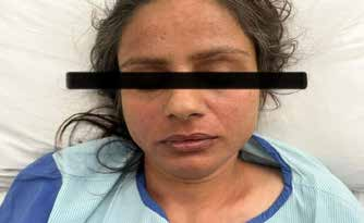
Figure 1: Malar rash.
-
Erythema was present over the distal interphalangeal joints of all fingers in both hands as shown in Figure 2, consistent with the patient’s complaint of painful, red fingers.
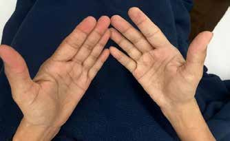
Figure 2: Painful erythema over digits.
Investigations
Laboratory investigations
-
Her baseline lab investigations are shown in Table 1.
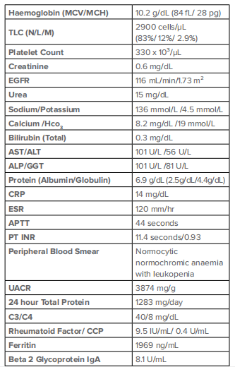
Table 1: Baseline laboratory investigations.
Abbreviations : ALP: Alkaline phosphatase; ALT: Alanine aminotransferase; APTT: Activated partial thromboplastin time; AST: Aspartate aminotransferase; CCP: Cyclic citrullinated peptide; CRP: C-reactive protein; C3: Complement component 3; C4: Complement component 4; D-dimer: Fibrin degradation product; DRVVT: Dilute Russell viper venom time; EGFR: Estimated glomerular filtration rate; ESR: Erythrocyte sedimentation rate; Ferritin: Iron-storage protein; GGT: Gamma-glutamyl transferase; HCO₃: Bicarbonate; IgA: Immunoglobulin A; INR: International normalized ratio; MCH: Mean corpuscular haemoglobin; MCV: Mean corpuscular volume; N: Neutrophil; L: Lymphocyte; M: Monocyte; PT: Prothrombin time; RF: Rheumatoid factor; TLC: Total leucocyte count; UACR: Urine albumin-to-creatinine ratio.
-
Connective tissue disease (CTD) profile – Her CTD Profile for autoimmune work up is shown in Table 2.
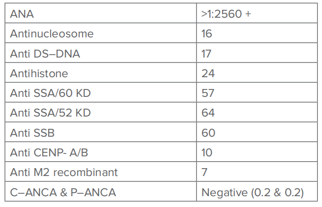
Table 2: Connective tissue disease (CTD profile) profile
Abbreviations : ANA: Antinuclear antibody; Anti-CENP-A/B: Anti-centromere protein A/B antibodies; Anti-dsDNA: Anti-double-stranded DNA antibody; Anti-M2 recombinant: Anti-mitochondrial M2 recombinant antibody; Anti-SSA/52 kD: Anti-Sjögren's-syndrome-related antigen A, 52 kilodalton; Anti-SSA/60 kD: Anti-Sjögren's-syndrome-related antigen A, 60 kilodalton; Anti-SSB: Anti-Sjögren's-syndrome-related antigen B antibody; C-ANCA: Cytoplasmic anti-neutrophil cytoplasmic antibody; P-ANCA: Perinuclear anti-neutrophil cytoplasmic antibody
Radiological investigations
-
All the radiological investigations done are shown in Table 3.
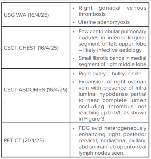
Table 3: Radiological investigations done for patient.
Abbreviations : CECT: Contrast-enhanced computed tomography; FDG: Fluorodeoxyglucose; IVC: Inferior vena cava; PET-CT: Positron emission tomography–computed tomography; USG W/A: Ultrasonography of whole abdomen.
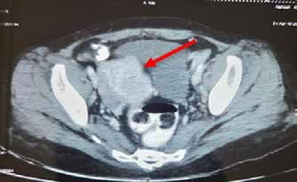
Figure 3: Right ovarian venous thrombosis.
Histopathological diagnosis:
-
Biopsy from right cervical lymph node (Figure 4): Showing non-granulomatous necrotising lymphadenitis.
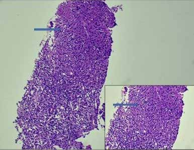
Figure 4: Right cervical lymph node biopsy
-
Renal biopsy (Figure 5): All glomeruli showed diffuse thickening of glomerular basement membrane with sub -epithelial spikes and pinholes.
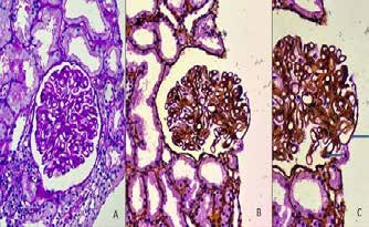
Figure 5: Renal biopsy: a) PAS stain (20X), b) silver stain (20X), c) Silver stain (40X).
-
Renal biopsy with direct immunofluorescence (Figure 6): sy with direct immunofluorescence (Figure 6): Showing peripheral and mesangial deposits with “Full House effect”.
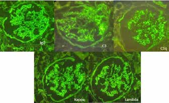
Figure 6: Renal biopsy with direct immunofluorescence.
Diagnosis
Based on the clinical presentation, laboratory investigations, immunological profile, and radiological findings, the patient was diagnosed with:
- Mixed connective tissue disease (MCTD ) with features overlapping:
- SLE: Positive ANA (>1:2560), anti-dsDNA, anti-nucleosome, anti- SSA/SSB antibodies, oral ulcers, malar rash, leukopenia, and nephrotic-range proteinuria with hypocomplementemia (low C3 and C4)
- Polymyositis: Proximal muscle weakness with mildly elevated creatine kinase (CK)
- Right GVT, confirmed on ultrasound and contrast-enhanced CT abdomen secondary to APS, supported by vascular thrombosis and positive beta-2 glycoprotein antibody
- Lupus nephritis Grade 5 with “Full house” effect – histopathologically confirmed.
Discussion
GVT represents an exceedingly rare but potentially serious vascular complication that can present diagnostic challenges due to its nonspecific clinical manifestations. In this case, the patient's persistent fever for five months without an identifiable cause initially classified her condition as FUO, which eventually led to the discovery of right GVT in the setting of MCTD with features of secondary APS.
GVT is an uncommon clinical entity with a reported incidence of approximately 0.08% in studies utilising computed tomography imaging for diagnosis.1
Traditionally, GVT has been most frequently associated with the postpartum period, following hysterectomy, or after lymphadenectomy for gynaecological malignancies, with up to 80% of cases occurring in these settings.1 The predominance of right-sided involvement (59% of cases in one study) has been attributed to anatomical factors including the longer length of the right gonadal vein compared to the left, and an increased absence of competent valves in the right gonadal vein.1 However, GVT occurring in the context of autoimmune disorders, particularly MCTD, represents an unusual presentation that warrants attention.
The hypercoagulable state observed in autoimmune disorders provides a mechanistic explanation for thrombotic events such as GVT in our patient. Active inflammation, a hallmark of connective tissue diseases, creates a prothrombotic environment characterized by upregulation of tumour necrosis factor-α (TNF-α) and activation of endothelial cells.2 This inflammatory milieu increases tissue factor expression (a natural procoagulant) while simultaneously downregulating protein C (a natural anticoagulant).2 Additionally, endothelial activation promotes platelet aggregation, further contributing to thrombus formation.
The risk of venous thromboembolism is particularly elevated during periods of increased disease activity, with studies suggesting that the highest risk occurs within the first year of disease onset.2 This temporal relationship aligns with our patient's presentation, as her thrombotic complication manifested during the active phase of her newly diagnosed MCTD.
MCTD, first described by Sharp et al. in 1972, represents an autoimmune disorder with overlapping features of systemic sclerosis, SLE, and polymyositis.3 With an estimated annual incidence of 1.9-2.1 cases per 100,000 adults and a female predominance (female-to-male ratio of 3.3:1), MCTD remains a relatively rare condition.3
While once perceived as a mild and treatable condition, MCTD can pose significant morbidity and mortality risks, particularly when complicated by vascular events.3 The thrombotic risk in MCTD appears to be mediated through multiple mechanisms, including endothelial dysfunction, inflammation-induced hypercoagulability, and the possible presence of antiphospholipid antibodies, as observed in our patient.
APS may occur as a primary disorder or secondary to systemic autoimmune diseases, particularly SLE and MCTD. Antiphospholipid antibodies are frequently detected in patients with SLE, with lupus anticoagulant found in approximately one-third of cases and anti-beta 2 glycoprotein-1 antibodies in about 20%. These antibodies demonstrate a strong association with thromboembolic events in patients with connective tissue diseases.
In our case, the patient exhibited clinical and laboratory features consistent with secondary APS (elevated beta-2 glycoprotein IgA levels and significantly elevated D-dimer), which likely contributed to the development of GVT. The recognition of APS in this setting has significant therapeutic implications, as it influences the choice of anticoagulant therapy. Vitamin K antagonists are generally preferred over direct oral anticoagulants for secondary thromboprophylaxis in patients with APS.
FUO, defined as a temperature exceeding 38.3°C (101.0°F) persisting for at least three weeks without an established aetiology after thorough clinical evaluation, represents a diagnostic conundrum that requires systematic investigation. When FUO occurs in the context of autoimmune disorders, the diagnostic complexity increases due to the multisystemic nature of these conditions and the variable manifestations of disease activity.
In our patient, the prolonged fever initially dominated the clinical picture, obscuring the underlying GVT. This case underscores the importance of comprehensive vascular imaging in patients with persistent unexplained fever, particularly those with clinical features suggestive of autoimmune disease. The combination of ultrasonography, contrast-enhanced computed tomography, and positron emission tomography proved instrumental in establishing the diagnosis of GVT and characterising the extent of systemic involvement.
The management of GVT in the setting of MCTD with secondary APS involves a multifaceted approach addressing both the thrombotic complication and the underlying autoimmune disorder. Anticoagulation therapy forms the cornerstone of GVT management, typically administered for a duration of three to six months.1 However, in patients with persistent antiphospholipid antibody positivity, extended or indefinite anticoagulation may be necessary to prevent recurrent thrombosis.
For the underlying MCTD, treatment typically includes immunomodulatory agents such as corticosteroids, hydroxychloroquine, and other disease-modifying antirheumatic drugs tailored to the specific clinical manifestations. The management of secondary APS requires particular attention to anticoagulation strategies, with vitamin K antagonists generally preferred over direct oral anticoagulants due to superior efficacy in preventing recurrent thrombosis in this patient population.
According to the Kidney Disease: Improving Global Outcomes (KDIGO) 2024 guidelines, a renal biopsy is essential in patients with SLE presenting with proteinuria ≥500 mg/day or unexplained rise in serum creatinine to classify lupus nephritis (LN) and guide therapy. However, in practice— especially in resource-limited settings or unstable patients—empirical treatment may be initiated when classical serological and clinical features are present.4
The updated KDIGO 2024 and American College of Rheumatology (ACR) 2024 guidelines recommend a “triple therapy” approach for active Class III/IV LN.4,5
- Glucocorticoids (preferably initiated with (IV) pulse methylprednisolone and tapered to ≤5 mg/day over 6 months)
- Mycophenolate mofetil (MMF)
- Belimumab or a calcineurin inhibitor (such as voclosporin)
Hydroxychloroquine is universally advised for all patients with LN, unless contraindicated, due to its renoprotective and immunomodulatory benefits.
From an Indian perspective, several randomised controlled trials have demonstrated comparable efficacy of low-dose cyclophosphamide and MMF as induction therapies for LN. A landmark Indian study by Bharati et al. 2019 compared two steroid regimens and demonstrated improved outcomes with a lower steroid burden, aligning with the KDIGO emphasis on minimising long-term glucocorticoid exposure.6
For the management of APS in this patient, vitamin K antagonists (VKAs) such as warfarin are preferred over direct oral anticoagulants (DOACs), particularly in those with arterial thrombosis or high-risk antibody profiles.
This case highlights several important clinical lessons. First, it illustrates the need for heightened awareness of thrombotic complications in patients with MCTD, even in the absence of traditional risk factors for venous thromboembolism. Second, it emphasises the value of comprehensive vascular imaging in the evaluation of FUO, particularly when associated with localised abdominal pain. Finally, it underscores the importance of screening for antiphospholipid antibodies in patients with connective tissue diseases who develop thrombotic events, as this has significant implications for management.
Future research directions should focus on elucidating the precise mechanisms underlying the thrombotic risk in MCTD, developing risk stratification tools to identify patients most susceptible to vascular complications, and optimising anticoagulation strategies for this unique patient population.
Conclusion
This case represents an unusual presentation of GVT manifesting as FUO origin in a patient with newly diagnosed MCTD and features of secondary APS. It highlights the complex interplay between autoimmunity, inflammation, and thrombosis, while emphasising the importance of comprehensive evaluation in patients with prolonged unexplained fever. Recognition of this rare association can facilitate prompt diagnosis and appropriate management, potentially preventing serious complications of untreated venous thromboembolism in this vulnerable patient population.
Aryan Arora, Prateek Bhanu, Sandeep Budhiraja, Anisha Manocha. An Unusual Case of Gonadal Venousa
Thrombosis Presenting as Fever of Unknown Origin. MMJ. 2025, June. Vol 2 (2).
References
- Alsharif S, Subahi A, Shirah B, et al. Incidental gonadal vein thrombosis diagnosed using computed tomography imaging: A single-centre, retrospective, cohort study. Cureus. 13(6): e15741.
- Lee JJ, Pope JE. A meta-analysis of the risk of venous thromboembolism in inflammatory rheumatic diseases. Arthritis Res Ther. 2014;16(5):435.
- Mohammed M, Mohammed R. Analysis of mixed connective disorder: a case report. Int J Res Med Sci. 2024;12(5):1754-8.
- Kidney Disease: Improving global outcomes (KDIGO) lupus nephritis work group. KDIGO 2024 clinical practice guideline for the management of lupus nephritis. Kidney Int. 2024;105(1S):S1-69.
- Sammaritano LR, Askanase A, Bermas BL, et al. 2024 American College of Rheumatology (ACR) Guideline for the Screening, Treatment, and Management of Lupus Nephritis. Arthritis & Rheumatology. 2025 Mar 24.
- Bharati J, Rathi M, Ramachandran R, et al. Comparison of two steroid regimens in induction therapy of proliferative lupus nephritis: A randomised controlled trial. Indian J Nephrol. 2019;29(5):373-5.