

Abstract:
When anaemia does not respond to vitamin and iron supplements and no genitourinary or gastrointestinal cause of blood loss is found, the patient requires a bone marrow examination to exclude rare causes like myelodysplastic syndrome (MDS). However, even the bone marrow examination and usual cytogenetic testing,including karyotyping and fluorescence in situ hybridisation (FISH), may be inconclusive. In such cases, where there is a diagnostic dilemma, next-generation sequencing (NGS) may be a ray of hope. Here, we report a few cases where bone marrow aspiration, biopsy, and cytogenetics were inconclusive, and NGS helped in re-classifying these patients as MDS. NGS served as a critical bridge between clinical suspicion and definitive diagnosis when other modalities failed.
Key words: Refractory Anaemia, Myelodysplastic Syndrome, Cytogenetics, Karyotyping, Next-Generation Sequencing, Diagnostic Dilemma.
Introduction
Unexplained anaemia is found in > 15% of individuals aged over 65 years and increases by > 50% in people over 80 years. Consequently, it is a major diagnostic challenge for all general practitioners.1 Bone marrow examination is the most common test performed for the diagnosis of anaemias or cytopenias when other causes like nutritional deficiencies, drug-induced causes, heavy metal toxicity, etc. are excluded. In some of these patients, even a bone marrow examination and cytogenetics by karyotyping or fluorescence in situ hybridisation (FISH) are normal and may not be diagnostic. Patients with cytopenias but without sufficient dysplastic changes or myelodysplastic syndrome (MDS)-defining cytogenetic alterations are considered to have idiopathic cytopenia of unknown significance (ICUS), and patients with cytopenias and with MDS-defining genetic alterations are considered to have clonal cytopenias with unknown significance (CCUS).2 Next-generation sequencing (NGS) can help in such cases and has revolutionised the diagnosis of MDS.
Case Report
Case Report 1
An 80-year-old hypertensive male presented with progressive anaemia over the last 3 months. His haemoglobin had decreased from 12 g/dL to 10 g/dL. White blood cell counts and platelets were normal. His mean corpuscular volume (MCV) was 84 fL, and the peripheral blood film did not show any immature or dysplastic cells. The reticulocyte count was 0.4%, while iron and vitamin B12 profiles were normal. He had a history of myocardial infarction 3 years prior, treated with coronary stenting, and was on aspirin. He was a vegetarian, nonalcoholic, and denied a history of melena or bleeding. He had received intravenous iron and oral vitamin B12 supplements without improvement. Initially reluctant to undergo invasive testing, he eventually consented to bone marrow aspiration, biopsy, and genetic testing (karyotyping, FISH, and NGS). Bone marrow revealed pure red cell aplasia (PRCA) without dysplastic changes in the myeloid or megakaryocytic series. Evaluation for Parvovirus B19 (antibodies and deoxyribonucleic acid [DNA] polymerase chain reaction [PCR]) and a positron emission tomography-computed tomography (PET-CT) scan were normal. Cytogenetics (karyotyping and MDS FISH) were reported to be normal; however, NGS revealed a DNA methyltransferase 3 alpha (DNMT3A) gene mutation. While the DNMT3A mutation is rare in patients presenting solely with PRCA, its presence is a significant indicator of an increased risk of MDS and progression to acute myeloid leukaemia (AML).
Case Report 2
A 65-year-old male was planned for total knee replacement for osteoarthritis in bilateral knees. On pre-anaesthetic evaluation, he was found to have pancytopenia with haemoglobin 9 g/dL, white cell count 3400/µL, and platelet count 50,000/µL. MCV was 102 fL, and the reticulocyte count was 1.4%. His surgery was deferred, and he was referred to the haematology clinic for further evaluation. He reported using alternative medicine for knee pain for 6 months. He was a non-smoker and non-alcoholic with no history of fever, weight loss, or jaundice. Peripheral blood film revealed macrocytic anaemia without dysplastic cells or blasts. The iron and vitamin B12 profiles were normal, and ultrasound of the abdomen did not reveal any abnormality. Bone marrow showed variable cellularity, ranging from 5% in some areas to 90% in other areas, without significant dyspoietic changes. The heavy metal profile was normal. Karyotyping and the MDS FISH panel were normal, but NGS revealed tumour protein p53 (TP53) gene variant. He was diagnosed as MDS with a TP53 mutation, and because his cytopenias progressed, he was started on azacitidine-based therapy.
Case Report 3
A 62-year-old male with hypertension on amlodipine and telmisartan had noticed a decrease in his haemoglobin over a period of 6 months. He had noticed a fall in haemoglobin from 13 g/dL to 12 g/dL and then 11 g/dL over a period of 6 months; otherwise, his haemoglobin had always been more than 14 g/dL prior to this. He did not have any other active complaints. He was nonvegetarian and an occasional alcoholic. His peripheral blood film showed normocytic normochromic anaemia with normal differential white cell counts and platelets. Iron, vitamin B12, and folate profiles were normal, and there was no history of alternative drugs, melena, or jaundice. After initially refusing, he consented to a bone marrow test when haemoglobin dropped to 9.5 g/dL. Bone marrow biopsy showed dyserythropoiesis with 25% ring sideroblasts. The cytogenetic and MDS FISH panel were normal, but NGS for the myelodysplastic syndrome panel showed a splicing factor 3b subunit 1 (SF3B1) mutation. He was given options of erythropoietin and luspatercept, and was started on erythropoietin injections, to which he responded with rise in haemoglobin to 12 g/dL after 8 weeks.
Case Report 4
A 62-year-old male with hypertension on amlodipine and telmisartan had noticed a decrease in his haemoglobin over a period of 6 months. He had noticed a fall in haemoglobin from 13 g/dL to 12 g/dL and then 11 g/dL over a period of 6 months; otherwise, his haemoglobin had always been more than 14 g/dL prior to this. He did not have any other active complaints. He was nonvegetarian and an occasional alcoholic. His peripheral blood film showed normocytic normochromic anaemia with normal differential white cell counts and platelets. Iron, vitamin B12, and folate profiles were normal, and there was no history of alternative drugs, melena, or jaundice. After initially refusing, he consented to a bone marrow test when haemoglobin dropped to 9.5 g/dL. Bone marrow biopsy showed dyserythropoiesis with 25% ring sideroblasts. The cytogenetic and MDS FISH panel were normal, but NGS for the myelodysplastic syndrome panel showed a splicing factor 3b subunit 1 (SF3B1) mutation. He was given options of erythropoietin and luspatercept, and was started on erythropoietin injections, to which he responded with rise in haemoglobin to 12 g/dL after 8 weeks.
Case Report 5
A 65-year-old female was planned for cholecystectomy for cholelithiasis. There was no other significant past medical history. Her pre-surgery evaluation revealed haemoglobin of 9.5 g/dL with normal white cell and platelet counts. Her peripheral blood smear and nutritional workup (iron, ferritin, vitamin B12, folate) were normal. Bone marrow revealed trilineage haematopoiesis with mild megaloblastic changes. Karyotyping and FISH for MDS were normal, and NGS (MDS panel) showed tet methylcytosine dioxygenase 2 (TET2) and additional sex combs-like 1 (ASXL1) mutations. She is currently on regular follow-up, maintaining haemoglobin between 9.5 to 10.5 g/dL without symptoms for the last 9 months.
The key clinical and laboratory features of the cases are summarised in Table 1.
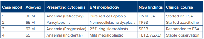
Table 1: Summary of cases.
Abbreviation: ASXL1: Additional Sex Combs-Like 1; DNMT3A: Deoxyribonucleic Acid Methyltransferase 3 Alpha; ESA: ErythropoiesisStimulating Agents; M: Male, F: Female, BM: Bone Marrow, NGS: Next-Generation Sequencing; SF3B1: Splicing Factor 3b Subunit 1; TET2: Tet Methylcytosine Dioxygenase 2; TP53: Tumour Protein p53.
Discussion
Anaemia is the most common disease in the world. It is prevalent among all age groups. The a etiopathogenesis of anaemia varies with the age of the patient. In children, worm infestation leading to iron loss from the gut is the most common cause. In females, menorrhagia and associated low iron intake are the most common causes. In adults and the elderly, there could be multiple causes of anaemia, including bleeding from haemorrhoids, alcohol intake, ingestion of non-steroidal anti-inflammatory drugs (NSAIDs) and aspirin leading to gastrointestinal ulcers, and colonic neoplasms. Most of these cases have a known pathology, and treatment is directed towards that cause. However, there is a group of refractory anaemia where the patient needs more than a routine evaluation to find out the cause of anaemia, and such patients need referral to a clinical haematologist. These patients have refractory anaemia, or cytopenias of undetermined significance, and such patients are usually elderly. MDS patients can present with anaemia or cytopenias and require bone marrow examination and genetic testing for the diagnosis.The patients presented here were interesting cases of anaemia who were refractory to treatment with haematinics, and even the bone marrow biopsy and cytogenetics by karyotyping and FISH did not reveal any abnormality. These individuals had normal peripheral blood values or only mild cytopenias that do not fulfil the diagnostic criteria for MDS. However, NGS clinched the diagnosis and guided further management. As with many other malignancies, MDS predominantly affects the elderly population with a median age at diagnosis of 60–70 years. Cytopenias and dysplasia in ≥ 10% of cells from one lineage are the sine qua non for the diagnosis of MDS.2 Diagnosis of MDS in patients with nonspecific morphological changes can be difficult. NGS can identify at least one somatic mutation in > 90% of patients with MDS.2,3 The introduction of NGS has facilitated the diagnosis of early stages of MDS and has increased our understanding of the genetic changes associated with the development and progression of MDS.4,5Supportive mutation information may be particularly helpful in cases with borderline morphologic dysplasia that complicate the use of cytopenias to establish an MDS diagnosis. The common NGS mutations found in MDS include SF3B1, TET2, ASXL1, serine and arginine rich splicing factor 2 (SRSF2), runt-related transcription factor 1 (RUNX1), DNMT3A, enhancer of zeste 2 polycomb repressive complex 2 subunit (EZH2) and TP53.6 DNMT3A mutations occur early in the course of MDS and suggest an early genetic event in leukaemogenesis. Patients with DNMT3A mutations have a worse overall survival and more rapid progression to AML.6 TP53 mutations in MDS are associated with very aggressive disease. Determination of variant allele frequency (VAF), usually > 25%, has been set for the diagnostic assessment to distinguish MDS from clonal haematopoiesis of indeterminate potential (CHIP) or CCUS.7 CHIP is said to be present when a healthy individual lacks haematological malignancy or clonal disorder, but carries a genetic mutation. Patients who have cytopenias without typical findings of MDS but have clonal mutations (by cytogenetics or NGS) are referred to as CCUS. These cases often represent a diagnostic "grey zone" where traditional morphology and gold-standard tests reach their limits. The mutation spectrum of CCUS patients is similar to mutations seen in MDS.6,7 There is no defined treatment of CCUS and patients with symptomatic cytopenias should be treated in clinical trials and those with MDS will require definitive treatment.
Conclusion:
When the diagnosis of anaemia or cytopenia is challenging, and the bone marrow and routine cytogenetics are not diagnostic of MDS, then NGS can help in resolving the diagnostic dilemma. NGS should be routinely incorporated into the diagnostic workup for refractory cytopenias to ensure accurate diagnosis, early intervention, risk stratification, and the implementation of personalised management strategies, and such patients should be referred to a clinical haematologist.
Sanjeev Kumar Sharma, Anamika Bakliwal, Anil Handoo. Bridging the Gap in the Diagnosis of Refractory
Anaemia by Next-Generation Sequencing. MMJ. 2026, March. Vol 3 (1).
References
- Bach V, Schruckmayer G, Sam I, et al. Prevalence and possible causes of anemia in the elderly: a cross-sectional analysis of a large European university hospital cohort. Clin Interv Aging.2014;9:1187–96.
- Valent P, Orazi A, Steensma DP, et al. Proposed minimal diagnostic criteria for myelodysplastic syndromes (MDS) and potential pre-MDS conditions. Oncotarget. 2017;8:73483–500.
- Bonadies N, Bacher VU. What role can next-generation sequencing play in myelodysplastic syndrome care? Exp Rev Hematol. 2019;6:379–82.se: A case report with review of the literature. Am J Surg Pathol. 2006;30:1330–6.
- Madaci L, Farnault L, Abbou N, et al. Impact of next-generation sequencing in diagnosis, prognosis and therapeutic management of acute myeloid leukemia/myelodysplastic neoplasms. Cancers. 2023;15(13):3280.
- Abdel-Wahab O, Figueroa ME. Interpreting new molecular genetics in myelodysplastic syndromes. Hematology Am Soc Hematol Educ Program. 2012;2012:56–64.
- Öz Puyan F, Alkan S. The progress of next generation sequencing in the assessment of myeloid malignancies.Balkan Med J. 2019;36(2):78–87.
- Brauninger A, Blau W, Kunze K, et al. Targeted next-generation sequencing is a sensitive tool for differential diagnosis of myelodysplastic syndromes in bone marrow trephines. J Mol Diagn. 2018;20:344–54.