

Abstract:
Plantar heel pain is most commonly attributed to plantar fasciitis or calcaneal spurs. Rarely, extra-osseous ossified lesions arising within the plantar soft tissues may present with similar symptoms and pose a diagnostic challenge. We report a rare case of symptomatic heterotopic ossification occurring entirely within the plantar soft tissue of the heel, without any attachment to the calcaneum. Imaging revealed a well-defined ossified lesion within the plantar soft tissue. Surgical excision was performed due to persistent symptoms. Histopathological examination confirmed heterotopic ossification with mature lamellar bone and no evidence of malignancy. This case highlights the importance of considering rare extra-osseous ossified lesions in the differential diagnosis of chronic plantar heel pain.
Key words: Heterotopic Ossification, Plantar Heel Pain, Soft Tissue Ossification, Heel Mass.
Introduction
Plantar heel pain is a frequently encountered orthopaedic complaint, most commonly caused by plantar fasciitis, calcaneal spur, or degenerative soft-tissue pathology.1 In contrast, ossified lesions arising within the plantar soft tissues are exceedingly rare and often misdiagnosed due to their nonspecific presentation.
Heterotopic ossification (HO) refers to the formation of mature lamellar bone in soft tissues outside the normal skeletal system.2 While commonly reported around large joints following trauma, surgery, or neurological injury, HO occurring in the plantar soft tissues of the foot is extremely uncommon, with only isolated case reports available in the literature.
We present a rare case of symptomatic HO arising entirely within the plantar soft tissue of the heel, without attachment to the calcaneum, and provide a focused review of similar reported cases.
Case Report
A 66-year-old female presented with pain and swelling over the plantar aspect of the right heel for several months. The pain was insidious in onset, gradually progressive, and aggravated by prolonged standing and walking. The patient described a sensation of “walking on a hard object.” There was no history of trauma, surgery, infection, or systemic illness.
Clinical examination
Local examination revealed a firm, well-defined swelling measuring approximately 2 × 2 cm over the plantar posteromedial aspect of the heel. The lesion was mildly tender and mobile, not fixed to the underlying calcaneum. The overlying skin was normal. Ankle and subtalar joint movements were full and painless. Distal neurovascular status was intact.
Investigations
Plain radiography
Plain radiographs of the right foot demonstrated a well-defined ossified mass located within the plantar soft tissues of the heel, with no cortical continuity or attachment to the calcaneum.
The calcaneal cortex appeared intact (Figure 1).
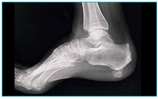
Figure 1: Pre-operative X-ray: Plain radiograph of the right foot showing a well-defined ossified mass in the plantar soft tissues of the heel, without cortical continuity or attachment to the calcaneum.
Magnetic resonance imaging (MRI) MRI revealed a well-circumscribed ossified lesion measuring approximately 1.9 × 1.3 cm within the plantar soft tissue of the heel. The lesion showed signal characteristics similar to mature bone and was surrounded by fibrous tissue. There was no involvement of the calcaneum, plantar fascia origin, or adjacent musculotendinous structures, and no aggressive features were identified (Figure 2).
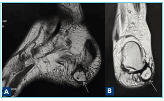
Figure 2A and B: Magnetic resonance imaging (MRI) images: MRI of the right heel showing a well-circumscribed ossified lesion within the plantar soft tissues, without involvement of the calcaneum, plantar fascia, or adjacent structures.
Management Given the persistent pain and functional limitation despite conservative measures, surgical excision was planned (Figure 3A–G). A medial plantar approach was used. Intraoperatively, a well-encapsulated ossified mass was identified within the plantar soft-tissue plane. The lesion was clearly separate from the calcaneum and plantar fascia and was excised completely (Figure 3E).
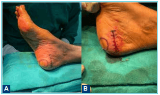
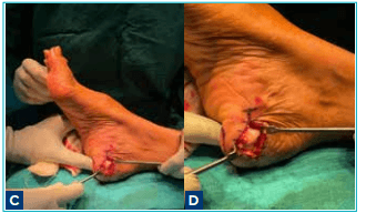
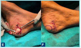
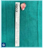
Figure 3A-G Intraoperative process: Preoperative clinical photograph showing the marked area of maximal tenderness over the plantar heel; intraoperative images demonstrating exposure and complete excision of a wellcircumscribed heterotopic ossified mass from the plantar soft tissues without attachment to the calcaneum; excised specimen; and postoperative photographs showing wound closure.
Histopathological examination
Microscopic examination demonstrated a wellcircumscribed lesion composed of mature lamellar bone with central fatty marrow spaces, surrounded by fibrous tissue. No cellular atypia or malignant features were identified, confirming the diagnosis of heterotopic ossification (Figure 4).
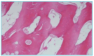
Figure 4: Histopathological examination: Photomicrograph showing mature lamellar bone with fatty marrow spaces, surrounded by fibrous tissue, without atypia, consistent with heterotopic ossification.
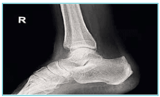
Figure 5: Post-operative X-ray: Postoperative plain radiograph of the right foot demonstrating complete excision of the plantar ossified mass, with no residual lesion visible.
Outcome and follow-up
The postoperative period was uneventful. The patient reported complete resolution of heel pain and returned to normal ambulation. No recurrence was noted on followup (Figure 5).
Discussion
Extra-osseous ossified lesions of the plantar heel are rare and can mimic common causes of heel pain, such as plantar fasciitis or calcaneal spurs. The differential diagnosis includes heterotopic ossification, myositis ossificans, extra-osseous osteochondroma, tumoral calcinosis, and malignant soft tissue tumours.3
Unlike calcaneal spurs or osteochondromas, which demonstrate continuity with the parent bone, heterotopic ossification is characterised by ossification within soft tissues without skeletal attachment. MRI is particularly valuable in confirming the extra-osseous location and excluding aggressive pathology.
Previously reported rare similar cases
Only a limited number of similar cases involving ossified lesions in the plantar or hindfoot soft tissues have been reported:
- Danya et al. reported a case of heterotopic ossification involving the hindfoot soft tissues, completely isolated from the underlying bone and confirmed by histopathology.4
- Trankovskiy et al. described ossification within the plantar soft tissues in the region of the calcaneus, presenting with pain during ambulation and a palpable plantar mass, similar to the present case.5
- Singh et al. reported an extra-osseous osteochondroma arising from the superficial fascia of the heel, presenting as a well-defined ossified plantar mass without attachment to the calcaneum.6
- Earlier reports have also documented soft tissue osteochondromas of the heel pad, emphasising that well-circumscribed ossified lesions can arise within plantar soft tissues independent of the calcaneum. 7
These reports, together with the present case, emphasise that extra-osseous ossified lesions of the plantar heel, although rare, are well-documented entities and should be included in the differential diagnosis of chronic plantar heel pain associated with a firm mass.
Consent
The patient provided informed written consent for publication of this case report and accompanying images.
Conflict of interest
The authors declare that they have no conflict of interest.
Funding
No financial support was received for this study
Conclusion:
Heterotopic ossification occurring entirely within the plantar soft tissue of the heel, without attachment to the calcaneum, is extremely rare. Awareness of this entity is essential to avoid misdiagnosis. Imaging and histopathological evaluation are crucial for accurate diagnosis, and surgical excision offers excellent symptomatic relief with low risk of recurrence.
Heterotopic ossification within the plantar soft tissues of the heel is an extremely rare cause of chronic plantar heel pain and should be considered in patients with persistent symptoms unresponsive to conservative treatment.
Dilveer Brar, Vishal Sharma, Kheman Grover. Symptomatic Heterotopic Ossification in the Plantar Soft
Tissue of the Heel: A Rare Cause of Chronic Plantar Heel Pain. MMJ. 2026, March. Vol 3 (1).
References
- Buchbinder R. Plantar fasciitis. N Engl J Med.2004;350(21):2159–66.
- Kaplan FS, Glaser DL, Hebela N, et al. Heterotopic ossification. J Am Acad Orthop Surg. 2004;12(2):116–25.
- Tyler P, Saifuddin A. The imaging of myositis ossificans. Semin Musculoskelet Radiol. 2010;14(02),201–16.
- Danya F, Marco B, Valentino C, et al. Case report: unusual heterotopic ossification of the hindfoot. Front Surg. 2022;9:917560.
- Trankovskiy SE, Grabovsky MB, Alpatov VN, et al. A clinical case of ossification in soft tissues of the plantar surface in projection of the calcaneus. Russ J Pediatr Surg. 2024;28(6):602–6.
- Zhu S, Zeng J, Zhang Z, et al. Extraosseous osteochondroma of superficial fascia layer of the heel: A case report and review of literature. Medicine (Baltimore). 2022;101(49):e32014.
- Singh R, Jain M, Siwach R, et al. Soft-tissue osteochondroma of the heel pad: a case report and review of literature. Foot Ankle Surg. 2010;16(3):e76–e78