

Abstract:
Serum sickness (SS) is an immune-complex–mediated illness that frequently occurs in patients after polyclonal antibody therapy, such as thymoglobulin. Although SS has been described secondary to thymoglobulin therapy in adults, the administration of horse antithymocyte globulin (ATG) has been associated with SS in 1% to 10% of cases. Early recognition and accurate diagnosis are key for managing thymoglobulin-induced SS, as treatment is highly effective in achieving good outcomes.
Key words: Antithymocyte globulin, ATG, Neutropenia, Serum Sickness, SS
Introduction
Severe acquired aplastic anaemia (AA) is a life-threatening disorder characterised by pancytopenia and bone marrow hypocellularity.1 T-cell-mediated destruction of haematopoietic progenitor and stem cells is implicated in the pathogenesis of most cases. Overall, 60%–70% of patients with AA achieve a haematological response after a single course of immunosuppressive therapy (IST). The most effective IST regimen consists of horse antithymocyte globulin (hATG) in combination with ciclosporin A, which has been shown to be superior to either agent alone or to combinations other than hATG with ciclosporin.2,3 ATG is a heterologous antiserum derived from animals immunised with human lymphocytes. Most large clinical trials in AA have utilised hATG, which is therefore considered the standard for AA treatment. As foreign proteins, antithymocyte globulin (ATG) preparations can elicit strong immune responses in humans.4 The formation of circulating immune complexes between foreign antigens and host antibodies may result in tissue deposition, leading to serum sickness (SS), which typically occurs around 7–10 days after initiation of ATG therapy.4
Case Report
A 50-year-old female presented with complaints of generalised weakness and decreased appetite. There was no history of fever or bleeding from any site.
A complete blood count (CBC) showed haemoglobin (Hb) of 3.8 g/dL, total leukocyte count (TLC) of 5,570/uL, platelet count of 50,000/uL, and neutrophils of 36%. Bone marrow examination was suggestive of pancytopenia with severe neutropenia, and bone marrow biopsy revealed < 10% cellularity. Paroxysmal nocturnal haemoglobinuria (PNH) testing was negative. She received a total of seven units of blood transfusion over two months at her local centre.
She was started on ciclosporin, a thrombopoietin receptor agonist, and danazol; however, danazol was discontinued after 15 days. After two months of therapy, she came to our centre, where investigations showed Hb of 7 g/dL, TLC of 6,810/uL, platelet count of 33,000/uL, and neutrophils of 43%. Liver and kidney function tests were normal, and viral markers were negative. Since she did not show any significant improvement, she was started on hATG (Thymogam) at a dose of 40 mg/kg/day for four days, along with ciclosporin 4.5 mg/kg/day and eltrombopag 150 mg daily.
Prior to ATG infusion, premedication with antihistamines, hydrocortisone, and antipyretics was administered. During the first day of ATG infusion, she developed fever with rigors without hypotension, for which antipyretics and hydrocortisone were repeated. She tolerated the remaining three days of ATG infusion without complications and was discharged on Day 5. At discharge, she was continued on prednisolone 1 mg/kg/day along with other supportive medications. Twelve days after intravenous (IV) thymoglobulin therapy, she presented with chills, lip swelling, myalgias, right eye conjunctival haemorrhage, rash over both legs, and bilateral sole pain with itching (Figure 1A and B).
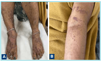
Figure 1A and B: Initial presentation of serum sickness.
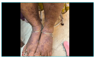
Figure 2: On Day 3 of steroid therapy.
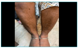
Figure 3: On Day 6 of steroid therapy
She was diagnosed with SS secondary to IV ATG and was started on IV methylprednisolone 120 mg stat, followed by prednisolone 2 mg/kg/day for three days, with subsequent tapering. By Day 3 and Day 6 of steroid therapy, she showed marked symptomatic improvement (Figures 2 and 3) and is continuing on regular outpatient follow-up.
Discussion
ATGs are antibodies used for immunosuppression against human T cells and are typically derived by injecting human thymocytes into animals such as horses or rabbits.5 ATG is believed to protect allografts by reducing inflammatory damage, modulating allorecognition processes, and attenuating the immune response, while increasing sensitivity to oral maintenance IST.6
The administration of rabbit or horse ATG has been associated with SS in approximately 1%–10% of cases.4,7,8 Risk factors include older age, higher levels of heterologous protein, type of ATG preparation, prior exposure to the antigen or source animal, and a history of hypergammaglobulinaemia or cryoglobulinaemia.4 SS is characterised by fever, lymphadenopathy, pruritic rash, polyarthralgia, and polyarthritis, typically developing 7–10 days after exposure to the exogenous antigen.6 A maculopapular or urticarial rash is usually the earliest manifestation. In severe cases, immunecomplex–mediated small-vessel vasculitis may lead to glomerulonephritis and tissue injury.6 Angioedema may also be present. Polyarthritis commonly involves large joints but may occasionally affect the spine or temporomandibular joint. The clinical presentation can be nonspecific and may mimic acute infections or rheumatologic conditions, leading to delays in diagnosis. SS is primarily a clinical diagnosis, and treatment should not be delayed while awaiting laboratory confirmation.6
SS is generally self-limiting and resolves once the inciting antigen is cleared. Due to its rarity, current treatment recommendations are based on clinical experience and case reports. Discontinuation and avoidance of re-exposure to the offending agent are essential. Mild cases may be managed symptomatically with non-steroidal anti-inflammatory drugs (NSAIDs) for fever and arthralgia, antihistamines, and topical corticosteroids for pruritic rash.6 Severe cases — especially those with disabling symptoms, haemodynamic instability, glomerulonephritis, or vasculitis — require high-dose oral or IV corticosteroids. These are typically administered for three days, followed by a rapid taper, with symptom resolution expected within 10 ± 2 days.6
Conclusion:
Severe acquired AA remains a life-threatening condition in which IST with hATG and ciclosporin represents the standard of care for patients who are not candidates for stem cell transplantation.1,2 While ATG-based regimens are highly effective, they are associated with immune-mediated adverse effects such as SS4 Prompt clinical recognition and early initiation of high-dose corticosteroids resulted in rapid symptom resolution and a favourable outcome.6
Krishna Kant Singh. Delayed Serum Sickness after Immunosuppressive Therapy with Horse
Antithymocyte Globulin in Severe Aplastic Anaemia: A Case Report. MMJ. 2026, March. Vol 3 (1).
References
- Scheinberg P, Young NS. How I treat acquired aplastic anemia. Blood. 2012;120(6):1185–96.
- Rosenfeld S, Follmann D, Nunez O, et al. Antithymocyte globulin and cyclosporine for severe aplastic anemia: association between hematologic response and long-term outcome. JAMA. 2003;289(9):1130–5.
- Scheinberg P, Nunez O, Weinstein B, et al. Horse versus rabbit antithymocyte globulin in acquired aplastic anemia. N Engl J Med. 2011;365:430–8.
- Suwansrinon K, Jaijareonsup W, Wilde H, et al. Sexand age-related differences in rabies immunoglobulin hypersensitivity. Trans R Soc Trop Med Hyg. 2007;101:2006.
- Bacigalupo A, Broccia G, Corda G, et al. Antilymphocyte globulin, cyclosporin, and granulocyte colony-stimulating factor in patients with acquired severe aplastic anemia (SAA): a pilot study of the EBMT SAA Working Party. Blood. 1995;85(5):1348–53.
- McPhee SJ, Papadakis MA, Rabow MW. Current Medical Diagnosis & Treatment 2011. 50th Edition. New York: McGrawHill; 2010.
- Boothpur R, Harding KL, Skelton RM, et al. Serum sickness after treatment with rabbit antithymocyte globulin in kidney transplant recipients with previous rabbit exposure. Am J Kidney Dis. 2010;55:141.
- Shah S, Jain P, Shah K, et al. Immunosuppressive therapy for aplastic anemia: a single-center experience from western India. Ann Hematol. 2019;98(1):41–6.