 WhatsApp Us (24/7)
 +91 926 888 0303 (24/7)


Abstract:
Hydatid disease (echinococcosis) is a parasitic infection caused by Echinococcus granulosus or Echinococcus multilocularis. While the liver is the most common site of infection, intracranial hydatid cysts are rare, accounting for less than 2% of cases. This case report describes a 43-year-old male presenting with a history of recurrent generalised tonic-clonic seizures and a single episode of seizure accompanied by a headache. Imaging, including computed tomography (CT) and magnetic resonance imaging (MRI), revealed a ruptured intracranial hydatid cyst, a rare complication of this parasitic infection. The report discusses the diagnostic challenges, the critical role of imaging in identifying such cysts, and the clinical management of the condition. Prompt diagnosis with the help of imaging and appropriate interventions is essential in managing ruptured intracranial hydatid cysts to prevent fatal outcomes.
Key words: Hydatid Cyst, Intracranial Hydatid Cyst, Ruptured Intracranial Hydatid Cyst, MR Spectroscopy, Contrast Enhanced MRI, Constructive Interference in Steady State MRI, Diffusion Weighted MRI, Susceptibility Weighted MRI.
Introduction
Hydatid disease, also known as echinococcosis, is a parasitic infection caused by the tapeworm Echinococcus granulosus or Echinococcus multilocularis in the larval stage. The faecal-oral route of infection infects humans when they come into contact with dogs or eat food tainted with the parasite's ova. South America, Australia, the Middle East, and some regions of North Africa are far more likely to have it than North America and Europe1. Although the disease most frequently affects the liver, it can also occasionally impact the brain. Intracranial hydatid cysts account for less than 2% of call cases, making them extremely rare2. These cysts develop through the haematogenous spread of the parasite, with the brain serving as an unusual secondary site of infection.
When an intracranial hydatid cyst ruptures, the results can be fatal. Acute neurological symptoms, such as seizures, focal deficits, and signs of elevated intracranial pressure, may be present in patients. Imaging techniques, particularly CT and MRI, play a vital role in identifying these cysts and their complications. This case report highlights a ruptured intracranial hydatid cyst, focusing on the imaging findings, surgical management, and the broader clinical implications of such a rare condition.
Case Report
A 43-year-old man presented to the emergency department with an episode of seizure following a headache lasting one day. He had a history of 3-4 episodes of generalised tonic-clonic seizures over the past 2 months. There was no history of fever, limb weakness, diplopia, or decreased vision. Additionally, there was no significant past medical or surgical history.
Clinical Findings
On neurological examination, the patient was conscious and oriented. The Glasgow Coma Scale (GCS) score was 4 for eye opening, 5 for verbal response, and 6 for motor response (E4V5M6). Bilateral pupils measured 3 mm and were reactive to light. Muscle power was 5/5 in all four limbs. The rest of the systemic examination was normal.
Radiological findings
Revealed well-defined bi-lobed T2 hyperintense cystic lesion in the left temporo-occipital lobe with surrounding vasogenic oedema (Figures 1a, b, c). A few tiny susceptibility foci were seen along the walls of both the cystic components, likely representing calcifications (Figure 1d). The axial 3D constructive interference in steady state (CISS) image revealed focal communication between the two cystic components, suggestive of a ruptured parent hydatid cyst with resultant daughter cyst formation (Figure 1e). Thin smooth peripheral contrast enhancement along both the cystic components, with evidence of increased relative cerebral blood volume and blood flow was seen along the periphery of the lesion (Figures 2a, b, c). On magnetic resonance (MR) spectroscopy, a small peak was noted at 2.37 ppm, likely suggesting a pyruvate peak (Figure 2d, red arrow), along with elevated choline and lactate (Figure 2e).
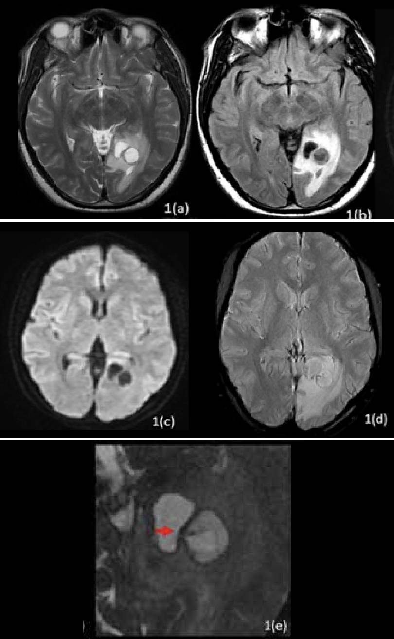
Figure 1: (a) Well-defined bi-lobed T2 hyperintense cystic lesion in the left temporo-occipital lobe with surrounding vasogenic oedema. (b) Medially located cystic component shows complete signal suppression on Fluid-Attenuated Inversion Recovery (FLAIR), whereas the laterally located component shows predominant central signal suppression on FLAIR. (c) No significant diffusion restriction is seen. (d) A few tiny susceptibility foci are seen along the walls of both the cystic components, likely representing calcifications. (e) Axial 3D Constructive Interference in Steady State (CISS) image reveals focal communication between the two cystic components, along with a thin linear membranous structure protruding from the laterally located cystic component (parent cyst) via the defect into the medial cystic component (daughter cyst). Findings suggest rupture of the parent hydatid cyst with resultant daughter cyst formation.
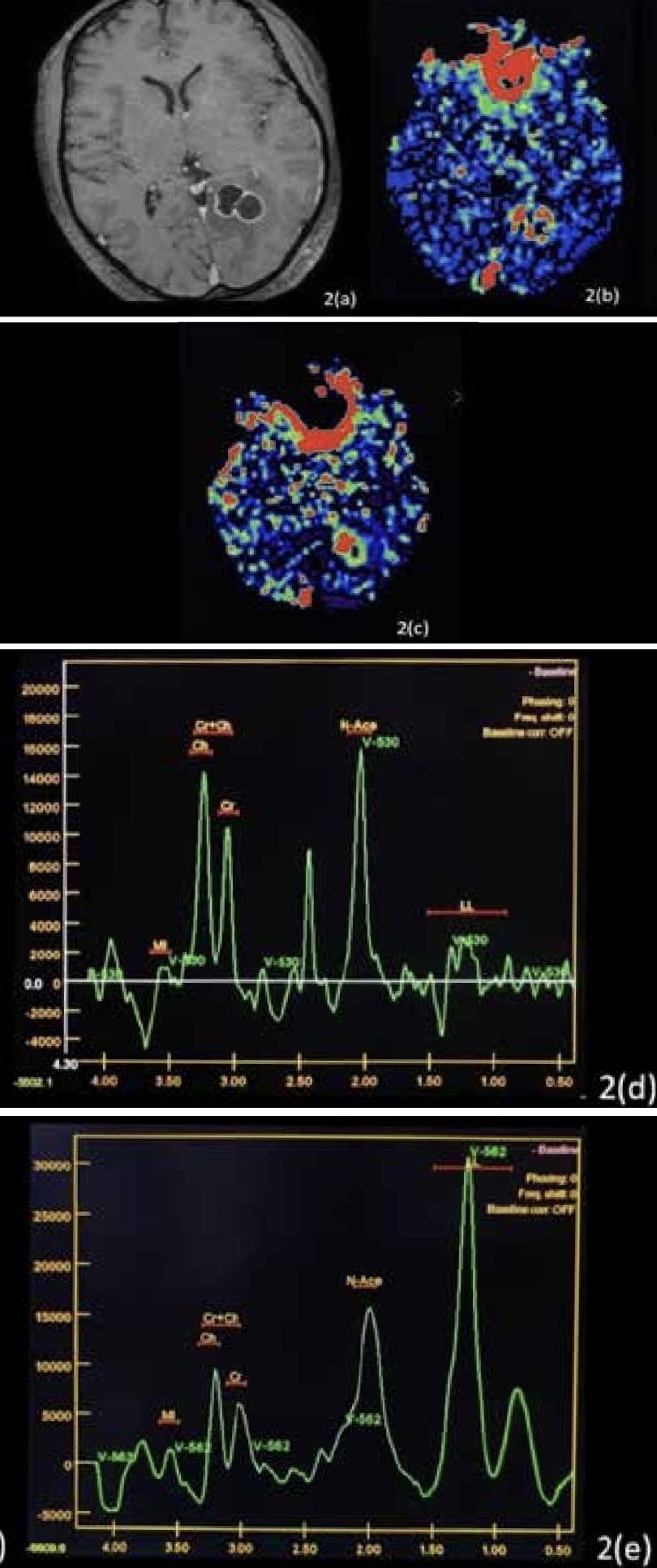
Figure 2: (a) Reveals thin smooth peripheral contrast enhancement along both the cystic components. (b) Evidence of increased relative cerebral blood volume (c) Blood flow seen along the periphery of the lesion. On MR spectroscopy: (d) a small peak is noted at 2.37 ppm likely suggesting a pyruvate peak (red arrow). (e) Elevated choline as well as lactate.
The possibility of a ruptured hydatid cyst was considered in view of the radiological findings. The presence of communication between the two cystic components and the thin linear membranous structure was highly suggestive of a hydatid cyst. Also, a pyruvate peak is considered specific for hydatid cysts. However, in view of the increased relative cerebral blood flow and volume, a differential diagnosis of neoplastic aetiology was also considered.
Management
The patient was treated with corticosteroids to manage cerebral oedema and anticonvulsants to control seizures. After a neurosurgical evaluation, a left parieto-occipital craniotomy with gross total excision of the lesion was performed. During surgery, an intra-axial, firm, cystic lesion with defined margins and mild vascularity was revealed. The surgical team meticulously removed the cyst and its contents. The tissue was sent for histopathological examination, which confirmed the diagnosis of hydatid disease, showing a few entrapped hooklets and calcareous bodies with surrounding foreign body giant cell reaction. No definite protoscolices or laminated membrane were found.
Discussion
Hydatid disease is caused by Echinococcus granulosus or Echinococcus multilocularis, and brain involvement is very rare, with incidence of less than 2% in cases.2 The parasite’s oncospheres migrate to the brain, with the supratentorial location being the most common, through the terminal branches of middle cerebral artery, usually in the temporo-parieto-occipital region.3
A ruptured intracranial hydatid cyst is an even rarer event that can lead to severe complications. The primary cysts are fertile because they have scolices and brood capsules; hence, rupture of primary cysts can result in recurrence.4 Leakage of cyst content causes an inflammatory reaction, worsening vasogenic oedema and contributing to seizures, focal neurological deficits, and raised intracranial pressure as observed in this case.
A spherical, well-defined, smooth, thin-walled, homogenous cystic lesion with a fluid density comparable to that of the cerebrospinal fluid (CSF) is seen on both CT and MRI, with or without septations or calcification. Both T1-weighted and T2- weighted images typically display a ring of low signal intensity around the cyst wall. Most instances show compression of the ventricles and midline structures; however, surrounding oedema and rim enhancement are usually absent in untreated or uncomplicated cases.5,6Lactate, alanine, and pyruvate are detected in the lesion by MR spectroscopy. Pyruvate is very specific to hydatid cyst. 7,8
The definitive treatment of cerebral hydatid cysts is primarily surgical. The goal remains to completely excise the cyst without any spill to avoid recurrence. Cyst rupture during surgery may occur in 28% of the cases. Cyst rupture into the subarachnoid space may lead to widespread dissemination, followed by a severe anaphylactic response, in addition to a higher recurrence rate.9,10 Albendazole can be used both pre- and post-operatively to sterilise the cyst, lower the risk of anaphylaxis, and decrease the tension on the cyst wall, which lowers the frequency of recurrence and the danger of spilling during surgery. In our case, total excision of the lesion was possible without any complications.11
Radiological differential diagnosis: The imaging features in this case helped distinguish hydatid disease from other possible diagnoses:
- Cystic tumors (e.g., glioblastoma or craniopharyngioma): These typically show irregular or nodular peripheral enhancement on post-contrast imaging. The solid component also shows diffusion restriction along with significant elevation of choline peak on MR spectroscopy.
- Brain abscesses: Abscesses often demonstrate restricted diffusion on diffusion-weighted imaging (DWI) and are associated with ring-enhancing lesions. The absence of diffusion restriction in our case ruled out this possibility
Conclusion
This case report emphasises the rarity of ruptured intracranial hydatid cysts. Imaging features, especially the presence of daughter cysts, are key to diagnosis. The diagnosis is confirmed by histopathological investigation, and surgical removal continues to be the cornerstone of treatment. Clinicians are reminded by this case to consider hydatid disease when making a differential diagnosis for cystic brain lesions, particularly in areas where it is endemic. Early detection and treatment are essential to improving outcomes for this potentially fatal illness.
Shashank Raj, Prem Kumar Ganesan, Anchal Singh. Ruptured Intracranial Hydatid Cyst. MMJ. 2025,
March. Vol 1 (5).
References
- Tuzun Y, Kadioglu HH, Izci Y, et al. The clinical, radiological and surgical aspects of cerebral hydatid cysts in children. Pediatr Neurosurg. 2004;40:155-60.
- bbassioun K, Rahmat H, Ameli NO, et al. CT in hydatid cyst of the brain. J Neurosurgery. 1985;62:781-2.
- Ciurea AV, Fountas KN, Coman TC, et al. Long-term surgical outcome in patients with intracranial hydatid cyst. Acta Neurochir (Wien). 2006;148:421-6.
- Gupta S, Desai K, Goel A. Intracranial hydatid cyst: A report of five cases and review of literature. Neurol India. 1999;47:214-7. 5. Bükte Y, Kemaloglu S, Nazaroglu H, et al. Cerebral hydatid disease: CT and MR imaging findings. Swiss Med Wkly. 2004;134:459-67.
- Polat P, Kantarci M, Alper F, et al. Hydatid disease from head to toe. Radiographics 2003;23:475-94.
- Kohli A, Gupta RK, Poptani H, et al. In vivo proton magnetic resonance spectroscopy in a case of intracranial hydatid cyst. Neurology. 1995;45(Pt 1):562-4.
- Jayakumar PN, Srikanth SG, Chandrashekar HS, et al. Pyruvate: an in vivo marker of cestodal infestation of the human brain on proton MR spectroscopy. J Magn Reson Imaging. 2003;18:675- 80.
- Najjar MW, Rajab Y, El-Beheiri Y. Intracranial hydatid cyst. Dilemma in diagnosis and management. Neurosciences (Riyadh). 2007;12:249-52.
- Onal C, Unal F, Barlas O, et al. Long-term follow-up and results of thirty pediatric intracranial hydatid cysts: half a century of experience in the Department of Neurosurgery of the School of Medicine at the University of Istanbul (1952–2001). Pediatr Neurosurg. 2001;35:72-81.
- Horton RJ. Albendazole in treatment of human cystic echinococcosis: 12 years of experience. Acta Trop. 1997;64:79- 93.