 WhatsApp Us (24/7)
 +91 926 888 0303 (24/7)


Abstract:
The craniovertebral junction (CVJ) is the most complex part of the axial skeleton, residing between the skull and the upper cervical spine. The maximum mobility of the neck is provided by the first and second cervical vertebrae (C1-C2), making it more prone for dislocation. Rotatory atlantoaxial dislocation (AAD) is rare, and when it occurs in combination with a large head and wasting of neck muscles, it presents a complex problem for correction. Here, we present a similar case which was successfully managed with C1-C2 spinal fusion, without causing any deficits to the patient and significantly improved the patient’s quality of life.
Key words: Craniovertebral Junction (CVJ) Anomaly, C1-C2 Fixation, Congenital Spinal Anomaly, Atlantoaxial Subluxation.
Introduction
The craniovertebral junction (CVJ) is the most complex part of the axial skeleton, residing between the skull and the upper cervical spine. Congenital, developmental, and acquired bony abnormalities result in compression and distortion of the neural structures, the vertebrobasilar vascular system, and the cerebrospinal fluid (CSF) channels. The clinical significance of these abnormalities has only been recognised recently, and mistaken diagnoses have led to delayed treatment and, at times adverse outcomes.1
Classification of atlanto-axial dislocations (AAD)
Atlanto-axial dislocations (AADs) may be classified into four types depending on the direction and plane of the dislocation, namely anteroposterior, rotatory, central, and mixed dislocations.
- Antero-posterior dislocation
- Mobile dislocation occurs in one plane and one direction. This is due to laxity of the transverse ligament. As a result, the second cervical vertebra (C2) dislocates posteriorly during flexion and realigns (reduces) in extension. Its anterior movement is prevented by the intact anterior arch of the atlas.2,3
- Hypermobile dislocation occurs in one plane and two directions. When os odontoideum is present, the C2 vertebral body movements cannot be restricted by the transverse ligament and anterior arch of atlas. Consequently, the C2 body dislocates in both directions within the sagittal plane—posteriorly under the transverse ligament during flexion and anteriorly under the arch of atlas during extension. It is important to identify this hypermobile variety before surgery, as standard intubation practices can pose a serious risk. During intubation, anaesthetists generally extend the patient’s neck, which can result in anterior dislocation of C2 and potential spinal cord injury. To prevent this, fibre-optic intubation is recommended, with a cervical collar in place to maintain stability. Flexion or extension of the neck should be strictly avoided during intubation.3,4
- Central dislocation
- The opposing facetal surfaces of the normal C1-C2 joints are typically horizontal and parallel in the sagittal plane. However, if these joints are oriented obliquely in the sagittal plane, then the C2 body tends to slip upwards due to the weight of the patient's head during flexion movements. Therefore, bilateral sagittal plane obliquity of these joints results in the telescoping of the C2 body into the C1 ring, leading to central dislocation. This telescoping invariably causes posterior movement of the C2 vertebra. In this condition, C1 is assimilated with the occiput, and there is fusion between the second and third cervical vertebra (C2-C3). Many neurosurgeons and radiologists mistakenly report this condition (after examining only sagittal views) as dolichoodontoid and basilar invagination (BI). In reality, it is the fusion of the C2-C3 bodies that causes the C2 body and odontoid process to be misidentified as dolichoodontoid. Furthermore, this radiological finding does not represent BI; rather it is central dislocation of C2. On axial view of a computed tomography (CT) scan, one can observe the C2 body positioned behind the anterior arch of the atlas (C1), along with posterior dislocation of C2.
- Central dislocation
Rotatory dislocation typically occurs in a single plane (axial) and in one direction only, either to the right or to the left. This results from the incompetence of the alar ligament. It usually occurs in children and presents as the classical ‘Cocked Robin’ position of the head.5,6
- Mixed dislocation
This dislocation involves displacement in two planes and directions: (a) central and posterior - central dislocation may be associated with posterior dislocation in many cases; (b) posterior and rotatory dislocation - a combination of posterior displacement along with rotation.
However, from the surgical point of view, these are divided into two categories: reducible and irreducible. Posterior fusion is the treatment of choice for rotatory AAD, using the transarticular screw fixation method). Often, irreducible AAD is due to inadequate extension in a dynamic X-ray study, which may also result from muscle spasm. If the anatomy at the occipito-atlantoaxial region (O-C1-C2; O: occiput, C1: atlas, C2: axis) appears normal on X-ray, the dislocation should be reducible. In cases where congenital anomalies at O-C1-C2 and irreducible AAD are observed on flexion/extension studies of the cervical spine, the C1-C2 joints should be evaluated using a CT. If the C1-C2 joint facet surfaces are normal, the AAD should be reducible with cervical traction or during surgery by mobilising the joints.
The commonly used classification system for rotatory AAD is Fielding’s classification, as described in Figure 1 and Table 1.7
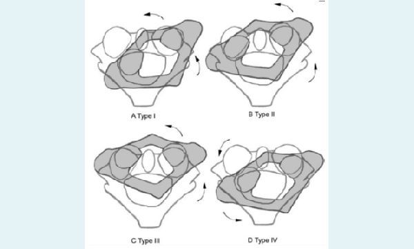
Figure 1: Fielding classification of rotatory atlantoaxial dislocation (AAD).
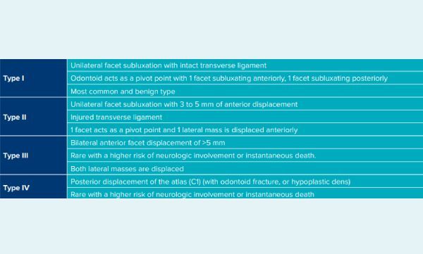
Figure 2: Fielding classification of rotatory atlantoaxial dislocation (AAD).
Discussion
Our patient was an 8-year-old boy with a Fielding Type I rotatory AAD. His neck deformity had progressively worsened, and by the time he presented to us, he had severe wasting of the neck muscles (Figure 2). At birth, he had undergone surgery for hydrocephalus and had received a ventriculoperitoneal shunt. The power in his upper and lower limbs was 5/5, with no features of myelopathy. The patient was ambulant on his own, however, due to the neck tilt, he was unable to carry out his daily activities.
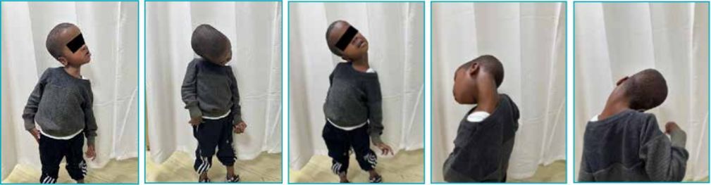
Figure 2: Preoperative images of the patient showing the neck deformity.
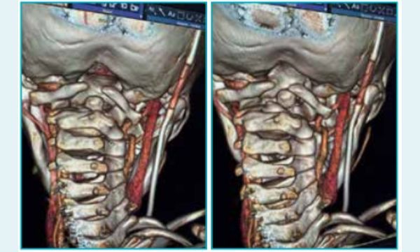
Figure 3: 3-dimensional reconstructed images of craniovertebral junction with head position held in supine position, showing reducibility of the C1-C2 joint.
As seen in Figure 2, the patient had a gross neck deformity that hampered his daily activities. The deformity reduced spontaneously when the patient was in a supine position. A preoperative CT scan of the CVJ was taken in the supine position, showed spontaneous reduction of the rotatory AAD. It also revealed that the left vertebral artery was atrophic and that the posterior arch of C1 was deficient.
The patient underwent C1- C2 transpedicular fixation with Goel’s screws and plate system. The most important step of the surgery required achieving good positioning in anatomically neutral position with cervical traction using Gardner-Wells tongs. The C1-C2 joint space was then opened, and intraoperative reduction was achieved. Joint fusion was achieved by introducing bone chips into the C1-C2 joint space. The screw-plate assembly was placed under continuous C-arm guidance.
Figure 5 illustrates the patient’s positioning in the operating theatre, while Figure 6 shows the C-arm images after achieving the desired C1 C2 fixation.
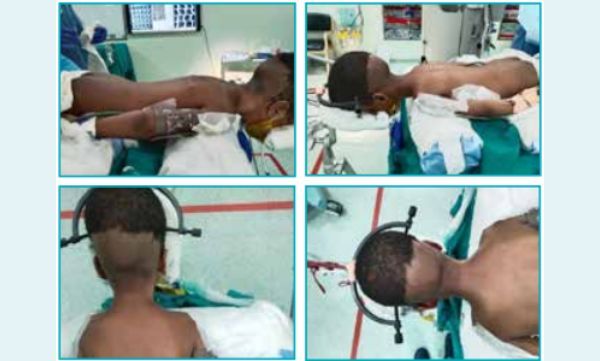
Figure 4: Positioning of the patient was done with cervical traction to reduce the atlantoaxial dislocation (AAD).
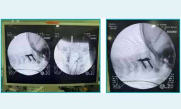
Figure 5: Intraoperative C-arm images after final fixation.
In the postoperative period, the patient was initially kept in a Philadelphia collar, but due to severe neck muscle wasting, the neck was not as stable as required. Therefore, a custommade Sternal Occipital Mandibular Immobiliser (SOMI) brace was applied to maintain the neck in the correct posture. Neck immobilisation in the postoperative period is as essential as intraoperative fixation. It helps facilitate the fusion of the C1-C2 joint, thus permanently stabilising it. No additional neurological deficits were noted in the postoperative period. Figure 5 shows the immediate postoperative status of the patient, while Figure 6 shows follow up after 4 weeks. The SOMI brace was further modified to meet the needs of neck immobilisation.
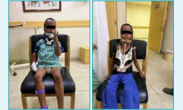
Figure 6: Immediate postoperative image showing straightening of neck. Sternal Occipital Mandibular Immobiliser (SOMI) brace was applied to stabilise the neck.
The patient achieved a reasonable correction of deformity along with CVJ stability. Isometric neck exercises were gradually introduced. Currently, the patient is able to walk unaided, and the neck deformity has significantly reduced.
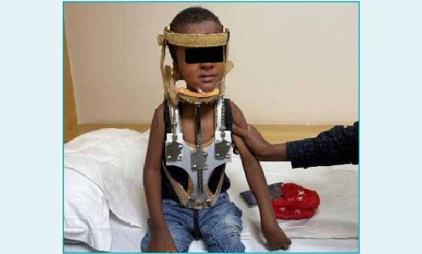
Figure 7: Follow up images. Few customised modifications were done to the Sternal Occipital Mandibular Immobiliser (SOMI) brace to better support the neck.
Conclusion
Complex CVJ anomalies require individualised treatment to achieve optimum outcomes. Multiple options are available for AAD, but the preferred treatment remains posterior fixation and stabilisation of the C1-C2 joint. The individual anatomy and associated complexities should be considered together to guide the treatment choice.
Rohit Bansil, Akash Singh, Nitin Chore. Management of Complex Craniovertebral
Junction (CVJ) Anomaly. MMJ. 2025, March. Vol 1 (5).
References
- Menezes AH. Craniovertebral junction anomalies: diagnosis and management. Semin Pediatr Neurol. 1997;4(3):209-23.
- Salunke P, Behari S, Sharma MS, et al. Pediatric congenital atlantoaxial dislocation: Focusing on the differences between the irreducible and reducible varieties. J Neurosurg Pediatrics. 2006;104(2 Suppl):115-22
- 17. Behari S, Bhargava V, Nayak S, et al. Congenital reducible atlantoaxial dislocation: Classification and surgical considerations. Acta Neurochirurgica (Wien). 2002;144:1165-77
- Behari S, Jain VK, Phadke RV, et al. C1-2 rotary subluxation following posterior stabilization for congenital atlantoaxial dislocation. Neurol India. 2000;48:164-9.
- Pang D, Li V. Atlantoaxial rotatory fixation: Part 1-Biomechanics of normal rotation at the atlantoaxial joint in children. Neurosurgery. 2004;55:614-26.
- Amini B Fielding and Hawkins classification of atlantoaxial rotatory subluxation. Case study, Radiopaedia.org (Accessed on 17 Jan 2025.) https://doi.org/10.53347/rID-42320
- Behari S, Bhargava V, Nayak S, et al. Congenital reducible atlantoaxial dislocation: Classification and surgical considerations. Acta Neurochirurgica (Wien). 2002;144:1165-77.