 WhatsApp Us (24/7)
 +91 926 888 0303 (24/7)


Abstract:
Thrombolysis during cardiopulmonary resuscitation (CPR) is always a dilemma while treating massive pulmonary embolism (PE). However, cardiac arrest due to massive PE can benefit from thrombolysis during CPR, and resuscitation may need to be prolonged for up to an hour. Here we present the case of a 50-year-old woman with massive PE who had cardiac arrest while receiving an alteplase infusion. She survived after receiving an additional bolus of tenecteplase during CPR, which lasted 50 minutes.
Key words: Pulmonary Embolism, Cardiac Arrest, Thrombolysis.
Introduction
Cardiac arrest due to pulmonary embolism (PE) has a mortality rate of up to 70% within the first hour of presentation and an overall mortality rate of up to 95%.1,2 Although there are no clear guidelines for thrombolysis in cardiac arrest due to PE, findings from the French National Cardiac Arrest registry recommend thrombolysis and demonstrate a survival benefit.3 However, a single bolus dose may not be sufficient, especially during cardiac arrest. There is limited experience in administering a second bolus dose, especially with a different thrombolytic agent, during cardiopulmonary resuscitation (CPR). Here we present a case report of massive PE leading to cardiac arrest, successfully treated with an initial bolus of alteplase followed by a second bolus of tenecteplase, along with nearly 50 minutes of CPR. The patient survived without any neurological deficit.
Case Report
A 50-year-old woman with chronic hypertension presented with an episode of giddiness in the morning, followed by progressive breathlessness and non-specific bilateral lower limb pain. She was referred from a local hospital with a diagnosis of acute coronary syndrome after receiving a loading dose of Ecosprin and Clopilet, along with an injection of enoxaparin (40mg), as her high-sensitivity troponin I was positive.
On arrival at the emergency department, the patient was tachypnoeic (respiratory rate [RR] - 34) but maintaining a saturation of 94% on room air, with a heart rate (HR) of 100/min and blood pressure (BP) of 100/60 mmHg. An electrocardiogram (ECG) was suggestive of right ventricular strain with right bundle branch block (RBBB). The ECG also showed the classic S1Q3T3 pattern of PE. A 2-dimensional (2D) echocardiographic screening revealed a right atrial clot with right ventricular distension. Urgent computed tomography (CT) pulmonary angiography revealed a saddle thrombus in the main pulmonary artery, extending into the right and left pulmonary artery and segmental branches (Figure 1).
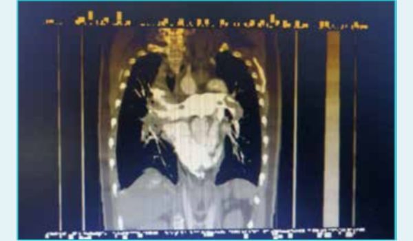
Figure 1: Computed tomography (CT) scan showing bilateral pulmonary embolism with large saddle-shaped thrombus extending into right and left pulmonary arteries.
The patient was shifted to the cardiac intensive care unit (ICU) with high-flow oxygen. She was hypotensive (88/36 mmHg); hence, a fluid bolus and a noradrenaline infusion were initiated. A central line was secured, and thrombolysis was started with an injection of alteplase (15 mg bolus followed by 85 mg over 2 hours), along with noradrenaline. As she was fully alert, bilateral positive airway pressure (BiPaP) support was provided. A venous blood gas (VBG) analysis was suggestive of high lactate levels (8.3 mmol/l) and a bicarbonate level of 13.5 mmol/L, as shown in Table 1.
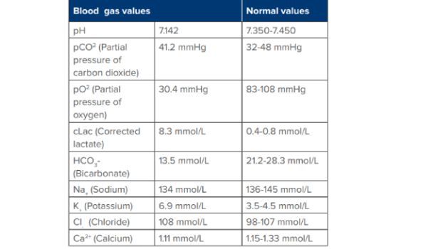
Table 1: Severe lactic acidosis with hypoxaemia postcardiopulmonary resuscitation (CPR).
The patient’s condition deteriorated further within 20 minutes of receiving intravenous (IV) alteplase and progressed to sudden pulseless electrical activity (PEA) with loss of consciousness. Immediate CPR was initiated, and the airway was secured with a cuffed oral endotracheal tube. Intermittent organised rhythm was observed during CPR, but it remained pulseless. The patient continued in PEA despite 5 minutes of CPR with a noradrenaline infusion, 1 mg adrenaline boluses, and a 500 mL bolus of IV normal saline (NS). Hence, a 50 mg bolus of alteplase was administered from the ongoing infusion, and high-quality CPR was continued.
The patient continued to be in cardiac arrest for another 20 minutes, during which 100 mL of sodium bicarbonate, 1 litre of NS, and 10 mL of calcium gluconate were administered along with adrenaline boluses. A vasopressin infusion was also initiated. Due to persistent PEA and massive PE, another bolus of thrombolytic agent —20 mg of tenecteplase—was administered. The patient was then started on a heparin infusion at 1000 IU per hour following a 5000 IU bolus.
After a total of 50 minutes of sustained efforts, the patient achieved return of spontaneous circulation (ROSC). An arterial line and a Foley’s catheter were inserted with due precautions, and noradrenaline and vasopressin was tapered as the BP improved. Two hours after ROSC, the patient started obeying simple commands. The arterial blood gas (ABG) results are shown in Table 2.
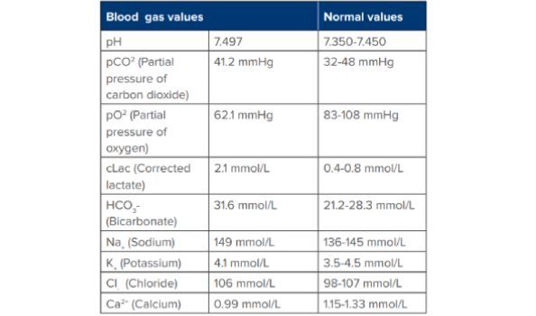
Table 2: Complete resolution of lactic acidosis in 15 hours.
A furosemide infusion was also started due to pulmonary oedema. An injection of glutathione was administered to prevent ischaemic hepatitis. Lactate levels had risen to 17 mmol/L post-CPR but were corrected overnight. A lower limb venous Doppler scan revealed right femoral and popliteal deep vein thrombosis (DVT). The patient was extubated after 2 days and was discharged home three days later.
Discussion
There is no clear guideline for thrombolysis in cardiac arrest secondary to PE. Even though some studies report no mortality benefit of thrombolysis during cardiac arrest, a recent study comparing tenecteplase with other thrombolytics has shown improved survival.4,5
The bolus dose of tenecteplase offers pharmacokinetic advantages compared to prolonged infusion of alteplase in cardiac arrest caused by PE. Our case also highlights the benefit of tenecteplase bolus, especially when administered through a central venous line during CPR. Additionally, the case reinforces the merit of prolonged high-quality CPR in cardiac arrest due to PE. A double bolus of thrombolytics may be useful without a significant risk of bleeding; however, this requires further confirmation in studies and case reports.
Conclusion
In conclusion, this case underscores the importance of prolonged CPR efforts for cardiac arrest due to massive PE. It also demonstrates the successful use of double-bolus thrombolysis during CPR. A similar strategy can be used by acute-care physicians managing cardiac arrest due to massive PE.
Sushil Gadekar, Abdul Ansari, Rushabh Shah. Incredible Survival Post Cardiac Arrest Due to Massive
Pulmonary Embolism with Double Bolus Thrombolysis and Prolonged Cardiopulmonary Resuscitation.
References
- Kucher N, Rossi E, De Rosa M, et al. Massive pulmonary embolism. Circulation. 2006;113(4):577-582.
- Koroneos A, Koutsoukou A, Zervakis D, et al. Successful resuscitation with thrombolysis of a patient suffering fulminant pulmonary embolism after recent intracerebral haemorrhage. Resuscitation. 2007;72(1):154-157.
- Javaudin F, Lascarrou J-B, Bastard QL, et al. Thrombolysis During Resuscitation for Out-of-Hospital Cardiac Arrest Caused by Pulmonary Embolism Increases 30-Day Survival. CHEST Journal. 2019 Aug 2;156(6):1167–75.
- Alshaya OA, Alshaya AI, Badreldin HA, et al. Thrombolytic therapy in cardiac arrest caused by cardiac etiologies or presumed pulmonary embolism: An updated systematic review and meta-analysis. Res Pract Thromb Haemost. 2022;6(4):e12745.
- Daniell JM, Mccormick J, Nasreen I, et al. Comparison of Tenecteplase Versus Alteplase for the Treatment of Pulmonary Embolism and Cardiac Arrest with Suspected Pulmonary Embolism. J Intensive Care Med. 2025;40(2):200-206.