 WhatsApp Us (24/7)
 +91 926 888 0303 (24/7)


Abstract:
Robotic-assisted ureteric reimplantation in the setting of post-surgical ureterovaginal fistula, remains challenging due to difficulties in ureteric identification in the absence of tactile feedback. Indocyanine green (ICG) is a water-soluble dye that can be identified using near infra-red fluorescence (NIRF). We describe our experience with the intraureteral instillation of ICG for identifying the ureter under NIRF during robotic-assisted ureteric reimplantation in patients with ureterovaginal fistulae. This was a retrospective study of 8 patients who underwent robotic-assisted ureteric reimplantation after a diagnosis of ureterovaginal fistula following pelvic surgery between November 2022 and November 2023. Informed consent was obtained from all patients for the off-label use of ICG. All patients underwent regular follow-up postoperatively for a mean period of 12 ± 1.44 months, with none having developed any clinical or radiological signs of failed repair. Thus, the intraureteral instillation of ICG allows precise localisation of the ureter, facilitating accurate dissection, which in turn helps reduce perioperative morbidity.
Key words: Ureterovaginal Fistula, Robotic-Assisted Ureteric Reimplantation, Indocyanine Green (ICG).
Introduction
Ureterovaginal fistulae may occur as a complication of gynaecological procedures, other pelvic surgeries, pelvic radiation therapy and pelvic trauma. The risk of ureteric injury during hysterectomy for benign pathology is 0.2%.1 The risk following radical hysterectomy has been variably reported to be between 1.3% and 2.4%.2 The pelvic ureter or distal one-third of the ureter is the segment most commonly injured during gynaecological procedures, and this is the sole site where injury may result in ureterovaginal fistulae. Any ureteric trauma that exposes the ureteral lumen or causes delayed necrosis of its wall, leading to subsequent urinary extravasation, may lead to a fistula. The passage of urine along non-anatomical planes into the vaginal incision results in continuous urinary incontinence, usually between 1 and 4 weeks post-surgery
The disruption of normal tissue planes and peri-ureteric inflammation and fibrosis may make identifying the ureter during surgical reconstruction challenging. Successful repair depends on the complete excision of the fibrotic ureteric segment while preserving as much healthy ureter as possible, followed by the creation of a watertight, tension-free ureterovesical anastomosis.
Laparoscopic and more recently, robotic-assisted reconstruction are increasingly being used for managing these cases. However, the lack of tactile feedback in the current-generation robotic platforms presents another limitation, necessitating visual identification of the ureter.
The intraluminal instillation of indocyanine green (ICG) dye into the ureter facilitates its identification under near-infrared fluorescence (NIRF). Its good tissue penetration and excellent safety profile make it an ideal real-time contrast agent for intraoperative visualisation.5 Here, we present our experience with the intraluminal use of ICG for ureteric identification during robotic upper urinary tract reconstruction in patients with ureterovaginal fistulae.
Materials and Methods
Our study was a retrospective review of eight patients who underwent surgery following a diagnosis of ureterovaginal fistula between November 2022 and November 2023. All procedures were performed by a single surgeon using the Da Vinci Xi System (Intuitive Surgical, Sunnyvale, CA). Consent had been obtained from all patients for the off-label use of ICG to aid ureteric identification.
The mean age of the patients was 48.5 years. All had previously undergone abdominal or laparoscopic hysterectomy for benign or malignant uterine conditions.
Preoperative evaluation included computed tomography (CT) urography or magnetic resonance (MR) urography, depending on the patient’s renal function parameters and urine culture results. Functional status evaluation was performed in two cases with gross hydroureteronephrosis. Three patients underwent preoperative percutaneous nephrostomy (PCN) tube placement due to sepsis at the time of initial presentation.
After the induction of anaesthesia, 10 ml of ICG mixed with 10 ml of distilled water was instilled via the PCN tube, in the three patients who had undergone preoperative PCN tube placement. In the remaining five cases, ultrasound-guided puncture of Figure 1: Endoview showing the area where the left ureter is presumed to be at. Table 1: Demographics of patient population, intraoperation parameters, and post operation outcomes. Abbreviations: BMI: body mass index; DUB: dysfunctional uterine bleeding; EBL: estimated blood loss; OR: operating room; PCN: percutaneous nephrostomy; TAH+BSO: total abdominal hysterectomy with bilateral salpingo-oophorectomy; TLH: total laparoscopic hysterectomy. Figure 2: Endoview showing the left ureter exhibiting fluorescence in FireflyTM mode. the ipsilateral renal collecting system was done using an 18G initial puncture (IP) needle, followed by instillation of the ICG solution. After port placement, the patient was placed in the Trendelenburg position. A 0-degree telescope was used, and the instruments included fenestrated bipolar forceps, monopolar scissors and ProGrasp forceps.
Following port placement, the peritoneum was incised, and the bowel was reflected off the area of interest. At this stage, the NIRF-Firefly mode was utilised, allowing precise delineation of the viable ureter’s course. The distal ureter was mobilised by sharp dissection. Next, the bladder was mobilised, and psoas hitch sutures were placed using 2-0 Vicryl, as shown in Figures 1 and 2. Mobilisation required division of the contralateral superior vesical pedicle in four cases. Ureteroneocystostomy was performed over a 6 Fr double-J stent in an end-to-end watertight fashion using 4-0 Vicryl sutures.
Stent removal was performed after 6 weeks. CT urography was conducted at 3 months and a Diethylenetriamine Pentaacetic Acid (DTPA) renogram was done at 6 months.
Discussion
ERCP is highly effective in detecting and treating choledocholithiasis. However, there is a significant risk of short-term complications that must be considered when carrying out the procedure. Outside of the high-volume referral centres, failed biliary cannulation can occur in up to 10% of cases.
European Society of Gastrointestinal Endoscopy has defined difficult biliary cannulation as one of the following: more than five contacts with papilla when trying to cannulate, more than 5 minutes spent to cannulate after visualisation of papilla, or more than one inadvertent cannulation or opacification of pancreatic duct.3 Difficult biliary cannulation results in increased risk of post-ERCP pancreatitis. In instances of initial failure to achieve deep biliary access, alternative techniques may be employed. These include: access papillotomy, rendezvous technique or endoscopic ultrasound-based methods.4,5 Pavlides M. et al, in his study had suggested a role of repeat ERCP after a few days of failed initial precut sphincterotomy for biliary cannulation with success in 82% of such cases.
Rendezvous ERCP is a well-defined procedure with use of percutaneous transhepatic biliary drainage (PTBD), however we used T-tube as an access for the guidewire to complete the procedure. Hence, T-tube may be considered as additional, economical and less invasive option in patients who have undergone a surgery with T-tube placement.
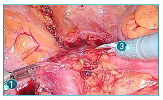
Figure 1: Endoview showing the area where the left ureter is presumed to be at.
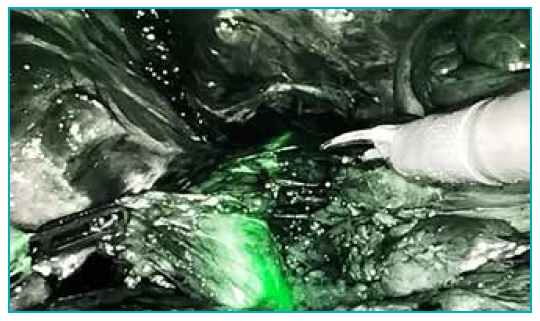
Figure 2: Endoview showing the left ureter exhibiting fluorescence in FireflyTM mode.
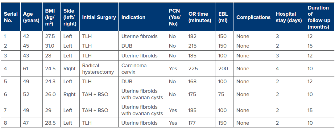
Table 1: Demographics of patient population, intraoperation parameters, and post operation outcomes. Abbreviations: BMI: body mass index; DUB: dysfunctional uterine bleeding; EBL: estimated blood loss; OR: operating room; PCN: percutaneous nephrostomy; TAH+BSO: total abdominal hysterectomy with bilateral salpingo-oophorectomy; TLH: total laparoscopic hysterectomy.
Results
Intra-ureteral instillation of ICG was performed in eight patients with ureterovaginal fistulae, at the time of robotic-assisted reconstruction. The mean patient age was 48.5 years, and mean basal metabolic index (BMI) was 27.35 kg/m2. The mean operating room time was 189 ± 13.9 minutes, the mean estimated blood loss was 128.12 ± 28.44 ml, and the length of hospital stay was 2.67 ± 0.57 days. There were no intraoperative complications in any case. The mean duration of follow up was 12 ± 1.44 months and none of the patients exhibited any clinical or radiological features of failed repair. Patient demographics are shown in Table 1.
Discussion
ICG was developed for near-infrared photography in 1955 by Kodak laboratories. FDA approval for its clinical use was announced in 1959.5 Approved applications include the assessment of liver function and hepatic blood flow, cardiac output and ophthalmic angiography. ICG is a water-soluble dye that becomes excited by light in the near-infrared (NIR) spectrum (820 nm), resulting in fluorescence. This is visualised using specific optical systems.6 The Da Vinci Xi system is equipped with a specific software for NIRF detection named FireflyTM (Novadaq Technologies, Mississauga, ON)
The safety profile of ICG for intravenous use is excellent, with anaphylactic reactions being rare.8 Presently, ICG is used in urology for distinguishing normal renal parenchyma from tumour tissue during partial nephrectomy,9 during selective renal arterial clamping,10 and for sentinel lymph node identification during radical prostatectomy.
Robotic-assisted ureteric reimplantation in the setting of ureterovaginal fistula following pelvic surgery presents difficulties in ureteric identification due to the lack of tactile feedback in the current-generation robotic platforms. The need to preserve the viable segments of the ureter to facilitate a tension-free ureteroneocystostomy makes accurate dissection around the terminal ureter—while preserving its blood supply—essential. Significant intrapelvic scarring may be seen in such patients due to prior surgery and due to urine extravasation along nonanatomical planes. Thus, identification of the ureter becomes difficult, and dissection may compromise its vascular supply or cause trauma to pelvic vasculature or nerves, increasing the perioperative morbidity.
Several techniques have been used to aid ureteric identification, including the use of methylene blue,12 placement of guidewires, and fibreoptic lighted ureteral stents.13 However, lighted stents have been associated with thermal damage to the urothelium, whereas guidewires may cause flaps to form in the urothelium.
Precise identification and excision of scar tissue are also integral aspects of successful ureteric reimplantation. NIRF with ICG greatly facilitates this process, likely by binding to intraluminal tissue proteins in the viable ureteric walls.
Regarding our surgical techniques, the preparation of the ICG solution, ultrasound-guided puncture of the pelvicalyceal system (PCS), and instillation of the dye into the PCS increase operative room time by 20 to 30 minutes. However, considering the potential benefits of precise ureteric delineation and accurate dissection, we believe this increase is an acceptable trade-off.
The limitations of our study include the small sample size and the lack of control group in whom ICG-guided NIRF was not used. Additionally, the intra-ureteric instillation of ICG remains an off-label use, with its adverse effect profile yet to be fully elucidated. Waheedu.
Conclusion
Intraureteral ICG instillation & NIRF facilitate precise delineation of the ureter and accurate identification of areas of interest, greatly increasing the ease of performing robotic-assisted ureteric reimplantations in patients with ureterovaginal fistulae. While the technique is reproducible & easy to perform, increased operating room time is a drawback. Additionally, larger-scale studies are required to further validate this technique.
Waheedu Zzaman, Ashik Suresh, Abhinav Veerwal. Use of Indocyanine Green During Robotic-Assisted Interventions for Ureterovaginal Fistulae: A Single-Centre Experience.
MMJ. 2025, March. Vol 1 (5).
References
- Brummer THI, Jalkanen J, Fraser J, et al. FINHYST, a prospective study of 5279 hysterectomies: complications and their risk factors. Hum Reprod Oxf Engl. 2011;26(7):1741-51.
- Likic IS, Kadija S, Ladjevic NG, et al.Analysis of urologic complications after radical hysterectomy. Am J Obstet Gynecol. 2008;199(6):644.e1-3.
- Mandal AK, Sharma SK, Vaidyanathan S, et al. Ureterovaginal fistula: summary of 18 years’ experience. Br J Urol. 1990;65(5):453-6.
- Lee Z, Simhan J, Parker DC, et al. Novel Use of Indocyanine Green for Intraoperative, Real-time Localization of Ureteral Stenosis During Robot-assisted Ureteroureterostomy. Urology. 2013;82(3):729-33.
- Alander JT, Kaartinen I, Laakso A, et al. A review of indocyanine green fluorescent imaging in surgery. Int J Biomed Imaging. 2012;2012:940585.
- Esposito C, Settimi A, Del Conte F, et al. Image-guided pediatric surgery using indocyanine green (ICG) fluorescence in laparoscopic and robotic surgery. Frontiers in Pediatrics. 2020;8:314.
- Pathak RA, Hemal AK. Intraoperative ICG-fluorescence imaging for robotic-assisted urologic surgery: current status and review of literature. Int Urol Nephrol. 2019;51(5):765-71.
- Olsen TW, Lim JI, Capone A Jr, et al. Anaphylactic Shock Following Indocyanine Green Angiography. Arch Ophthalmol. 1996;114(1):97.
- Tobis S, Knopf JK, Silvers CR, et al. Near Infrared Fluorescence Imaging After Intravenous Indocyanine Green: Initial Clinical Experience With Open Partial Nephrectomy for Renal Cortical Tumors. Urology. 2012;79(4):958-64.
- Bjurlin MA, Gan M, McClintock TR, et al. Near-infrared Fluorescence Imaging: Emerging Applications in Robotic Upper Urinary Tract Surgery. Eur Urol. 2014;65(4):793-801.
- van der Poel HG, Buckle T, Brouwer OR, et al. Intraoperative Laparoscopic Fluorescence Guidance to the Sentinel Lymph Node in Prostate Cancer Patients: Clinical Proof of Concept of an Integrated Functional Imaging Approach Using a Multimodal Tracer. Eur Urol. 2011;60(4):826-33.
- Verbeek FPR, van der Vorst JR, Schaafsma BE, et al. Intraoperative Near Infrared Fluorescence Guided Identification of the Ureters Using Low Dose Methylene Blue: A First in Human Experience. J Urol. 2013;190(2):574-9.
- Low RK, Moran ME. Laparoscopic use of the ureteral illuminator. Urology. 1993;42(4):455-7.