 WhatsApp Us (24/7)
 +91 926 888 0303 (24/7)


Abstract:
Peripartum cardiomyopathy (PPCM) is a rare but severe form of heart failure that develops between the last month of pregnancy and five months postpartum, characterised by a left ventricular ejection fraction (LVEF) of less than 45%. The condition often mimics normal pregnancy symptoms, leading to delayed diagnosis and complications. This case report discusses a 34-year-old woman with a twin pregnancy following in vitro fertilisation (IVF) and pregnancy-induced hypertension, who developed PPCM at 35.2 weeks of gestation. Her rapid progression to severe symptoms necessitated intensive care and multidisciplinary intervention. This report is unique due to the combination of a twin IVF pregnancy, an underlying hypertensive disorder, and critical peripartum complications that were managed successfully. It highlights the unmet need for timely diagnosis and interprofessional collaboration in managing PPCM.
Key words: Peripartum Cardiomyopathy, Twin Pregnancy.
Introduction
Peripartum cardiomyopathy (PPCM) is an idiopathic cardiomyopathy associated with heart failure secondary to left ventricular (LV) systolic dysfunction near the end of pregnancy. Many aetiological processes have been suggested, including viral myocarditis, an abnormal immune response to pregnancy, excessive prolactin secretion, and prolonged tocolysis.1 This disorder carries a high mortality rate.
The current diagnostic criteria for PPCM include cardiac failure in a previously healthy woman occurring in the last month of pregnancy or within 5 months postpartum; absence of a determinable aetiology for the cardiac failure; absence of demonstrable cardiac disease prior to the last month of pregnancy; and echocardiographic evidence of diminished LV systolic function. Its clinical outcome varies, ranging from complete recovery to death. PPCM is a diagnosis of exclusion
Case Report
A 34-year-old, second gravida with a twin pregnancy following in-vitro fertilization (IVF) and pregnancy-induced hypertension was admitted via the emergency department at 35.2 weeks of gestation with a short history of cough, shortness of breath. and leg swelling for four to five days. No previous history of heart disease noted. Her first pregnancy was uneventful with normal vaginal delivery. Following admission, she was initially planned for conservative management and underwent investigations. However, after an ultrasonography (USG) scan, she developed severe breathing difficulty and cyanosis, necessitating immediate transfer to the intensive care unit (ICU). A multidisciplinary approach was adopted, involving a physician, cardiologist, pulmonologist, and the anaesthesia team. Non-invasive ventilation was initiated, and a 2-dimensional echocardiogram (2D echo) revealed a left ventricular ejection fraction (LVEF) of 20% with global LV hypokinesia.
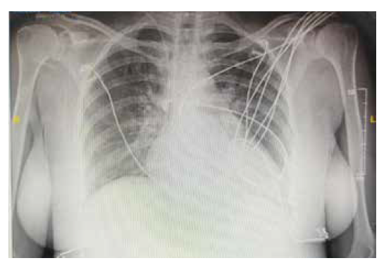
Figure 1: Pre-delivery X-ray – suggestive of left ventricular dysfunction. The X ray was performed predelivery when the patient was symptomatic with breathing difficulties and revealed an enlarged heart.
After stabilising the maternal condition, USG Doppler (Figures 2 and 3) was rechecked for the twins. In view of the maternal condition, a decision was made to proceed with an emergency lower segment caesarean section (LSCS). Both twins were delivered safely and shifted to the neonatal ICU (NICU). The patient was intubated prior to LSCS and kept on ventilator support for 24 hrs. She was then gradually weaned off the ventilator and transferred from the ICU to the ward. She was discharged after five days in a stable condition. A 2D echo before discharge showed an LVEF of 45% (Figure 4). Both babies were also discharged with their mother in stable condition.
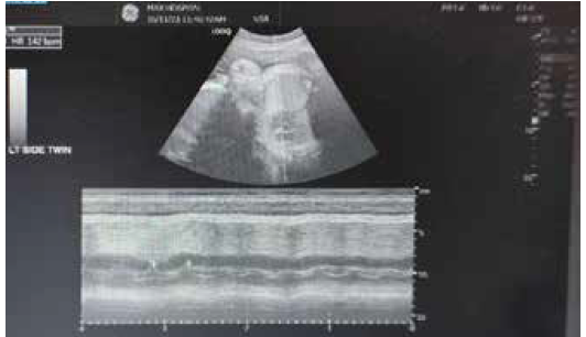
Figure 2: Ultrasound done for twin pregnancy – Baby 1.
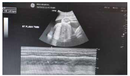
Figure 3: Ultrasound done for twin pregnancy – Baby 2.
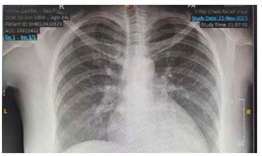
Figure 4: Post-delivery X-ray showing marked improvement in the condition following medical management.
Discussion
PPCM is an idiopathic cardiomyopathy that presents with heart failure secondary to LV systolic dysfunction towards the end of pregnancy or in the months after delivery, in the absence of any other identifiable cause of heart failure. The exact mechanism of the disease is unknown; however, different hypotheses have been proposed regarding its aetiology, including viral myocarditis, nutritional deficiencies, autoimmunity, microchimerism, haemodynamic stresses, vascular dysfunction, hormonal insults, and underlying genetics.
A nationwide population-based study reported an incidence of 10.3 cases per 10,000 live births. The incidence increases with age, with a maximum incidence between 40 and 54 years.5 Data from South Asia suggest that PPCM affects between 1 in 837 and 1 in 1374 deliveries.6,7 Due to advancements in diagnostic techniques and increased awareness, the reported incidence of PPCM has increased from 8.5 to 11.8 in recent years.
PPCM is a diagnosis of exclusion, with most being diagnosed postpartum. Although the LV may not be dilated, the ejection fraction is nearly always reduced to below 45%. PPCM is more common in multiple gestations, advanced maternal age, and obesity. This may be due to a vascular insult, secondary to hormonal effects of advanced pregnancy, resulting in cardiomyopathy in women with an underlying genetic predisposition.
Enhancing healthcare team outcomes PPCM is a rare cause of cardiomyopathy that occurs during late pregnancy or the early postpartum period. This condition can be life-threatening and is characterised by significant LV dysfunction and heart failure. PPCM does not have a precisely defined aetiology. In 2010, the European society of Cardiology classified PPCM as an idiopathic cardiomyopathy. The diagnosis of PPCM requires a high index of suspicion and remains a diagnosis of exclusion. Its initial medical management is similar to that of other causes of heart failure with special attention to how the condition can affect the pregnancy. Additional therapeutic considerations include management of arrhythmias, anticoagulation therapy, and mechanical support. PPCM has special considerations for treatment due to side effects of medications that can cross the placenta and affect the foetus. An interprofessional approach is recommended for managing PPCM. Because of the high morbidity and mortality associated with the condition, prenatal nurses, obstetricians and nurse practitioners specialising in obstetrics must be well-informed about PPCM. Early referral to a cardiologist is crucial to improving mortality. In moderate-to-severe cases, an interprofessional team should manage the patient, often requiring ICU admission and close foetal monitoring via repeated ultrasound assessments. to assess the viability of the foetus. All patients should be counselled on the potential foetal toxicity of cardiac medications.
Finally, the patient should be educated to avoid future pregnancies if the ejection fraction fails to improve as recurrent PPCM increases the risk of mortality up to 50%. Despite optimal therapy, PPCM still carries a mortality rate of 10% to 20%.
Conclusion
In our case, the patient was in the prepartum phase and a prompt diagnosis and management led to successful outcomes for both the mother and the twins. Other causes of heart failure were evaluated, as PPM is a diagnosis of exclusion. Given the increased risk of recurrence in future pregnancies, the patient should be strongly advised against further pregnancies and closely monitored for her long-term cardiac function.
Ankita Chandna, Sanjay Raina, Naveen Bhamri, Inder Mohan Chugh, Puneet Sharma. Management of Peripartum Cardiomyopathy in Twin Pregnancy.
MMJ. 2025, March. Vol 1 (5).
References
- Triebel J, Clapp C, Martínez de la Escalera G, et al. Remarks on the prolactin hypothesis of peripartum cardiomyopathy. Front Endocrinol. 2017;8:77.
- Huang Y, Chen T, Zhang M, et al. Successful management of fatal peripartum cardiomyopathy in a young pregnant woman: A case report. Medicine. 2018;97(15):e0408.
- Brar SS, Khan SS, Sandhu GK, et al. Incidence, mortality, and racial differences in peripartum cardiomyopathy. Am J Cardiol. 2007;100(2):302-4.
- Ntusi NB, Mayosi BM. Aetiology and risk factors of peripartum cardiomyopathy: a systematic review. Int. J. Cardiol. 2009;131(2):168-79.
- Kolte D, Khera S, Aronow WS, et al. Temporal trends in incidence and outcomes of peripartum cardiomyopathy in the United States: a nationwide population based study. J Am Heart Assoc. 2014;3(3):e001056.
- Hasan JA, Qureshi A, Ramejo BB, et al. Peripartum cardiomyopathy characteristics and outcome in a tertiary care hospital. JPMA. J Pak Med Assoc. 2010;60(5):377.
- Pandit V, Shetty S, Kumar A, et al. Incidence and outcome of peripartum cardiomyopathy from a tertiary hospital in South India. Trop Doct. 2009;39(3):168-9.
- Mielniczuk LM, Williams K, Davis DR, et al. Frequency of peripartum cardiomyopathy. Am J Cardiol. 2006;97(12):1765-8.
- Dayoub EJ, Datwani H, Lewey J, et al. One-Year Cardiovascular Outcomes in Patients With Peripartum Cardiomyopathy. J Card Fail. 2018;24(10):711-715.
- Honigberg MC, Sarma AA. Pregnancy Among Survivors of Childhood Cancer: Cardiovascular Considerations. Curr Treat Options Cardiovasc Med. 2018;20(7):54.