 WhatsApp Us (24/7)
 +91 926 888 0303 (24/7)


Abstract:
Breast cancer holds the dubious distinction of being the most prevalent malignant tumour among women worldwide. The 5-year survival rate for patients with advanced or metastatic forms of the disease is estimated at a mere 23%, with approximately 20% of cases exhibiting an amplification of the human epidermal growth factor receptor 2 (HER2). HER2 overexpression was linked to a dismal prognosis prior to the advent of targeted HER2 therapies. The introduction of ado-trastuzumab emtansine (T-DM1) has emerged as one of the standard treatment options for HER2-positive breast cancer patients who experience a recurrence or progression of the disease. This case concerns a patient with recurrent metastatic breast cancer who achieved an unusually extended period of time without the disease progressing while on T-DM1 treatment. After experiencing disease progression on multiple treatment lines involving trastuzumab, the patient achieved a nearly complete clinical remission (cCR) with T-DM1 therapy. The availability of cost-effective biosimilars has expanded access to advanced biologic therapies, which were previously limited to a small segment of the population due to the cost barrier.
Key words: Juvenile, Fibro-Osseous, Aggressive, Histologic.
Introduction
The juvenile ossifying fibroma (JOF) is a rare benign neoplasm, classified within the group of fibro-osseous tumours according to the 2017 World Health Organisation (WHO) classification.1 It is considered as a separate entity from the conventional ossifying fibroma and is further divided into two histological subtypes: juvenile trabecular ossifying fibroma (JTOF) and juvenile psammomatoid ossifying fibroma (JPOF).2 It predominantly affects children but may occur in adults. The clinical trajectory of JOF is often belligerent, mimicking malignancy like osteosarcoma. So, it is important to accurately recognise JOF for adequate diagnosis and further treatment.
Case report
An 11-year-old boy presented with the primary complaint of swelling in the right maxillary region for the past year, which started following trauma and progressively increased in size over time. An incisional biopsy was conducted at another hospital where the diagnosis of giant-cell reparative granuloma (GCRG) was made. The slide was later reviewed at Max Super Speciality Hospital, Lucknow, and the diagnosis of JTOF was confirmed. Simultaneously, laboratory investigations were conducted, all of which were within normal limits. Radiological investigations revealed a well-demarcated radiolucent lesion showing intracortical osteolysis with characteristic sclerotic band and moderate cortical expansion. The patient later underwent surgery. Following the administration of general anaesthesia, a right gingival incision was made, and a flap was elevated. Osteotomy along with deroofing was conducted followed by excision and curettage of the necrosed bone (Figures 1a and 1b).
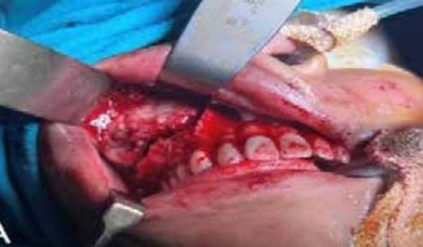
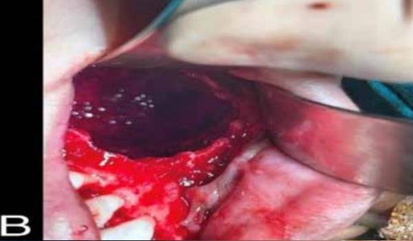
Figure 1a and 1b: Intraoperative findings and osteotomy with deroofing followed by excision and curettage.
Reconstruction and closure were then done with local mucosal flap using vicryl. Excised tissue was fixed in the 10% formalin and was sent to the Department of Laboratory Medicine for complete histo-morphological analysis. The patient was put on Ryle’s Tube (RT) feed at 200 ml every 3 hours and was advised to rinse with 0.2% chlorhexidine gluconate solution, along with intravenous medications to prevent post-operative discomfort. Sutures were removed after 10 days, following which the patient was on regular follow-up for 3 months. No recurrence was noted.
Multiple greyish-white to greyish-brown tissue pieces aggregating to 9.0 x 5.0 x 2.5 cm, and two flat bony tissue pieces were received in the laboratory. Sections revealed cell-rich fibrous tissue exhibiting spindle cell proliferation and bands of osteoid formation with occasional osteoblastic rimming (Figures 2a and 2b). The spindle cells were observed entrapping.
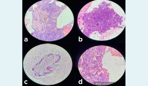
Figure 2: Juvenile trabecular ossifying fibroma. 2a: Osteoid formation with osteoblastic rimming and surrounding spindle cells (H and E x 20). 2b: Densely cellular fibrous tissue showing spindle cell-proliferation and spicules of woven bone (H and E x 20). 2c: Bony trabeculae forming lattice-like pattern (H and E x 20). 2d: Multiple locules surrounded by multinucleated giant cells resembling aneurysmal bone cyst (H and E x 40).
Surrounding irregular, interconnecting trabeculae of immature bone tissue containing coarse lacunae with plump osteocytes. The bony trabeculae were identified as anastomosing together forming a lattice-like pattern (Figure 2c). Multiple degenerative locules and cystic spaces lined by multinucleated giant-cell reminiscent of aneurysmal bone cyst were also identified (Figure 2d). Overall histopathological features were suggestive of JTOF
Discussion
Fibro-osseous lesions exhibit a melange of various processes in which the normal architecture of bone is replaced by fibrous tissue containing variable amount of mineralization. As per WHO classification of 2017; they are characterised as:1
- Ossifying fibroma
- JTOF
- JPOF
- Fibrous dysplasia
- Familial gigantiform cementoma
- Cemento-osseous dysplasia
- Osteochondroma
JOF seems to be a neoplastic outcome of the myxoid tissue precursor of cartilage and bone of paranasal sinuses. It is categorised into two broad categories: JPOF and JTOF, both manifesting as encapsulated lesions, infiltrating the adjacent bone, and usually arising outside the tooth-bearing areas of the craniofacial skeleton. The most distinguishing feature of JOF is its aggressive clinical behaviour and higher incidence in young adults. However, there is a marked difference between the two categories of JOF, and they can only be distinguished based on histological criteria. The trabecular variant (JTOF) usually occurs in 8-12 years of age (mostly in males>females) with the most common site of involvement being the maxilla followed by the mandible and sino-nasal region.3, 4 Whereas, the psammomatoid variant (JPOF) occurs between 16-33 years of age with the periorbital walls of paranasal sinuses as the most common site of involvement. Mandibular and extracranial involvement is rarely seen. Histologically, both subtypes have a similar stroma but JTOF is composed of densely cellular fibrous tissue containing immature bone and fibrillary osteoid in a trabecular pattern showing osteoblastic rimming while PJOF is also densely cellular but contains spherical or cementum-like lamellated calcifications (ossicles), the characteristic psammomatoid bodies.5
The presentation of both TJOF and PJOF is similar; however, the line of management is still contentious.6,7 Locally radical surgery is the management of choice; whilst preserving important adjacent structures.8 There is no standardised follow-up protocol, however long-term clinical and radiological monitoring is imperative to detect late recurrences.9 Recurrence rate is attributed to 30-56% secondary to incomplete excision and an aggressive nature of tumour.9 However, the case reported here was followed up for 3 months without recurrence, enlightening that thorough surgical resection, rather than conservative curettage, is therefore the preferred line of treatment. The patient was also counselled to attend follow-up appointments every six months until he reaches 18 years of age. In the event of any recurrence, management using a patient-specific implant will be implemented.
The primary aim of this case report is to highlight the key issue that, despite advancements in molecular analysis techniques, the classification, diagnosis, and management of fibro-osseous lesions remain challenging due to numerous clinical, histological, and radiographic overlaps. Hence, an interdisciplinary approach in the diagnosis and management of these lesions is highly recommended.
Conclusion
This case report represents the first documented instance from India of a patient experiencing a prolonged PFS benefit while receiving T-DM1 biosimilar treatment. The introduction of cost-effective biosimilar agents in India has made them the standard of care for patients who have exhausted early lines of treatment and for those who require longterm targeted therapy. It provides an opportunity to expand access to effective treatments and improve outcomes for HER2-positive breast cancer patients with CNS involvement. It also offers a more affordable option without compromising therapeutic efficacy, enabling patients to continue targeted therapy for extended periods. This not only enhances the potential for improved disease control but also contributes to maintaining a favourable quality of life for patients. By leveraging the benefits of cost-effective biosimilar agents, healthcare providers can optimise treatment options and ensure that patients receive the most appropriate and beneficial care available.
Zoya Qamar, Anju Shukla, Fatema Shareh Naqvi, Satinder Pal Singh Tulsi. Unboxing the Dilemma of a Fibro-
Osseous Lesion of Maxilla: A Case Report. MMJ. 2025, March. Vol 1 (5).
References
- WHO Classification of Head and Neck Tumors. 4th Edition IARC Press. 2017;203-260.
- Rosai and Ackerman’s Surgical Pathology. 11th Edition Elsevier. 2018;1:215-216.
- Mill’s and Sternberg’s Diagnostic Surgical Pathology. 7th Edition. Wolter Kluwer; 2022;1:999-1000.
- El-Mofty S. Psammomatoid and trabecular juvenile ossifying fibroma of the craniofacial skeleton: two distinct clinicopathologic entities. Oral Surg Oral Med Oral Pathol Oral Radiol Endod. 2002;93(3):296-304.
- Speight PM, Carlos R. Maxillofacial fibro-osseous lesions. Current Diagnostic Pathology. 2006;12(1):1-0.
- Rallis G, Schoinohoriti O, Krasadakis C, et al. Trabecular juvenile ossifying fibroma: updated review of the literature and report of an interesting case. J Otolaryng Head Neck Surg. 2017;3(010).
- Pandit N, Saini N, Kler S, et al. Juvenile trabecular ossifying fibroma. J Indian Soc Periodontol. 2014;18(2):232-235.
- Appiani MC, Verillaud B, Bresson D, et al. Ossifying fibromas of the paranasal sinuses: diagnosis and management. Acta Otorhinolaryngol Ital. 2015;35(5):355.
- Tolentino ES, Centurion BS, Tjioe KC, et al. Psammomatoid juvenile ossifying fibroma: an analysis of 2 cases affecting the mandible with review of the literature. Oral Surg Oral Med Oral Pathol Oral Radiol. 2012;113(6):e40-e45.