 WhatsApp Us (24/7)
 +91 926 888 0303 (24/7)


Abstract:
Alloimmunisation against red cell antigens is a critical challenge in transfusion medicine, especially in patients with prior antigen exposure. We report a case of a 71-year-old female with severe anaemia, multiple alloantibodies (Anti-C and Anti-e), and a recent transfusion history. The case highlights the complexities of identifying compatible donor units and underscores the importance of advanced diagnostic methods, such as solid-phase red cell adherence (SPRCA), phenotyping, and elution techniques, in emergency transfusion scenarios. Proactive inventory management and extended antigen matching are discussed as key strategies for reducing alloimmunisation risk and improving patient outcomes.
Key words: Alloantibody, Phenotyping, Alloimmunisation.
Introduction
Transfusion of red blood cells (RBCs) is a common lifesaving intervention.1 However, alloimmunisation against non-ABO antigens can complicate subsequent transfusions, leading to delays in finding compatible blood and increasing the risk of haemolytic transfusion reactions (HTRs).2,3 Alloantibodies, particularly those targeting the Rh and Kell systems, are clinically significant due to their high immunogenicity. The incidence of alloimmunisation is higher in populations with prior antigen exposure, such as transfusion-dependent patients.1 This report details the challenges of managing a transfusion in a patient with multiple alloantibodies in an emergency setting.
Case Report
Patient presentation: 71-year-old female presented to the emergency ward with severe anaemia (haemoglobin: 6.2 g/dL). Blood grouping, antibody screening, and crossmatching were initiated to facilitate the transfusion of two units of packed red blood cells (PRBCs).
Laboratory findings: The patient's laboratory findings indicated a blood group of A positive. The antibody screening results were positive, and crossmatching was incompatible with all tested units. Additionally, the direct antiglobulin test (DAT) was positive, and the autocontrol (A/C) also showed a positive result.
Antibody identification: Advanced serological workups, including solid-phase red cell adherence (SPRCA), revealed the presence of Anti-C and Anti-e antibodies. Elution studies confirmed these findings. Due to the high prevalence of C (87%) and e (98%) antigens in the Indian population, most donor units were incompatible.
Management
To identify compatible units, multiple panels (Cap R Ready ID, Panocell 16, and Panocell 10 enzyme panels) and extended crossmatching were performed. Eventually, two O Positive R2R2 units (C-, c+, e-, E+) were located and transfused successfully.
Discussion
Pathophysiology of alloimmunisation: Alloantibodies against RBC antigens develop after exposure to foreign antigens through transfusion, pregnancy, or organ transplantation. Anti-C and Anti-e antibodies belong to the Rh system, which is highly immunogenic. These antibodies can cause HTRs, complicating transfusion management and posing significant risks to patients.
Diagnostic challenges: The case highlights the critical role of antibody screening and identification in transfusion medicine:
1. Antibody screening: A sensitive test, such as SPRCA, is essential to detect clinically significant antibodies that may not be identified through crossmatching alone (Figure 1).1
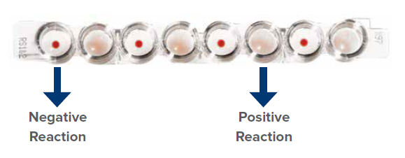
Image of Capture-R Ready Screen strip.
- In a positive test, indicator red cells cannot move to the bottom of the well because Anti-IgG-IgG complexes are formed on the antigen surface of the immobilised red cell membrane layer.
- In a negative test, indicator red cells can move easily to the bottom of the well and form a red cell button.
- The degree of red cell adhesion to the monolayer is the key indicator of positive and negative reactions (Figure 1).
2. Elution studies: his procedure helped confirm the antibodies bound to the patient’s RBCs, ruling out additional alloantibodies.
3. Phenotyping: Recent transfusions made phenotyping of the patient’s RBCs unreliable, necessitating alternative approaches for antibody identification.
4. Panel testing: : Using multiple panels (e.g., Panocell 16, Panocell 10) enables the detection of antibodies with broad reactivity, as seen in this case of Anti-C and Anti-e (Figure 2 and Figure 3). 82 The Max Medical Journal March 2025
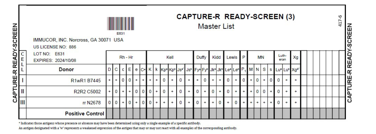
Figure 2: Antigram of Capture-R Ready Screen.
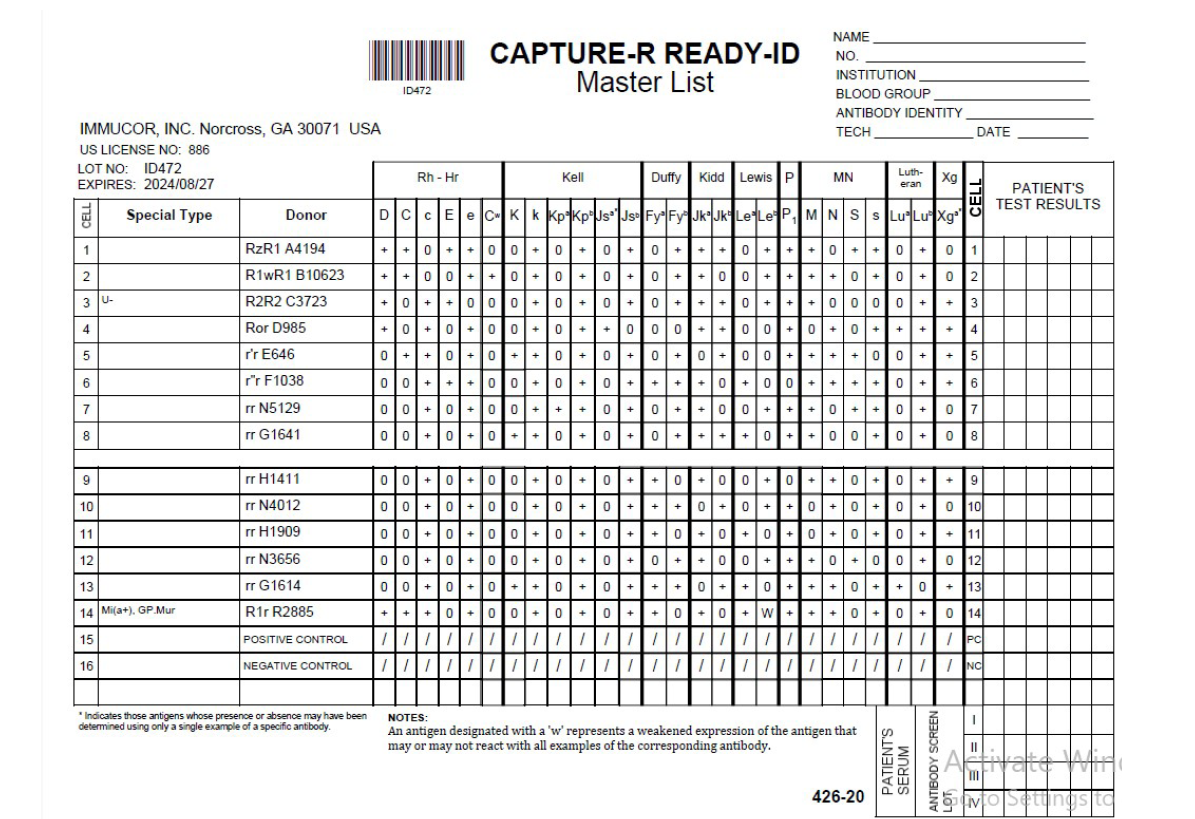
Figure 3: Antigram of Capture-R Ready-ID.
Management Strategies
- Inventory management and phenotyping
- Phenotyped donor pool: Routine Rh and Kell phenotyping ensures the availability of antigen-negative units for alloimmunised patients.
- Rare unit identification: Extended typing and strategic donor selection enabled rapid identification of compatible units, saving crucial time in an emergency
- Leukocyte-depleted blood: Reduces immunogenicity by minimising white cell contamination.
- Extended antigen typing: Characterizing antigen profiles of patients and donors can pre-emptively identify potential incompatibilities
- Advanced technology: SPRCA and molecular techniques enable precise and timely antibody detection, improving patient outcomes.
Prevention of alloimmunisation To reduce future alloimmunisation risks, it is critical to transfuse antigen-matched blood for at least the following systems: Rh (C, c, E, e) and Kell.3 Additional strategies include:
- Leukocyte-depleted blood: Reduces immunogenicity by minimising white cell contamination.
- Extended antigen typing: Characterizing antigen profiles of patients and donors can pre-emptively identify potential incompatibilities.
- Advanced technology: SPRCA and molecular techniques enable precise and timely antibody detection, improving patient outcomes.
Implications for practice: Most centres now recommend proactive matching for Rh and Kell antigens, even in the absence of antibodies, due to their high immunogenicity and involvement in HTRs.1,3 This approach is cost-effective in the long term, as it prevents the development of additional alloantibodies and associated complications.
Conclusion
This case illustrates the complexities of managing transfusions in a patient with multiple alloantibodies, emphasising the importance of advanced diagnostic techniques and strategic approaches in emergency transfusion medicine. The patient presented with severe anaemia, a positive antibody screen, and incompatible crossmatches, making the identification of suitable donor units a significant challenge. Through the use of highly sensitive methods such as SPRCA and comprehensive antibody identification panels, the antibodies Anti-C and Anti-e were identified, highlighting the utility of advanced technologies in complex serological cases.
The high prevalence of the C and e antigens in the Indian population (87% and 98%, respectively) posed additional challenges, as most donor units were incompatible.4 Despite these difficulties, the transfusion team successfully located two O Positive R2R2 units for the patient, underscoring the critical role of maintaining an antigen-typed donor inventory.5 Routine Rh and Kell phenotyping of donors proved invaluable in this situation, enabling the rapid identification of rare antigen-negative units, reducing delays, and improving patient outcomes.
The case underscores the importance of preventing alloimmunisation through proactive measures. Extended antigen typing for both patients and donors, along with the use of leukocyte-depleted red blood cells, can significantly reduce the risk of alloantibody formation.3 Matching for highly immunogenic antigens, such as those in the Rh and Kell systems, even in the absence of antibodies, should be standard practice to prevent future complications.
Furthermore, the case highlights the necessity of skilled personnel, access to advanced serological tools, and a wellmanaged blood inventory system in resolving complex transfusion challenges.5 By adopting a multidisciplinary approach and leveraging available resources effectively, it is possible to overcome even the most challenging transfusion scenarios.
In conclusion, this case emphasises that the key to successful transfusion management lies in a combination of advanced diagnostic capabilities, strategic inventory practices, and preventive measures. These efforts not only ensure timely and safe transfusion but also enhance patient care by minimising the risks associated with alloimmunisation and haemolytic transfusion reactions. This case serves as a reminder of the ongoing need for innovation and vigilance in transfusion medicine.
Sangeeta Pathak, Satish Kaushik, Ruchi Dubey. Case Report: A Complex Case of Alloimmunisation and Transfusion Management in a 71-Year-Old Female.
MMJ. 2025, March. Vol 1 (5).
References
- Harmening DM. Modern blood banking & transfusion practices. 1st Edition. Philadelphia: FA Davis; 2018
- Townsend CM, Beauchamp RD, Evers BM, et al. Sabiston textbook of surgery: The biological basis of modern surgical practice. 20th Edition. Philadelphia: Elsevier Saunders; 2018. 924-33.
- Conn JM, Goncalves MA, Mansour KA, et al. The mediastinal parathyroid. Am Surg. 1991;57:62-6.
- Russell CF, Edis AJ, Scholz DA, et al. Mediastinal parathyroid tumors: experience with 38 tumors requiring mediastinotomy for removal. Ann Surg. 1981;193:805-9.
- Hordijk M, Veen EJ, Schreinemakers Jennifer MJ. ICG-Guided Video-Assisted Thoracoscopic Surgery for Resection of an Ectopic Mediastinal Parathyroid Adenoma. J Endocr Surg. 2023;85-88
- Fischer JE. Fischers Mastery Of Surgery. 7th Edition. Philadelphia: Wolters Kluwer; 2019. 1610.
- Issacs KE, Belete S, Miller BJ, et al. Video-assisted thoracoscopic surgery for ectopic mediastinal parathyroid adenoma. BJS open. 2019;3:743-49.
- Naik D, Jebasingh KF, Ramprasath, et al. Video Assisted Thoracoscopic Surgery for excision of an ectopic mediastinal intra-thymic parathyroid adenoma. JCDR. 2016; 10(6):22-24.
- Amir K, Khan AZ, Rew D, et al. Video assisted thoracoscopic excision of mediastinal ectopic parathyroid adenomas: a UK regional experience. Ann Cardiothorac Surg. 2015; 4(6):527-34.
- Abdulsalam MS, Devanayagam S, Santosham, et al. Mediastinal parathyroid adenoma removal by Video Assisted Thoracoscopy Surgery. Ann Afr Med. 2021; 20(2) 150-53.
- Veronica CK, Mariana CT, Graciela A, et al. Thoracoscopic treatment of mediastinal ectopic parathyroid adenoma: a latinamerica experience case series and literature review. J Cardiothorac Surg. 2024;19:17.