 WhatsApp Us (24/7)
 +91 926 888 0303 (24/7)


Abstract:
Neurovascular conflict syndromes are hyperactive cranial nerve disorders caused by chronic contact between vascular structures and cranial nerves in the posterior fossa. Since the original description by Walter E. Dandy, neurovascular compression has been widely accepted as the underlying mechanism in conditions such as trigeminal neuralgia, hemifacial spasm, and glossopharyngeal neuralgia. This review summarises current knowledge of the anatomical basis and pathophysiological mechanisms of these disorders based on relevant literature. The root entry/exit (Obersteiner–Redlich) zone, where central myelin transitions to peripheral myelin, is particularly vulnerable to pulsatile vascular compression. This leads to focal demyelination and ephaptic transmission, resulting in abnormal cross-talk between nerve fibres and ectopic impulse generation. Mechanisms such as neuroplasticity and kindling further contribute to the hyperexcitability of cranial nerve nuclei, producing characteristic clinical manifestations. Radiological and intraoperative studies support the role of neurovascular compression; however, discrepancies between imaging and clinical findings indicate a multifactorial process involving both peripheral and central mechanisms. Emerging entities, including vagal rhizopathies and the role of venous compression, further expand the spectrum of these syndromes. Despite advances in imaging and microvascular decompression, the precise cellular mechanisms remain incompletely understood. This review highlights the interplay of anatomical susceptibility and neurophysiological changes in the pathogenesis of neurovascular conflict syndromes.
Key words: Neurovascular Conflict, Neurovascular Compression, Trigeminal Neuralgia, Hemifacial Spasm, Glossopharyngeal Neuralgia, Ephaptic Transmission, Demyelination, Cranial Nerves, Posterior Fossa, Obersteiner–Redlich Zone, Microvascular Decompression, Neuroplasticity.
The sensory root is frequently indented, lifted up or bent at an angle by the artery…This I believe is the cause of tic douloureux. — Walter E Dandy 1
Introduction
The pulsatile impulse generated by the heart in the cardiac cycle propagates the pressure-related events right up to the capillaries. Pulsatile propulsion of blood through blood vessels is responsible for its circulation and transport of oxygen and nutrients. Venous flow too has a waveform pattern, related partly to thoracic excursions leading to cyclical variations in intrathoracic pressure, thus regulating the venous return. Intracranially, the major trunks of the arteries and veins, and the trunks of major cranial nerves, course through the cerebrospinal fluid-filled subarachnoid space. Since Walter Dandy 1 first described the contact of a vessel loop with the trigeminal root in patients with trigeminal neuralgia, the theory of neurovascular compression has been widely accepted. It is believed that a close contact between a major artery and a nerve trunk over a prolonged period may cause changes in the nerve trunk, in the form of localised demyelination and initiate the phenomenon of ephaptic transmission (Figure 1).
While the theory of neurovascular compression and consequent ephapsis and its clinical consequences is widely accepted, a number of studies, hypotheses, and clinical and radiological observations have attempted to establish this association. This article reviews the data on the observations, likely pathological changes, and the pathophysiological mechanisms. A PubMed database search was performed for articles on pathophysiology and observations, using the key words trigeminal neuralgia, hemifacial spasm (HFS), glossopharyngeal neuralgia, vertigo, ephaptic transmission, neurovascular compression. A total of 60 articles and book chapters were selected based on direct relevance and quality of information provided that enhanced the current understanding of the pathophysiology of neurovascular compression. The articles and book chapters were reviewed, and the pathophysiology of neurovascular compression causing specific syndromes was analysed.
An abnormal volley of impulses to the target musculature or sensory receptors can follow such ephaptic transmission. Subsequently, there can be a rekindling phenomenon at the brainstem nuclei of the nerves concerned, leading to constant electric discharge and impulse transmission. As the cranial nerves enter/exit the brainstem in the posterior fossa, there is a change in the myelin sheath characteristics, central on the brainstem side and peripheral on the distal side. This change occurs close to the entry/ exit zone over a variable segment of the nerve, referred to as Obersteiner-Redlich zone. Extraneous contact with a vessel over this zone or close to the brainstem results in hyperactive, dysfunctional syndromes.
In the case of the trigeminal nerve, the response takes the form of trigeminal neuralgia in the region of distribution of one or more branches. In the case of the facial nerve, there are intermittent tonic-clonic facial movements of one half of the face, commonly known as HFS. Such disorders of ephaptic transmission can also be seen in space-occupying lesions (most of the time, in the posterior fossa) (Figure 2), which probably act by causing or accentuating existing anatomical conflict between an artery and the corresponding nerve. Demyelination, as in multiple sclerosis, can also cause ephaptic transmission, possibly by short-circuiting the impulse volleys. However, a contrarian observation involves the fact that the proximal part of the superior cerebellar artery (SCA), as it passes below and is separated from the posterior cerebral artery (PCA) by the oculomotor nerve. Nearly two-thirds of SCAs have a point of contact with the oculomotor nerve on the inferior surface of the nerve. The SCA passes near and frequently has points of contact with the oculomotor, trochlear, or trigeminal nerves. The oculomotor nerve may occasionally be constricted between the PCA and SCA. 2 Y et, the only manifestation of oculomotor nerve compression by a vessel or a posterior communicating artery aneurysm is nerve dysfunction in the form of oculomotor nerve palsy, and an optic nerve compressed by an ophthalmic segment aneurysm manifests as optic atrophy. Is the response to a pulsatile impulse different for different cranial nerves?
The Transitional Zone
Obersteiner and Redlich3 studied spinal nerves as they entered the spinal cord and described the transition zone (TZ) of myelin depletion. They interpreted this finding as a lack of myelin sheath for about 50 µm. It is now apparent that this depletion of myelin was due to a preservation artefact, and the myelin sheath of the central portion is thinner4 and more susceptible to injury.5 The TZ from central myelin to peripheral myelin still carries the label of Obersteiner-Redlich zone.
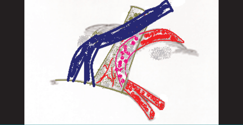
Figure 1: Sketch of trigeminal nerve compression by the superior cerebellar artery/Dandy’s vein at the entry zone.
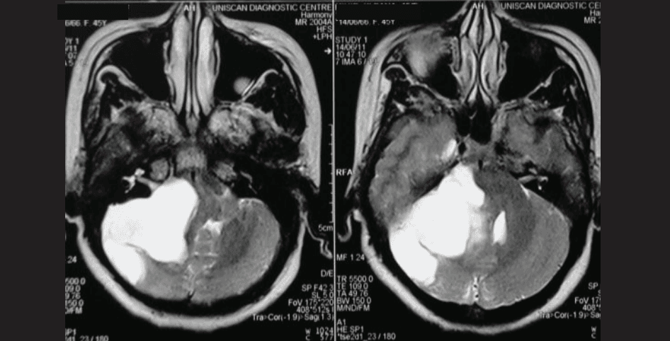
Figure 2: Magnetic resonance imaging (MRI) showing left cerebellopontine angle epidermoid in a patient with hemifacial spasm.
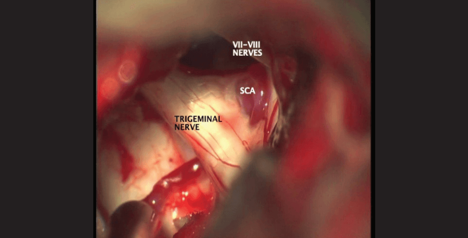
Figure 3: Intraoperative photograph of the trigeminal nerve being compressed by a loop of the superior cerebellar artery (SCA).
There are three types of fibrocollagenous supporting tissueassociated with each nerve:
- Endoneurium: A delicate, collagen-rich tissue surrounding each nerve fibre
- Perineurium: A thin sheath with aggregation of funiculi
- Epineurium: The fibroareolar tissue that surrounds the funiculi and forms the nerve trunk.
In the central nervous system (CNS), there is a lack of endoneurium, perineurium, and epineurium, and the axons are ensheathed by oligodendrocytes and follow a parallel course. The transition from a central myelin (from oligodendrocytes) to peripheral myelin (elaborated by Schwann cells) takes place at a variable distance after the nerve exits the brainstem, much like handing over the baton in a relay race. The sensory component of any nerve has a longer segment of central myelin than that in the motor root. 6 Guclu et al. 7 studied the relationship between the length and volume of the central myelin portion of these nerves and the incidence of corresponding syndromes of the cranial nerves. Central myelin is longer in case of facial nerve, 7 while it is wider in case of trigeminal nerve. It is very short in glossopharyngeal and vagus nerves, and these two nerves exit the brainstem as multiple rootlets, unlike the facial nerve which exits as a single nerve.
In its course to the brainstem, the cranial nerve splits into thinner rootlets, which further subdivide into smaller rootlets. The TZ occurs within each of these mini-rootlets, and centrally, the nerve bundles enter the brainstem as a mass of compact white matter.8 The CNS portion of the mini-rootlet is convex in shape. Thus, the TZ is arched, and there are two compartments within the mini-rootlet: a peripheral zone consisting of axons ensheathed in endoneurium, and a central compact track of white matter. There is a deficiency of central myelin in the area deficient in endoneurium, and myelin in this region is thin as compared to that within the brainstem. 4 The TZ is also the region where the endoneurial microvasculature exits to anastomose with the extraneural plexus, thus making the TZ relatively poorly vascularised, creating a locus minoris resistae (Achilles heel). 8
Pathophysiology
The current understanding of neurovascular compression is that a vessel is in close contact with a cranial nerve in the posterior fossa (Figures 3–5). Further, Jannetta 9 observed that these vessels cross the nerve at a right angle, and a vessel running parallel to the nerve does not cause clinical manifestations.
Mechanical ectopic excitation:
There is probably focal compression and demyelination in the course of adjacent axons, leading to a false synapse (ephapse).10 Nielsen further stated that there is bidirectional cross-transmission between fibres, decreased conduction velocity in “pre-ephaptic” motor fibres, focal slowing of conduction over the suspected site of compression, as suggested by increased latency of the R1 component of the blink reflex. There is also lateral spread of orthrodromic impulses, leading to increased amplitude of the blink reflex and occurrence of synkinetic responses in other facial muscles, as well as autoexcitation related to the passage of a single anti- or orthrodromic impulse, with lateral spread of current often resulting in a “cascade effect” that causes clinically evident tonic-clonic spasms.11 Ectopic excitation induced by hyperventilation, presumably due to a reduction in the concentration of extracellular calcium caused by respiratory alkalosis, can precipitate tonic-clonic events. All these observations fulfil the predictions made by the hypothesis that HFS is caused by abnormal conduction of impulses through the peripheral portion of the facial nerve. Rasminsky12 summarised these four events as:
- Mechanically induced or ectopic excitation
- Reflection of impulses: orthodromic and/or antidromic conduction
- Ephaptic excitation or “cross-talk” between axons
- After discharge or autoexcitation
It is possible that a demyelinated axon stimulates a myelinated one, instead of two demyelinated axons transmitting impulses. 13 Due to resistance (resulting from vascular compression) offered by the demyelinated axon, the impulse is directed and transmitted to an adjacent myelinated axon, stimulating muscles innervated by different axons. This phenomenon also explains synkinesis.
Neuroplasticity:
Neuroplasticity, or reorganisation of the system, explains some of the features of neurovascular compression. Extrapolating the observations from limb or digital amputees, it is observed that the topographic representation of it is altered after amputation. The deprived area of the somatosensory cortex then becomes responsive to adjacent skin areas. Chronic stimulation of the facial, trigeminal, cochleovestibular, and glossopharyngeal nerves results in reorganisation of nuclei of these nerves, and abnormal discharges, perceived as pain in the trigeminal distribution, and facial spasm in the facial nerve motor distribution. 14
Kindling:
The kindling theory states that a vascular loop compresses the nerve, causing demyelination and creating a focus of ectopic excitation. 15,16 Chronic stimulation causes reorganisation in the facial nerve nucleus in patients with HFS. 17 Another explanation proposed is aberrant regeneration in the facial nerves distal to the point of compression, in such a manner that some of the axons are misdirected to other targets. 18 In the trigeminal nerve, compression causes demyelination of inhibitory fibres. Tactile stimulation of the gum or alveolus causes increased activity of the trigeminal nucleus, increased discharge through the fibres, some of which may be demyelinated due to compression, and there is amplification of the impulse. The nuclear activity increases, and increased discharges are perceived as intense, intermittent, lancinating pain, with a background dull ache over the same region.
Each of the three neurovascular complexes in the posterior fossa includes one of the three cerebellar arteries, one of the three parts of the brainstem, one of the three cerebellar peduncles, one of the three cerebellar surfaces, one of the three fissures between the brainstem and cerebellum, and one of the three groups of cranial nerves. There may be a combination of two concurrent neurovascular conflicts, causing tic convulsif (trigeminal neuralgia with HFS). 19,20 The offending vessel in such cases is a tortuous vertebral artery, dissection of which should begin caudally and laterally. Tumours are also known to cause tic dolourouex or tic convulsif, probably by causing vessels to lie against the TZ.20-22 Rarely, supratentorial tumours can present with trigeminal neuralgia, presumably due to caudad displacement of the tentorium, causing abnormal neurovascular contact (Figure 6). Such contact might be reversed when the tumour is excised, and anatomy returns to normal.
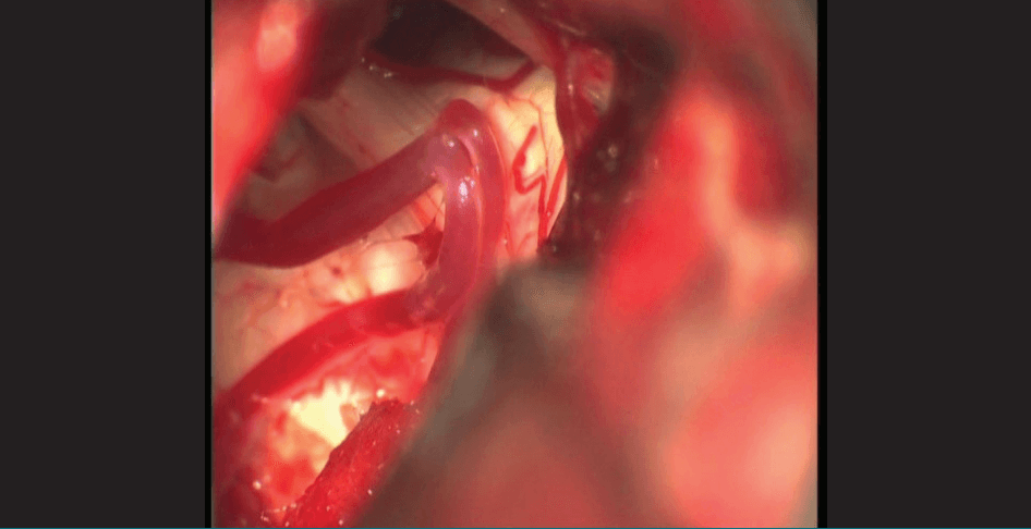
Figure 4: Intraoperative photograph of the trigeminal nerve after the loop of the superior cerebellar artery was freed from the brainstem side and axilla of the nerve.
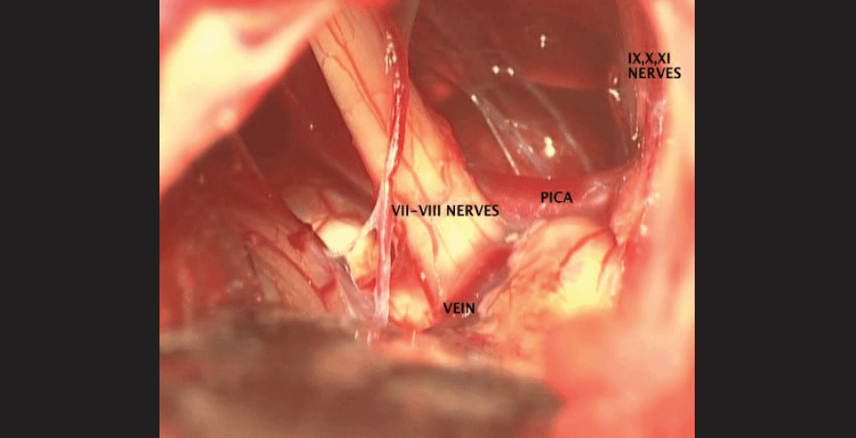
Figure 5: Intraoperative photograph of the cerebellopontine angle showing the facio-vestibulocochlear nerve complex compression by the anterior inferior cerebellar artery (AICA) and a vein at the entry/exit zone.
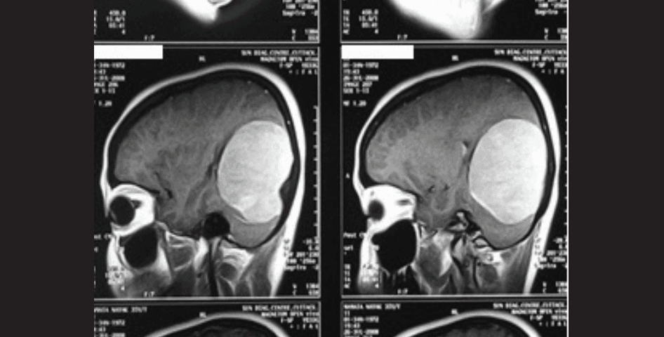
Figure 6: Sagittal T1-weighted contrast magnetic resonance imaging (MRI) of the brain showing a large tentorial meningioma (growing into the supratentorial compartment). The patient presented as ipsilateral trigeminal neuralgia as the sole symptom.
Histopathological Changes
Devor et al. 23 described structural and morphological changes seen in the trigeminal nerve as:
- Distortion
- Deviation
- Groove formation
- Nerve atrophy
- Axonal loss and demyelination
- Myelin abnormalities (dysmyelination)
- Presence of excess collagen
- Nerve atrophy 24
The microscopic findings of compressed segments ofhe trigeminal, facial, and glossopharyngeal nerves are remarkably similar, despite the variations in the site of biopsy and nerves involved. Beaver et al. 25 studied trigeminal nerve specimens from trigeminal nerve sections made for trigeminal neuralgia and demonstrated hypermyelination and demyelination. Demyelination at the point of contact was also reported in nerve specimens, obtained during microvascular decompression for HFS, on electron microscopy by Ruby and Jannetta. 26 The exact portion of the nerve compressed was biopsied, and the authors proposed that naked, demyelinated axons came into contact and effected ephaptic transmission. Hypertrophied myelin and Schwann cells were intermixed with axons. Brihaye et al.27 reported similar findings of demyelination in patients with glossopharyngeal neuralgia, who had compression due to an atheromatous vertebral artery. Ishii et al. 28 found demyelination and hypermyelination of the glossopharyngeal nerve in pharyngeal and cervical segments extracranially. In the case of the vestibulocochlear nerve, however, there has been no demonstrable demyelination. On the contrary, there is endoneurial fibrosis, 29 which is a plausible explanation for deafferentation.
According to another plausible theory proposed by Calvin et al.,30 the mechanism of trigeminal neuralgia involves a slightly injured nerve due to constant pulsations of a vessel adhered to the nerve. Amplification of neural activity and cross-transmission at the site of contact also results from a lack of input of A-fibres to the caudal trigeminal nucleus, a mechanism which could result in disinhibition of pain circuits and thereby result in the pain. Fromm and colleagues31,32 provided an explanation for trigeminal neuralgia with segmental inhibition, since the painful condition responds to baclofen, a drug that enhances segmental inhibition. Moreover, carbamazepine inhibits trigeminal neuralgia by acting centrally and is effective in HFS, indicating that it does not act at the site of vascular compression.
A combination of peripheral and central nervous system changes is involved in the aetiology of trigeminal neuralgia. There is an apparent disconnect between nociception and cortical perception of pain. There is reduced tactile and temperature sensation, indicating damage to small unmyelinated and large myelinated fibres, and abnormal temporal summation of pain, suggesting hyperexcitability. 33 With loss of inhibitory fibres, tactile stimuli on the face or gums cause increased activity in the trigeminal nucleus, resulting in increased discharge across the nerve to the effector organs. There is further amplification of impulses at the site of demyelination and reflection of these impulses back to the trigeminal nucleus. Summation of these impulses and the response of the nucleus results in severe episodic, lancinating pain over the distribution of the nerve. In the facial skin, there is abnormal neuronal activity, a higher rate of firing of axons of small-calibre fibres. Centrally, there is nociceptive modification of signal processing, N-methyl-D-aspartate (NMDA) activation with central hyperexcitability. 34
Gardner35 postulated that HFS occurs due to a reverberating circuit set up between afferent and efferent fibres at a point of facial nerve compression. He also advocated sectioning of the nervus intermedius in cases of failed facial nerve decompression. The underlying cause of compression is invariably an ectatic or aberrant blood vessel that compresses the facial nerve at its exit from the brainstem. 36 The root entry/exit zone has some distinct features: the nerve fibres are ensheathed by the arachnoid membrane only, without the epineurium, and there are no connective tissue septa traversing the individual fascicles. This region is also the TZ between central (oligodendroglia) and peripheral (Schwann cells) myelination. 37 All these features result in increased vulnerability and therefore susceptibility to stimuli such as compression, leading to lateral excitation of facial axons by ephaptic transmission 38 and hyperexcitability of facial motor neurons.39 The site of compression plays an important role in the pathogenesis of HFS. 5 The cessation of antidromic stimulation when the conflicting nerve and vessel are separated has been demonstrated by electromyography (EMG) monitoring, which returns to pathological activity if the vessel is replaced.40 Nielsen11 proposed abnormal conduction of impulses through the peripheral portion of the nerve with ectopic excitation and ephaptic transmission due to focal demyelination. Nielsen11 also observed electrophysiologically increased resistance in the para-axonal space, which is a prerequisite for ectopic/ephaptic excitation. Microvascular decompression decreases such resistance and facilitates remyelination, thus explaining the delayed benefits of microvascular decompression in HFS, and the severity of compression has no statistically significant difference on the outcome after microvascular decompression. 41
Not all cases of HFS have neurovascular contact. Aoki and Nagao42 described a case involving a 5-year history of HFS, in whom neurosurgical exploration was negative for any vascular contact or conflict. The authors merely dissected around the nerve complex, and symptoms were relieved after surgery. Conversely, Sunderland reported neurovascular contact between a redundant loop of the anterior inferior cerebellar artery (AICA) and the facial nerve in more than 60% of his autopsy cases, thus proving that the incidence of neurovascular conflict is higher than the reported incidence of HFS in the general population. 43
Ridder et al. reviewed neurovascular conflict of the vestibulocochlear nerve in patients with tinnitus. 44 They hypothesised, based on electrophysiological studies by auditory brainstem responses, that neurovascular conflict must occur in the intracranial portion of the nerve. Tinnitus may not be the result of demyelination, but rather due to desynchronisation of auditory impulses. The more synchronised the nerves fire, the higher the amplitude of the evoked potentials. Contact of a blood vessel with a nerve may alter neural conduction (decreased conduction velocity in some fibres or inactivate some fibres), decreasing the temporal coherence of the firing in the central segment of the auditory nerve, decreasing the amplitude of peak II, and clinically this may result in frequency-specific tinnitus. 45 The desynchronised signal transmission within the auditory nerve from a vascular conflict may lead to reorganisation of auditory nuclei in the auditory brainstem and auditory cortex by way of neuronal plasticity. 46,47 Clinically, patients suffer recurrent vertigo, disequilibrium, ataxia, and unilateral high-frequency sensorineural hearing loss, and spontaneous nystagmus on electronystagmography. 48 According to the explanation offered by Schwaber and Whetsell, 49 the onset follows an episode of vestibular neuronitis and adherence of the vessel to the vestibulocochlear nerve. There is demyelination of the nerve at the point of contact and deafferentation, leading to reorganisation of vestibular nuclei and discharges from these nuclei. It is likely that there are corresponding changes in the cortex, as well as in the interaction between the vestibulospinal system, ocular motility, visual pathways, and the reticular system. Motion and visual cues can precipitate or accentuate the symptoms due to abnormal discharges.
Degrees of nerve contact:
Anderson et al. 50 and Cheng et al. 51 described a variable degree of nerve and vessel contact in patients with neuralgia, ranging from no contact to severe deformation with or without nerve atrophy.
Patients with severe deformation tend to have a higher incidence of atrophy, which can be diagnosed preoperatively,51 and the zone of atrophy consistently correlates with focal demyelination.
Brainstem Surface Conflicts
Can a contact between an ectatic and aberrant artery and brainstem trigger features of neurovascular conflict (Figure 7A–C)? Such neurovascular conflicts have been reported to cause ipsilateral sensorimotor symptoms, hypertension, intractable hiccups, and painful HFS with spasm of masticatory and ocular motor muscles.52 Ipsilateral tongue wasting has also been observed (Figure 8). The hypothesis explaining multiple cranial nerve involvement is neurovascular conflict between a vascular loop or an ectatic vessel and neighbouring brainstem nuclei. The reversal of clinical manifestations after microvascular decompression supports this hypothesis. 52 Intrinsic brainstem lesions like diffuse glioma can also be associated with HFS (Figure 9).
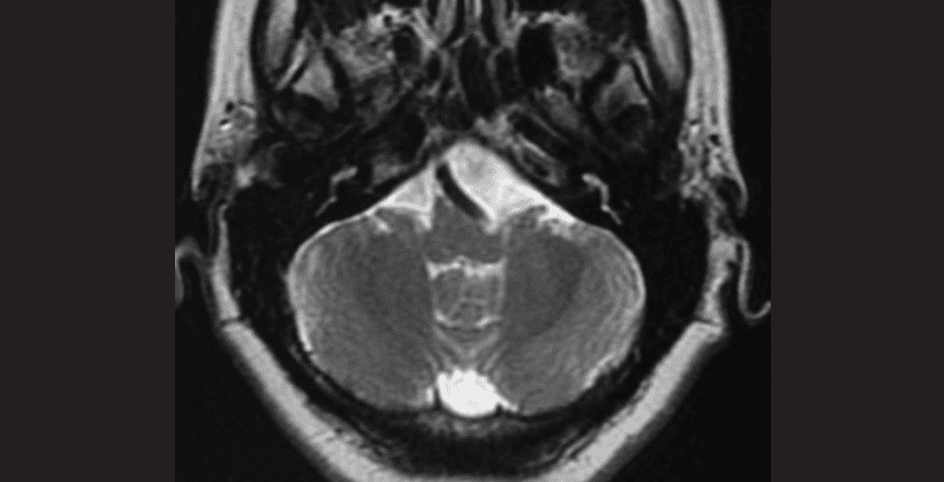
Figure 7A: Axial T2-weighted magnetic resonance imaging (MRI) showing an ectatic vertebral/basilar artery causing indentation on the brainstem.
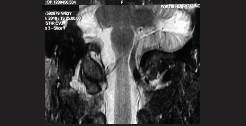
Figure 7B: Coronal T2-weighted magnetic resonance imaging (MRI) showing an ectatic vertebral/basilar artery causing indentation on the brainstem.
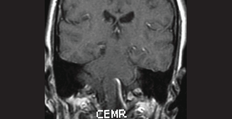
Figure 7C: Coronal T2-weighted magnetic resonance imaging (MRI) showing an ectatic vertebral/basilar artery in the patient with ipsilateral tongue wasting.
Venous Neurovascular Conflicts
Veins also play a part in neurovascular conflicts, which may be purely venous or in combination with an arterial conflict. Far from being rare, there are reports of neurovascular conflicts due to pure venous compression (Figures 10 and 11), and a significant number of these have localised arachnoiditis.53 Characterisation of venous compression is difficult, and many such cases may have atypical facial pain, with a background of continuous pain and paroxysmal fits. The incidence range is wide, varying from 6.1% to 68%, with an average of 25.3%.53 In another study, Dumot and Sindou54 reported their experience with 326 consecutive patients who underwent microvascular decompression from 2005 to 2013. Of these, 124 (38%) had a venous conflict, alone in 29 (8.9%) or an association with an artery in 95 (29.1%). There may be associated pathology like focal arachnoiditis and angulation of the nerve over the petrous apex.
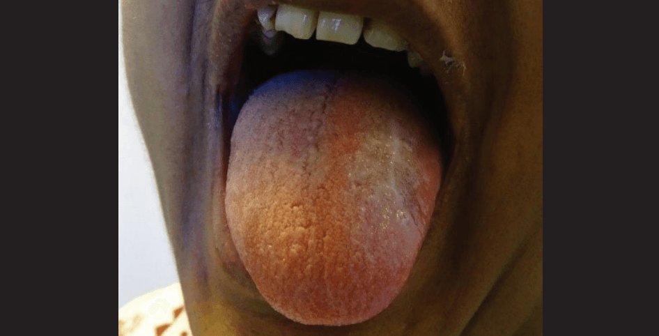
Figure 8: AIpsilateral tongue wasting in a patient.
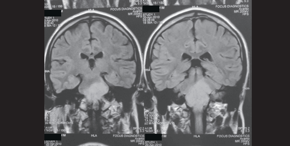
Figure 9: Magnetic resonance imaging (MRI) (T1-weighted contrast) with diffuse glioma of the brainstem in a patient who presented with hemifacial spasm.
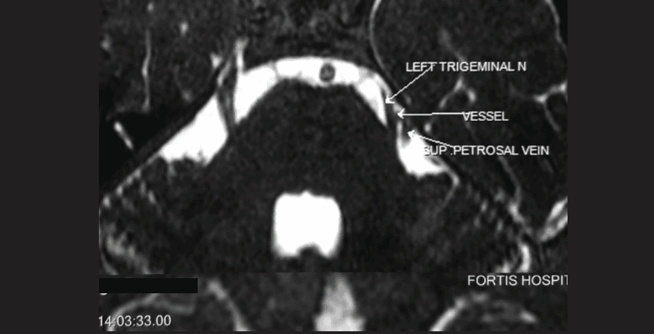
Figure 10: Magnetic resonance imaging (MRI) (T2-weighted axial) showing venous conflict with the trigeminal nerve.
Preoperative diagnosis of neurovascular conflict is essentially clinical, based on the characteristic pain profile of trigeminal nerve root compression and appearances in hemifacial clonic contractions. Diagnosis of involvement of other cranial nerves rests on intractable tinnitus and vertigo (vestibulocochlear nerve) and intermittent lancinating pain in the tonsillar fossa (glossopharyngeal neuralgia). Almost all cases of glossopharyngeal neuralgia are idiopathic,55 although some cases have been described due to a calcified stylohyoid ligament,56 nasopharyngeal and cerebellopontine angle tumours,57 or compression by an artery.27,58 These symptoms have to be evaluated by an otologist. Rarely, there may be conflict between the vertebral artery and the brainstem, or the hypoglossal nerve (Figures 7 and 8). Trigeminal neuralgia (after exclusion of atypical mimics of the same) is graded by severity, according to the Barrows Neurological Institute Grading into five grades (Table 1).
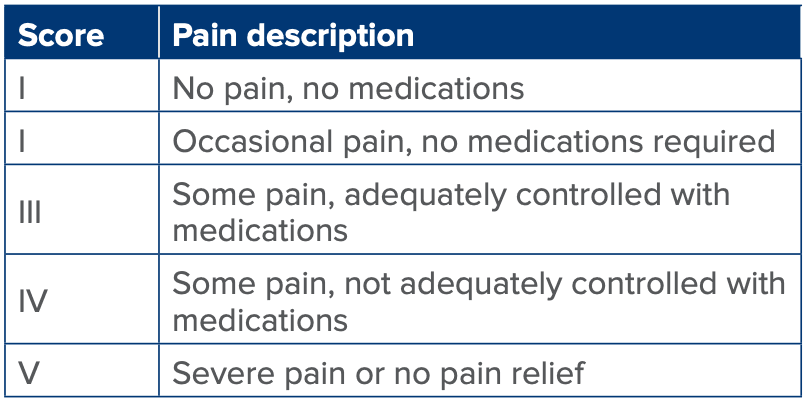
Table 1: Barrow Neurological Institute Pain Institute Score.
HELPS and VANCOUVER
Honey et al. (2025) described episodic vagal rhizopathies manifesting as hemilaryngopharyngeal spasm (HELPS) and chronic non-productive cough due to vagus-associated neurogenic cough occurring due to unilateral vascular encroachment of its root (VANCOUVER) secondary to compression of vagal rootlets (that supply the laryngeal musculature) by posterior inferior cerebellar artery (PICA) in the posterior fossa. These symptoms may be accompanied by glossopharyngeal neuralgia. There are symptom-free periods between such episodes. Patients present with a feeling of choking and incessant coughing and may have to be intubated or tracheostomised. These patients may present to the emergency room (ER) and be misdiagnosed as having gastroesophageal reflux or may be labelled as psychogenic. Magnetic resonance imaging (MRI) of the posterior fossa shows a neurovascular conflict between the ipsilateral PICA and the vagus nerve.59 Microvascular decompression of the vagal nerve is curative in these patients.
Neuroimaging and Operative Correlation
Detection of neurovascular conflict:
T2-weighted MRI study of the posterior fossa, along with T1-weighted contrast administration, is generally sufficient to demonstrate the neurovascular conflict (Figures 11–15). Leal et al.60 carried out three-dimensional (3D) time-of-flight magnetic resonance angiography (3D TOF MRA), and 3D T1- gadolinium enhanced MRI to observe the vessels related to nerve in 100 patients with trigeminal neuralgia. Image analysis of these 100 patients showed that 88 had vessels in relation to the nerve, and 12 did not. All 88 patients had neurovascular conflict observed at surgery, with no false positives. Of the 12 patients who did not have vessels related to nerves, nine did not have neurovascular conflict at the time of surgery, concluding that there were three false negatives. Thus, the sensitivity of MRI was 96.7%, and the specificity of 100%, when correlated to surgical findings. Ronak Jani et al.61 studied the images and operative findings around the trigeminal nerve in 27 patients undergoing surgery for HFS. In these patients with no symptoms of neuralgia, they found that 23 patients had intraoperative evidence of neurovascular compression of the trigeminal nerve, 18 out of whom had MRI evidence of an aberrant artery in the root exit zone of the trigeminal. Further, they observed that the remaining five patients, who did not have MRI evidence of neurovascular conflict for the trigeminal nerve, had intraoperative neurovascular conflict.
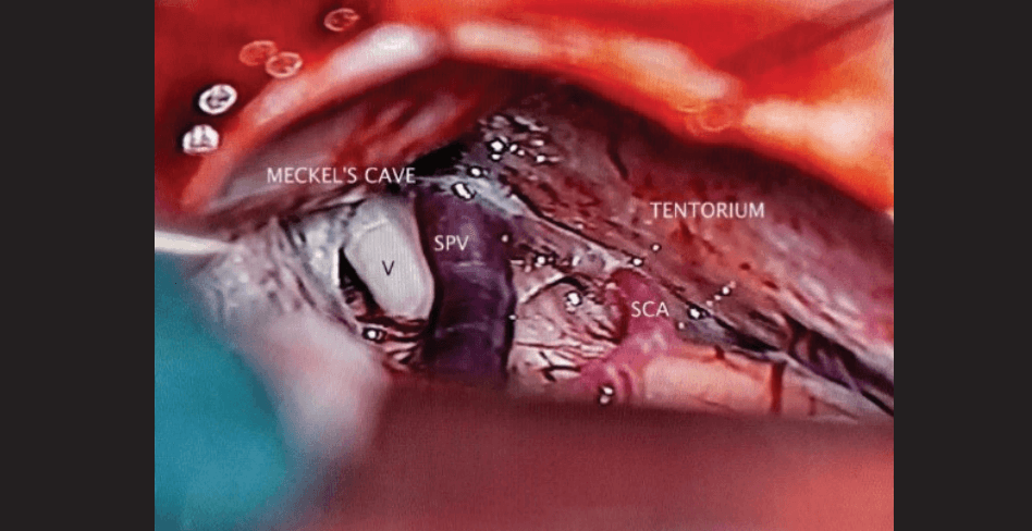
Figure 11A: Intraoperative photograph showing venous compression on the left trigeminal nerve.
Abbreviations: SCA: Superior Cerebellar Artery; SPV: Superior Petrosal Vein
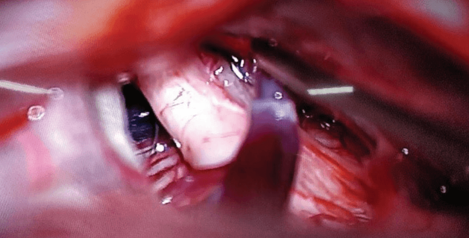
Figure 11B: Intraoperative photograph showing indentation on the trigeminal nerve due to venous compression.
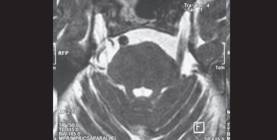
Figure 12: Magnetic resonance imaging (MRI) (T2-weighted axial) showing venous conflict with the trigeminal nerve.
Prediction of culpability of the vessel in neurovascular conflict:
In their study, the vessel was correctly predicted to be compressing the nerve in 91 out of 100 patients, with accurate prediction of the SCA, AICA artery and basilar artery, while prediction was moderate in venous compression.
Prediction of the site of compression on the surface of the nerve:
Image analysis correctly identifies the surface of the circumference of the nerve as the site of compression, while predicting the severity of compression.
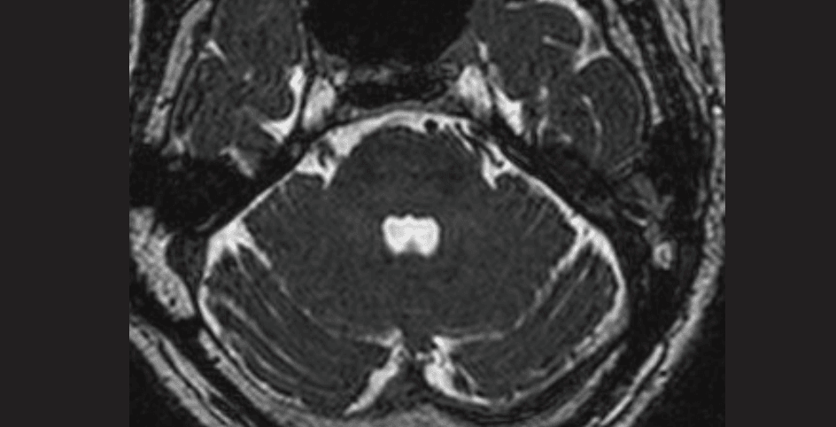
Figure 13: Magnetic resonance imaging (MRI) (T2-weighted axial) showing neurovascular conflict on the left side involving the trigeminal nerve.
Abbreviations: SCA: Superior Cerebellar Artery; SPV: Superior Petrosal Vein
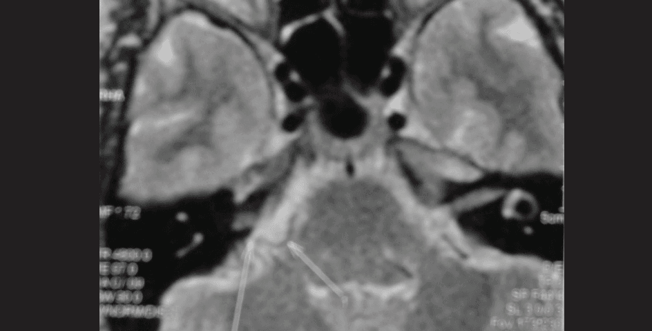
Figure 14: Magnetic resonance imaging (MRI) (T2-weighted) showing neurovascular conflict on the right side involving the facial nerve.
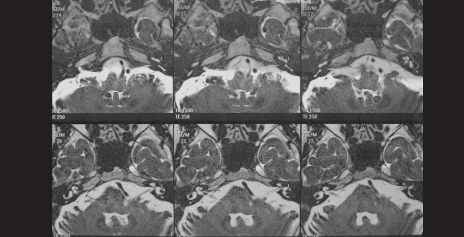
Figure 15: Magnetic resonance imaging (MRI) (T2-weighted axial) showing neurovascular conflict at VII–VIII entry/exit zone.
Conclusion
After nearly five centuries of clinical description and more than a century of surgical management, the pathogenesis of neurovascular compression syndromes is by no means completely settled. While imaging studies confirm neurovascular conflict in the majority of patients, the exact cellular and biochemical changes are unknown. Studies about the TZ and neuronal plasticity in response to ephapsis hold promise for unfolding the mystery of the changes that lead to clinical manifestations. Cochleovestibular manifestations and compression of the medulla by ectatic vessels also fall in the realm of the unknown. Recently described vagus nerve compression (HELPS and VANCOUVER syndromes) adds to the spectrum of neurovascular conflict lesions in the posterior fossa. Microvascular decompression, with its consistent and often long-lasting, permanent relief of symptoms, provides fair evidence of the culpability of TZ neurovascular conflict.
Harjinder S Bhatoe. The Neurovascular Conflict Syndromes: Part I: A Review of Anatomy and Pathophysiology. MMJ. 2026, June. Vol 3 (2).
DOI: XXXX_XXXX_XXXX_XXXX
References
- Dandy WE. Concerning the cause of trigeminal neuralgia. Am J Surg. 1934;24:447–
- Sunderland S. Neurovascular relations and anomalies at the base of the brain. J Neurol Neurosurg Psychiat. 1948;2:243–
- Kulkarni A. Complex Neurovascular Syndromes: Is the Compressing Vessel Alone the Culprit?. J Neurosci Rural Pract. 2022;13(2):283–
- Sunderland S. Cranial nerve injury. Structural and pathophysiological considerations and a classification of nerve injury. In: Samii M, Jannetta P (Eds). The cranial nerves. New York: Springer Verlag; 1981: p. 16–
- Jannetta PJ. Neurovascular compression in cranial nerve and systemic disease. Ann Surg. 1980;192:518–
- Tarlov IM. Structure of nerve root
- Guclu Bulent, Sindou Marc, Meyronet David, et al. Cranial nerve vascular syndromes of the trigeminal, facial and vagoglossopharyngeal nerves: comparative anatomical study of the central myelin portion and transitional zone; correlation with incidences of corresponding hyperactive dysfunctional syndromes. Acta Neurochir. 2011;153:2365–
- Berthold C-H, Carlstedt T, Corneliuson O. Anatomy of the nerve root at the central-peripheral transitional region. In: Dyck PJ, Thomas PK, Lambert EH, Bunge R (Eds). Peripheral Neuropathy. Philadelphia: WB Saunders; 1984. p. 156–
- Jannetta PJ. Microvascular decompression for hemifacial spasm. In: May M (Ed). The Facial Nerve. New York: Thieme Stratton; 1985. p. 499–508.
- Nielsen VK. Pathophysiology of hemifacial spasm. I. Ephaptic transmission and ectopic excitation. Neurology. 1984;34:418–
- Nielsen VK. Electrophysiology of hemifacial spasm. In: May M (Ed). The Facial Nerve. New York: Thieme; 1986. p. 487.
- Rasminsky M. Ectopic impulse generation in pathologic nerve fibers. In: Dyck PJ et al. (Eds). Peripheral Neuropathy. Philadelphia: WB Saunders; 1984. p. 911–
- Rasminsky M. Ephaptic transmission between single nerve fibers in the spinal roots of “dystrophic” mice. J Physiol. 1980;305:151–
- Schwaber Mitchell K. Vascular compression syndromes. In: Jackler Robert K, Brackmann Derald E (Eds). Neurotology. St Louis: Mosby; 1994. p. 881–904.
- Møller AR, Sen CN. Recordings from the facial nucleus in the rat. Signs of abnormal facial muscle response. Exp Brain Res. 1990;81(1):18–
- Møller AR, Jannetta PJ. Physiological abnormalities in hemifacial spasm studied during microvascular decompression operations. Exp Neurol. 1986;93:584–600.
- Møller AR, Jannetta PJ. On the origin of synkinesis in hemifacial spasm: Results of intracranial recordings. J Neurosurg. 1984;61:569–
- Digre KB, Corbett JJ. Hemifacial spasm: Differential diagnosis, mechanism and treatment. Neurology. 1988;49:151–
- Zhong Jun, Zhu Jin, Li Shi-Ting, et al. Microvascular decompression in patients with coexistent hemifacial spasm and trigeminal neuralgia. Neurosurgery. 2011;68:916–
- Desai K, Nadkarni T, Bhayani R, et al. Cerebellopontine angle epidermoid tumor presenting with “tic convulsif” and tinnitus: case report. Neurol Med Chir. 2002;42(4):162–
- Wakabayashi T, Tamaki N, Saroh H, et al. Epidermoid tumor presenting as painful tic convulsif. Surg Neurol. 1983;19(3):244–
- Ogansawara H, Oki S, Kohno H, et al. Tentorial meningioma and painful tic convulsif: case report. J Neurosurg. 1995;82(5):895–
- Devor M, Govrin-Lippmann R, Rappaport ZH. Mechanism of trigeminal neuralgia: an ultrastructural analysis of trigeminal root specimens obtained during microvascular decompression surgery. J Neurosurg. 2002;96:932–
- Leal PRL, Barbier C, Hennier M, et al. Atrophic changes in the trigeminal nerves of patients with trigeminal neuralgia due to neurovascular compression and their association with severity of compression and clinical outcomes. J Neurosurg. 2014;120:1484–
- Beaver DL, Moses HL, Ganote CE. Electron microscopy of the trigeminal ganglion. III. Trigeminal neuralgia. Arch Path. 1965;79:571–
- Ruby JR, Jannetta PJ. Hemifacial spasm. Ultrastructural changes in the facial nerve induced by neurovascular compression. Surg Neurol. 1975;4:369–
- Brihaye J, Périer O, Smulders J, et al. Glossopharyngeal neuralgia caused by compression of the nerve by an atheromatous vertebral artery. J Neurosurg. 1956;13:299–301.
- Ishii T. Glossopharyngeal neuralgia: surgical treatment and electron microscopic findings. Laryngoscope. 1976;86:577–
- Belal A, Ylikoski J. Pathologic significance of Meniere’s symptom complex: a histopathologic and electron microscopic study. Am J Otol. 1980;1(4):275–
- Calvin WH, Loeser JD, Howe JF. A neurophysiological theory for the pain mechanism of tic douloureux. Pain. 1977;3:147–
- Fromm GH. Pathophysiology of trigeminal neuralgia. In: Fromm GH, Sessle BJ (Eds). Trigeminal Neuralgia. Boston: Butterworth-Heinemann; 1991.
- Fromm GH, Chattha AS, Terrence CF, et al. Role of inhibitory mechanism in trigeminal neuralgia. Neurology. 1981;31:683–
- Levine David A, Argoff Charles E, Pilitsis Julie. Trigeminal neuropathic pain and anesthesia dolorosa. In: Kim Burchiell (Ed). Surgical management of pain. New York: Thieme; 2015. p. 180–
- Attal N. Neuropathic pain: mechanisms, therapeutic approach, and interpretation of clinical trials. Continuum (Minneap Minn). 2012;18(1):161–
- Gardner WJ. Cross talk – the paradoxical transmission of a nerve impulse. Arch Neurol. 1966;14:149–
- Girard N, Poncet M, Caces F, et al. Three-dimensional MRI of hemifacial spasm with surgical correlation. Neuroradiology. 1997;39:46–
- Nielsen VK. Electrophysiology of the facial nerve in hemifacial spasm: ectopic/ephaptic excitation. Muscle Nerve. 1985;8:545–
- Sanders DB. Ephaptic transmission in hemifacial spasm: a single fiber EMG study. Muscle Nerve. 1989;12:690–
- Martinelli P, Giuliani S, Ippoliti M. Hemifacial spasm due to peripheral injury of facial nerve: a nuclear syndrome? Mov Disord. 1992;7:181–
- Samii M, Günther Thomas, Iaconetta Giorgio, et al. Microvascular decompression to treat hemifacial spasm: Long term results for a consecutive series of 143 patients. Neurosurgery. 2002;50:712–
- Kim Joo Pyung, Chung Jong Chul, Chang Won Seok, et al. Outcomes of surgical treatment for hemifacial spasm associated with the vertebral artery: severity of compression, indentation and color change. Acta Neurochir. 2012;154:501–
- Aoki N, Nagao T. Resolution of hemifacial spasm after posterior fossa exploration without vascular compression. Neurosurgery. 1986;18:478–
- Sunderland S. Neurovascular relations and anomalies at the base of the brain. J Neurol Neurosurg Psychiatry. 1948;11:243–
- De Ridder D, Vanneste Sven, Adriaensens Ine, et al. Vascular compression of the cochlear nerve and tinnitus. A pathophysiological investigation. Acta Neurochir. 2012;154:807–
- De Ridder D, Heijneman K, Haarman B, et al. Tinnitus in vascular conflict of the eighth cranial nerve: A surgical pathophysiological approach. Prog Brain Res. 2007;166:401–
- Szmyd B, Sołek J, Błaszczyk M, et al. The Underlying Pathogenesis of Neurovascular Compression Syndromes: A Systematic Review. Front Mol Neurosci. 2022;15:923089.
- Muhlnickel W, Elbert T, Taub E, et al. Reorganization of auditory cortex in tinnitus. Proc Natl Acad Sci USA. 1998;95:10340–
- Schwaber MK, Hall JW. Cochleovestibular nerve compression syndrome: I. Clinical features and audiovestibular test findings. Laryngoscope. 1992;102(9):1020–
- Schwaber MK, Whetsell WO. Cochleovestibular nerve compression syndrome: II. Histopathology and theory of pathophysiology. Laryngoscope. 1992;102(9):1030–
- Anderson VC, Berryhill PC, Sandquist MA, et al. High resolution three-dimensional magnetic resonance imaging in the evaluation of neurovascular compression in patients with trigeminal neuralgia. A double-blind pilot study. Neurosurgery. 2006;58:666–
- Cheng Jian, Meng Jinli, Wenke Liu, et al. Nerve atrophy in trigeminal neuralgia due to neurovascular compression and its association with surgical outcomes after microvascular decompression. Acta Neurochir (Wien). 2017;159:1699–
- Idriceanu Tania-Mihaela, Sindou Marc. Painful spasms in facial, masticatory, and motor ocular muscles reversed after microvascular decompression of a neurovascular conflict at the brainstem. Acta Neurochir. 2017;159:1701–11
- Dumot Chloe, Brinzeu Andrei, Berthiller Julien, et al. Trigeminal neuralgia due to venous neurovascular conflicts: outcome after microvascular decompression in a series of 55 consecutive patients. Acta Neurochir. 2017;159:237–
- Dumot Chloe, Sindou Marc. Trigeminal neuralgia due to neurovascular conflicts from venous origin: an anatomical-surgical study (consecutive series of 124 operated cases). Acta Neurochir. 2015;157:455–
- Onofrio Burton M. Glossopharyngeal neuralgia. In: Wilkins RH, Rengachary SS (Eds). Neurosurgery Vol III. New York: McGraw-Hill; 1996:3969–
- Graf CJ. Glossopharyngeal neuralgia and ossification of stylohyoid ligament. J Neurosurg. 1958;16:448–
- Dandy W. The Brain. In: Lewis D (Ed). Practice of Surgery. Hagerstown MD: WF Prior; 1932. p. 201–
- Laha RK, Jannetta PJ. Glossopharyngeal neuralgia. J Neurosurg. 1977;47:316–
- Leal PRL, Harmeir Marc, Froment Jean Claude, et al. Preoperative demonstration of the neurovascular compression characteristics with special emphasis on the degree of compression, using high-resolution magnetic resonance imaging: a prospective study, with comparison to surgical findings, in 100 consecutive patients who underwent microvascular decompression for trigeminal neuralgia. Acta Neurochir. 2010;152:817–
- Ronak H Jani, Hughes Marlon A, Gold Michael S, et al. Trigeminal nerve compression without trigeminal neuralgia: Intraoperative vs Imaging evidence. Neurosurgery. 2019;84:60–
- Honey Christopher R. The vagal rhizopathies. Front Neurol. 2025;15:1513160.