 WhatsApp Us (24/7)
 +91 926 888 0303 (24/7)


Abstract:
Keyhole neurosurgery is a surgical technique that allows neurosurgeons to perform complex procedures through small “keyhole” incisions, often with the aid of specialised instruments and advanced imaging technology. Unlike traditional open surgery, which involves large incisions and significant manipulation and damage to surrounding tissue, minimally invasive neurosurgical (MIN) techniques minimise trauma to the patient's body and reduce the risk of complications. One of the primary benefits of keyhole neurosurgery is a reduced risk of post-operative complications, including less tissue damage, lower pain levels, shorter recovery time, reduced blood loss, lower infection rates, and a decreased risk of nerve damage. With the aid of advanced imaging and navigation technology, neurosurgeons can not only visualise but also locate the target area with greater accuracy, enabling them to perform highly complex procedures with increased safety and efficacy. This precision also allows for smaller incisions, which can further reduce the risk of complications and promote a faster recovery. Keyhole techniques can be used for a wide range of neurosurgical procedures, including brain tumours, spinal cord tumours, aneurysms, and other conditions. It is a highly promising approach to treating complex neurosurgical conditions. With continued advancements in technology and surgical techniques, it is likely that we will see an even greater adoption of MIN approaches in neurosurgery in the future. The current article focuses on these keyhole approaches used in the treatment of neurosurgical pathologies with the aim of achieving excellent surgical outcomes with minimal morbidity and risks to the patients.
Key words: Surgical Minimalism, Neurosurgery, Keyhole Neurosurgery, Supraorbital Keyhole, Keyhole Kawase.
Introduction
Skull base surgery has traditionally involved extensive, often invasive approaches designed to achieve maximal tumour resection or lesion access, such as transcranial or transfacial routes. These extensive approaches, including the pterional, fronto-orbitozygomatic, and subtemporal craniotomies, provided excellent exposure but were associated with significant morbidity, longer recovery times, and complex surgical corridors.
In recent decades, advances in imaging, instrumentation, and endoscopic techniques have catalysed a shift towards less invasive, "keyhole" approaches — minimally invasive corridors that access lesions through smaller craniotomies or even natural openings. These approaches aim to reduce morbidity while maintaining or improving surgical efficacy.
The concept of the keyhole approach emerged from neurosurgery's broader movement toward minimally invasive techniques. In skull base surgery, the development of the super- and supraorbital approaches, including the "eyebrow" and "eyelid" routes, exemplifies this trend. These approaches leverage natural corridors (e.g., cribriform plate, sphenoid sinus) or minimal bony openings to reach deep-seated lesions with less soft tissue disruption. These corridors — supraorbital, pterional, retrosigmoid, and endonasal — are strategically selected based on tumour location, morphology, and adjacent neurovascular anatomy.1 These tailored approaches enhance outcomes by reducing brain retraction, operative time, blood loss, and hospital stay — all while preserving oncologic safety even in large to giant intracranial tumours.1–3
Keyhole approaches often involve:
- Smaller, strategically placed craniotomies
- Use of endoscopic visualisation
- Neuro-navigation systems
- Specialised instrumentation
The Term “Keyhole”: Explanation
The term keyhole refers to viewing the entire room through a small keyhole in the room door, using multiple viewing angles. This is a much more elegant and minimally invasive means of looking into the contents of the room rather than opening the entire door at the hinges. A smaller incision has several definite advantages as compared to a larger traditional craniotomy; however, this remains a subject of large randomised controlled trials, and most of the patients prefer smaller incisions in view of the following putative advantages: 4-7
- Lesser damage to the cutaneous nerves and vascular supply of the scalp, hence less post- operative pain (leading to lesser post-operative hospital stay and analgesia requirement) and better wound healing
- Smaller incisions are mostly relatively simple and linear and require less time to open and close
- Better cosmesis
- Lesser exposure of the cortical tissue during the entire surgery with less attendant thermal damage
- Less muscular dissection and lesser chances of temporalis muscle atrophy in the post-operative period
- Repeat surgery is technically much easier with keyhole, with lesser area of the dura and brain tissue that is required to expose
- Wound healing is faster, and radiation can be given early in the post-operative period as compared to traditional craniotomies
The “Keyhole” Concept
Operating through a keyhole is not much different from operating through a larger incision. However, before proceeding with keyhole surgery, it is prudent to understand the keyhole concept and learn the basic principles of the keyhole approach. 4–7
The following are the properties of a keyhole:
- The term “keyhole” relates to a concept and not to any size. The neurosurgeons should learn to think about how to make their incisions as small as possible, as is necessary to achieve their surgical goals.
- The keyhole is immovable and should be meticulously planned before surgery. Therefore, neuro-navigation is mandatory to plan a keyhole placement. There is very little room for error in keyhole surgery, and mistakes are not forgiving.
- It is impossible to look at the back of the keyhole. The pathology located at the depths is best visualised; however, superficially located diseases are not very good for a keyhole (Figure 1).
- A single viewing trajectory is not enough to work through a keyhole. The view can be enhanced significantly by frequently changing the viewing angles of the microscope and the position of the patient during surgery (Figure 2).
- The best view through a keyhole is down the centre of the opening, which usually lies along the long axis of the lesion.
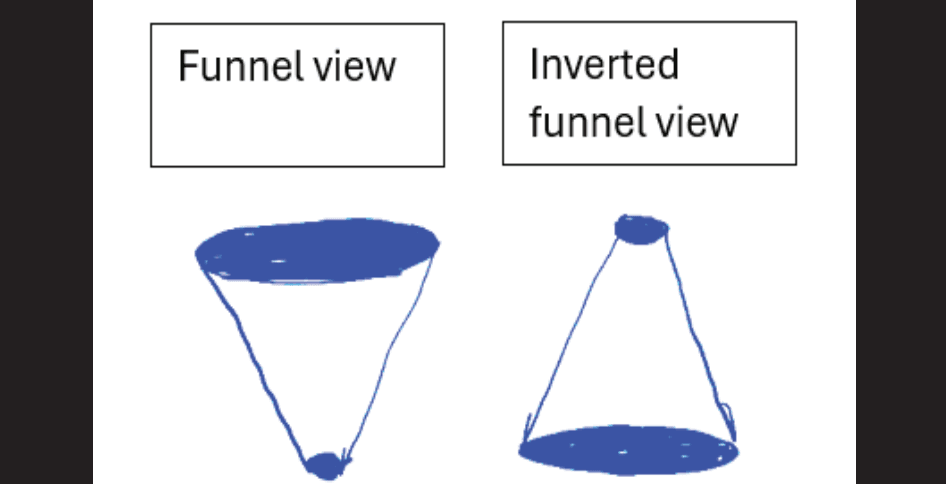
Figure 1: Schematic illustration showing the view through the microscope (funnel view) as opposed to the vision through a keyhole (reverse-funnel view).
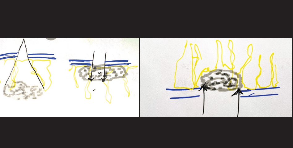
Figure 2: Schematic illustration showing the effectiveness of the keyhole in deeply situated lesions, as opposed to those that lie superficially.
Planning of a Keyhole
The thinking process and planning for a keyhole surgery are critical for the success of surgery and should start by carefully studying the radiographic images of the patient. The surgeon should be physically present in the operating room from the very beginning of the surgery, for the planning, execution and placement of the keyhole and should himself plan and execute these steps, rather than leaving them over to some resident or anyone else and showing up in the operating room after the opening of the dura. The margin of error is absolutely small in keyhole surgeries, and this is the most common reason this approach is not very popular among most of the neurosurgeons. 4–7
The basic principles in planning a keyhole are elaborated below and should be followed to avoid any problems during surgery.
- Cerebrospinal fluid (CSF) should be drained early after the dural opening. One must be extremely patient after opening the dura and the first step is the drainage of as much CSF as possible from the CSF spaces. This relaxes the brain, allowing surgery to proceed without retraction, even if it appears intimidating immediately after dural opening, particularly in the presence of brain bulge. Sometimes, especially in large tumours, one may electively choose to insert a lumbar drain before the surgery for draining the CSF either before opening the dura or during the surgery.
- Cisternal spaces and trans-sulcal corridors should be used whenever possible for approaching the lesion.
- The working trajectory is best along the long axis of the tumour. Hence, the keyhole is best placed in the area where the long axis of the lesion intersects the surface. This rule is obviously not followed when any eloquent or important area falls in the trajectory of the long axis (Figure 3A and B).
- The approach to any lesion is prioritised by the location and anatomical relationships of the most difficult-to-access portion of the lesion, because it is this critical part of the lesion that requires utmost careful planning and meticulous skills, rather than the majority of the lesion. Sometimes, it necessitates a change in the approach while at other times it requires the use of an endoscope. Hence, it is a good practice always to compartmentalise the lesion into an easily accessible portion and a difficult-to-access portion.
- The keyhole surgery is to be performed under super high microscopic magnification. Under this extreme magnification, the focal length adjustments need to be done at frequent intervals, especially with the changes in the angle of view. These focal length adjustments can be made by the assistant. The use of a mouthpiece is very handy in a keyhole approach and is a good microscope accessory to have.
- The use of an endoscope should be considered in every keyhole surgery, especially in skull base surgery. The endoscope complements the keyhole in viewing difficult to access areas of the lesion and increases the visibility without increasing the size of the exposure or without adding any extensive skull base approaches (e.g., orbitotomy) to the original approach.
Mini-pterional Craniotomy
The standard pterional craniotomy is the main workhorse for a majority of complex neurosurgical operations. The standard craniotomy entails a larger craniotomy and extensive muscle dissection with the disadvantages of temporalis muscle atrophy, poor cosmesis and jaw pain in the post-operative period (Figures 4–6).
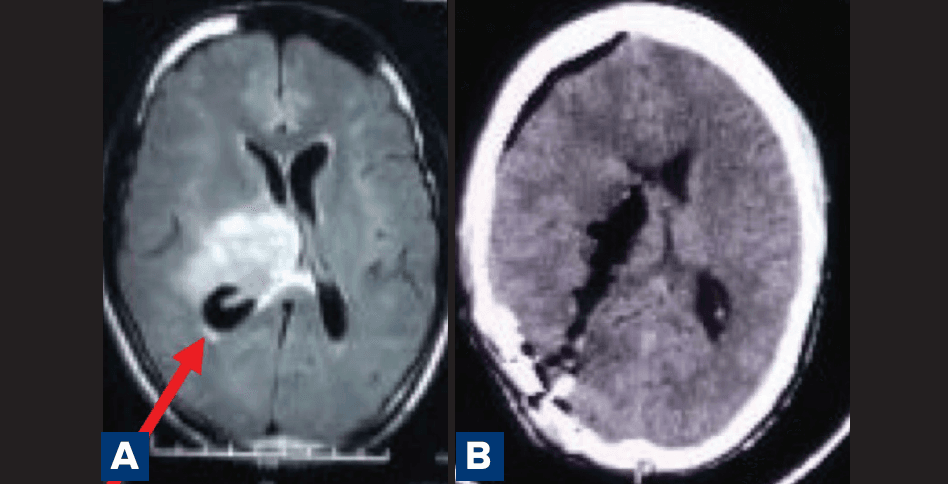
Figure 3: A. Pre-operative magnetic resonance imaging (MRI) of a patient showing right-sided thalamic glioma, demonstrating the working trajectory along the long axis of the tumour; B. Immediate post-operative computed tomography (CT) of the same patient showing gross total tumour resection following keyhole principles.
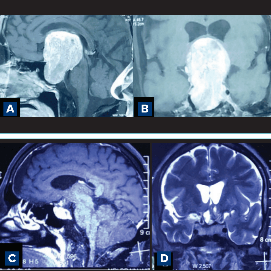
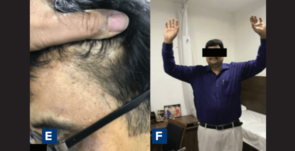
strong>Figure 4A–F: Mini-pterional craniotomy for a giant pituitary adenoma: A–B. Pre-operative sagittal and coronal contrast magnetic resonance imaging (MRI) of the patient showing a giant pituitary adenoma reaching right up to the third ventricle; C–D. Follow-up 2 years post-operative sagittal contrast and T2- weighted coronal MRI of the same patient. Note the presence of a small residual tumour in the sella, which is being followed up on a regular basis; E. Side and; F. Front profile photograph of the patient at follow-up.
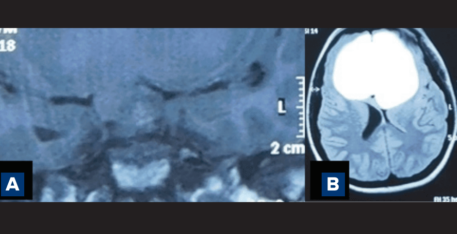
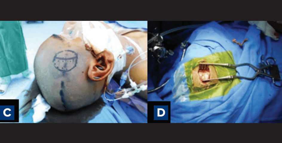
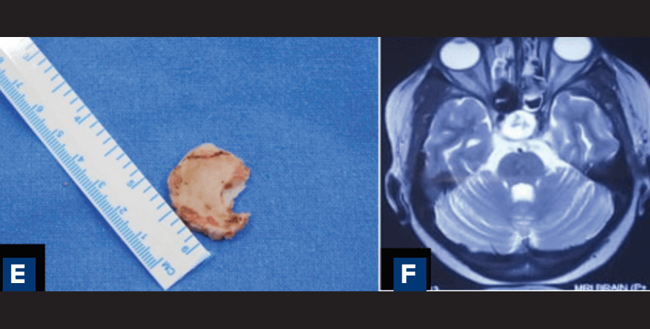
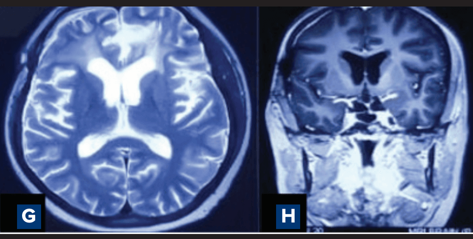
Figure 5A–H: Mini-pterional approach for a giant craniopharyngioma: A–B. Pre-operative coronal and axial magnetic resonance imaging (MRI) showing the lesion; C. The head positioned on a horseshoe headrest, turned 30 degrees contralaterally and slightly extended, with planning of the incision and exposure; D. Depicting the extent of exposure; E. Showing the size of the bone flap removed; F–H. Follow-up post-operative MRI at 2 years showing total excision and no recurrence.
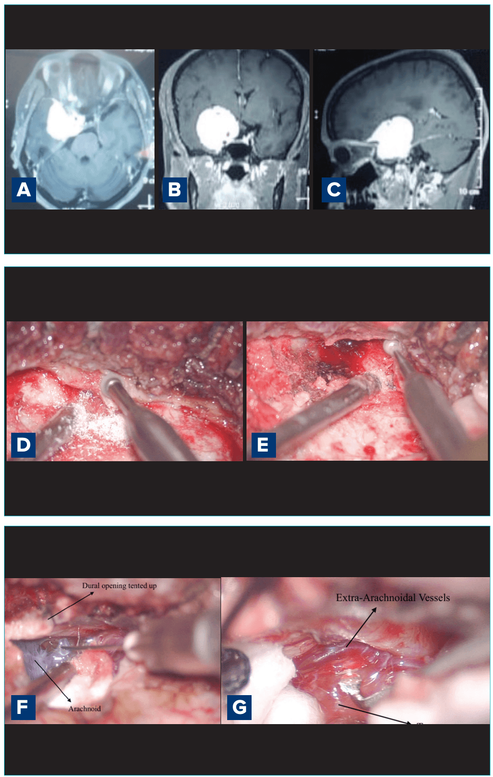
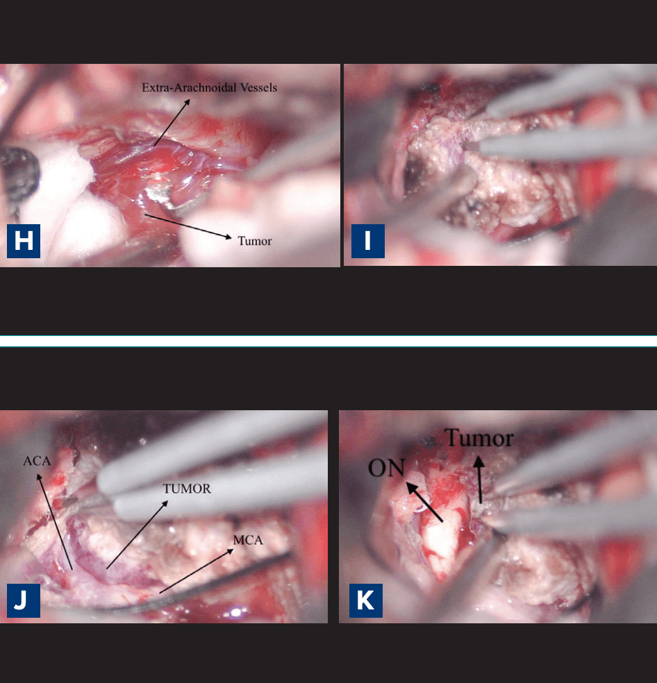
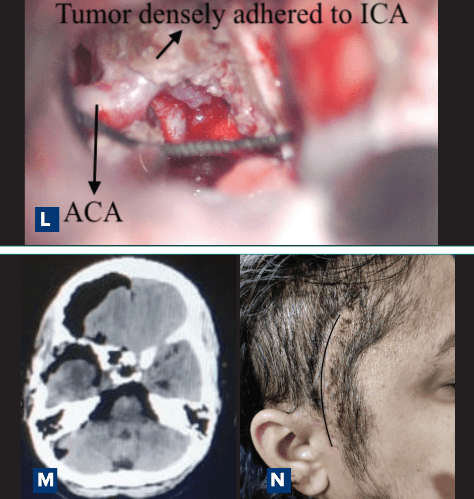
Figure 6A–N: Mini-pterional craniotomy for a right-sided medial sphenoid wing meningioma: A–C. Pre-operative contrast magnetic resonance imaging (MRI) showing the lesion; D. Extra-dural drilling of the temporal base to allow better visual access to the tumour; E. Extradural drilling of the outer sphenoid wing to increase the visual angle, for better visualisation of the tumour through the mini-pterional craniotomy; F. Draining of cerebrospinal fluid (CSF) by opening the arachnoid cistern. This step helps in gaining access to the tumour. Note that only a part of the overlying dura is incised and opened in line with the Sylvian fissure, so that the whole of the brain does not protrude out; G. Tumour visualisation and separation of the arachnoid layer over the tumour; H. Dissection of surrounding vessels from the tumour surface by arachnoid dissection to expose as much of the tumour surface through the narrow operating window as possible; I. Tumour debulking using bipolar dissection, scissors, and cavitron ultrasonic surgical aspirator (CUSA); J. After adequate debulking and arachnoid dissection, critical structures start to become visible. The image shows dissection of the arachnoid plane of the tumour from the anterior and middle cerebral arteries; K. Dissection and separation of the tumour from the optic apparatus; K–L. Near-total excision of tumour. A tiny bit of tumour densely adhered to the main trunk of the internal carotid artery (ICA) has been left; M. Immediate post-operative computed tomography (CT) scan; N. Patient in the follow-up showing excellent cosmesis of the mini-pterional craniotomy.
Abbreviations: ACA: Anterior Cerebral Artery; MCA: Middle Cerebral Artery; ON: Optic Nerve; ICA: Internal Carotid Artery
The mini-pterional craniotomy addresses these concerns associated with the standard pterional craniotomy by reducing the size of the skin incision, lesser muscle dissection and smaller craniotomy without compromising on the versatility of this approach. Figueiredo et al. described a modification of the pterional approach (PT), the mini-pterional craniotomy (MPT), and compared the anatomic exposure provided by these two approaches. These authors concluded that the MPT craniotomy provides comparable surgical exposure to that offered by the PT. The advantages of the MPT include reduction of tissue trauma and bony removal, a decrease in surgical time, and improved cosmetic outcomes.8
The skin incision is about 2.5–3 cm long and is a lazy-C shaped, being bisected by a line drawn from the sphenoid ridge. The underlying temporalis fascia is incised and reflected anteriorly with the scalp flap with tacking sutures. The underlying temporalis muscle is then incised and retracted using skin hooks. The electrocautery is not to be used here at all in the muscle dissection in order to avoid later muscle atrophy. The craniotomy is then fashioned, and the sphenoid ridge is drilled with a 2 mm diamond burr, if required. The dura is opened linearly along the Sylvian fissure, particularly in meningiomas, to minimise premature brain bulging prior to tumour removal. The Sylvian fissure is then opened in usual manner thereafter and the CSF letting has to be done patiently from the arachnoid cisterns to relax the brain and continue working even without the need for a retractor at most of the time during the surgical procedure. The closure is similar to that of a standard pterional craniotomy.
Supraorbital Keyhole Approach (SOKHA)
The eyebrow approach is a very versatile approach providing access to a number of differently located pathologies near the optic apparatus, carotid artery, anterior communicating artery, hypothalamus, inferior frontal lobe and proximal Sylvian fissure. It can also provide access to the olfactory groove, interhemispheric fissure, interpeduncular fossa, mesial temporal structures and the middle fossa (endoscope- assisted). The approach has excellent cosmetic and functional outcomes. SOKHA approach targets anterior and midline skull base tumours — olfactory groove meningiomas (OGM), tuberculum sellae meningiomas (TSM), craniopharyngiomas, and select pituitary adenomas. Via a small superciliary incision, a minimal craniotomy grants access to the anterior cranial fossa; angled micro-instruments and endoscopy extend visualisation around corners, enabling resection of even large tumours with the same safety, efficiency and lower complication rates. 9–13
The patient is positioned supine with head extendedso that the frontal lobe falls backwards with gravity away from the orbital floor, and turned slightly to the contralateral side (depending on the location of the pathology), so that it falls away from the temporal lobe. The skin incision is made at the upper border of the eyebrow, extending from just lateral to the supraorbital notch medially to the lateral edge of the eyebrow till the fronto-zygomatic suture. The use of electrocautery is to be minimal in this approach, as it has cosmetic implications for wound healing. The cuff of pericranium is then elevated on the frontal side and retracted down anteriorly towards the face, and the frontal flap is retracted with scalp hooks backwards. A key burr hole is then made laterally under the temporalis muscle, and a keyhole craniotomy is then made flush with the orbital roof, just lateral to the supraorbital nerve and frontal sinus. The concept of ‘cheating’ in the keyhole surgery cannot be better explained than in the eyebrow approach. The orbital margins are then drilled and bevelled, and the undulations in the orbital roof are made flat. This ‘cheating’ increases the angle of vision considerably and allows an uninterrupted line of vision to the pathology during the entire surgery, with minimal or no brain retraction. The dura is then opened in a C-shaped fashion and retracted inferiorly (Figure 7).
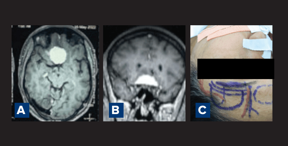
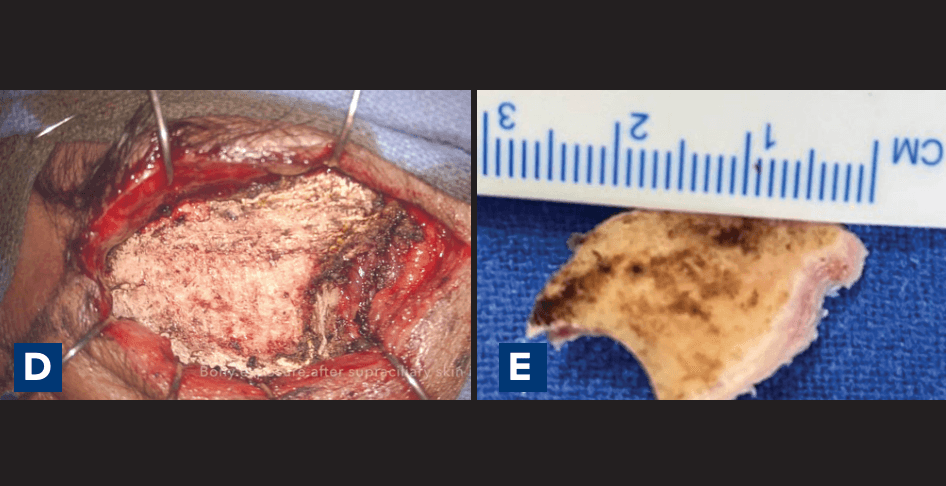
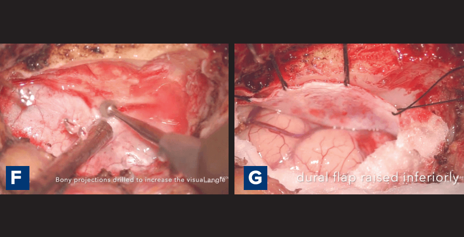
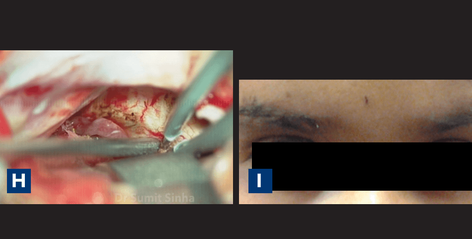
Figure 7A–I: Supra-orbital keyhole approach (SOKHA) for a planum meningioma: A–B. Pre-operative axial and coronal contrast magnetic resonance imaging (MRI) images showing the extent of the tumour; C. Illustrative incision marking and surface marking of the craniotomy to aid in pre-operative surgical planning. The head is positioned so that the malar eminence is the highest point, with the head slightly rotated contralaterally and slightly extended. The incision is preferably closer to the upper border of the eyebrow, and extends from slightly medial to the supra-orbital notch to the brow laterally; D. Demonstrating the bony exposure after the elevation of the cut end of the frontalis muscle and pericranial flap; E. Size of the bone flap; F. Showing the drilling of the bony projections in the orbital roof to increase the visual angle; G. Durotomy in a C-shaped fashion; H. Devascularisation of the tumour done by coagulating the attachment at the skull base; I. Post-operative photograph of another patient showing good cosmesis and a minimally visible scar.
The closure of the eyebrow incision must be meticulous to achieve excellent cosmetic outcomes. The frontal sinus, if opened, is carefully waxed and occluded with gel foam pieces. The pericranium is then approximated, and the frontalis muscle is sutured with 3-0 Vicryl sutures. The skin is then approximated using 3-0 nylon subcuticular sutures, which can be removed by pulling, on the 7th post-operative day.
A meta-analysis involving 2,191 tuberculum sellae meningiomas (TSMs) and 1,510 olfactory groove meningiomas (OGMs) in the endoscope-assisted supraorbital keyhole approach (eSKA) with microscopic transcranial approach (mTCA) and expanded endoscopic endonasal approach (EEA). The gross total resection (GTR) rates were 85% (TSM) — 85% (OGM) for eSKA, 90%/91% for mTCA, and 84%/83% for EEA.3 This suggests comparable efficacy, validating keyhole approaches for well-selected lesions.
Safety remains paramount. CSF leak rates were lowest in eSKA (1.6%–2.1%), followed by mTCA (1.6%–6.5%), and highest in EEA (9%–14%). 12 Mortality and arterial injury were consistently under 1% across approaches. Visual improvement for pre-operative deficits stood at ~66% (TSM) — 53% (OGM) with eSKA, lagging behind EEA (~82%/55%) yet approaching mTCA (~64%/46%).
A large single-centre study involving eyebrow SOKHA vs traditional frontotemporal/bifrontal craniotomies for OGMs was reported. Thirty-two supraorbital approach (SOA) and 25 traditional transcranial approach (TTA) patients were included. The mean extent of resection (EOR) was not significantly different by approach (TTA: 99.1% vs SOA: 98.4%, p = 0.91). Olfaction was preserved or improved at similar rates (TTA: 47% vs SOA: 43%, p = 0.99). The mean length of stay (LOS) was significantly shorter for SOA patients (4.1 ± 2.8 days) than for TTA patients (9.4 ± 11.2 days) (p = 0.002). The authors found an association between an increase in post-operative fluid-attenuated inversion recovery (FLAIR) cerebral oedema and TTA (p = 0.031). Quality of life (QOL) as assessed by the anterior skull base questionnaire (ASBQ) at last follow-up did not differ significantly between groups (p = 0.74). 14
Another retrospective study of 62 patients (64 operations) demonstrated 95% resection for meningiomas, 84% for craniopharyngiomas, 53% for pituitary adenomas, with no mortality and ~4.5% major complications. These findings confirm SOKHA's durable safety and efficacy, particularly for anterior skull base lesions.15
Subtemporal Keyhole Approach
The patient should be positioned with the head extended and turned maximally to the contralateral side, so that the temporal lobe falls away from the middle fossa floor. The mini-subtemporal craniotomy can be placed anteriorly for lesions of the middle fossa or anterior tentorial incisura, or slightly posteriorly for lesions of the lateral midbrain, tentorium or middle tentorial incisura. The exact location of the craniotomy has to be decided by using image guidance, placing it along the long axis of the lesion, extended to the surface or at least at the centre of the lesion.
The skin incision is a 2–3.5 cm skin incision running coronally and reaching up to the zygoma in case of an anterior craniotomy and running postero-superiorly or diagonally towards the pinna in the posterior approaches. The anterior craniotomies are made flush with the temporal base, while the posterior ones reach the transverse sigmoid sinus junction or are close to the middle fossa. The muscle is then split linearly along the direction of its fibres, retracted, and a 2–3 cm bone flap is then turned flush with the middle fossa floor. The vein of Labbe is to be protected during the subtemporal craniotomy. The mastoid air cells are taken care of by proper waxing if inadvertently opened. The dural opening is based inferiorly (Figures 8 and 9).
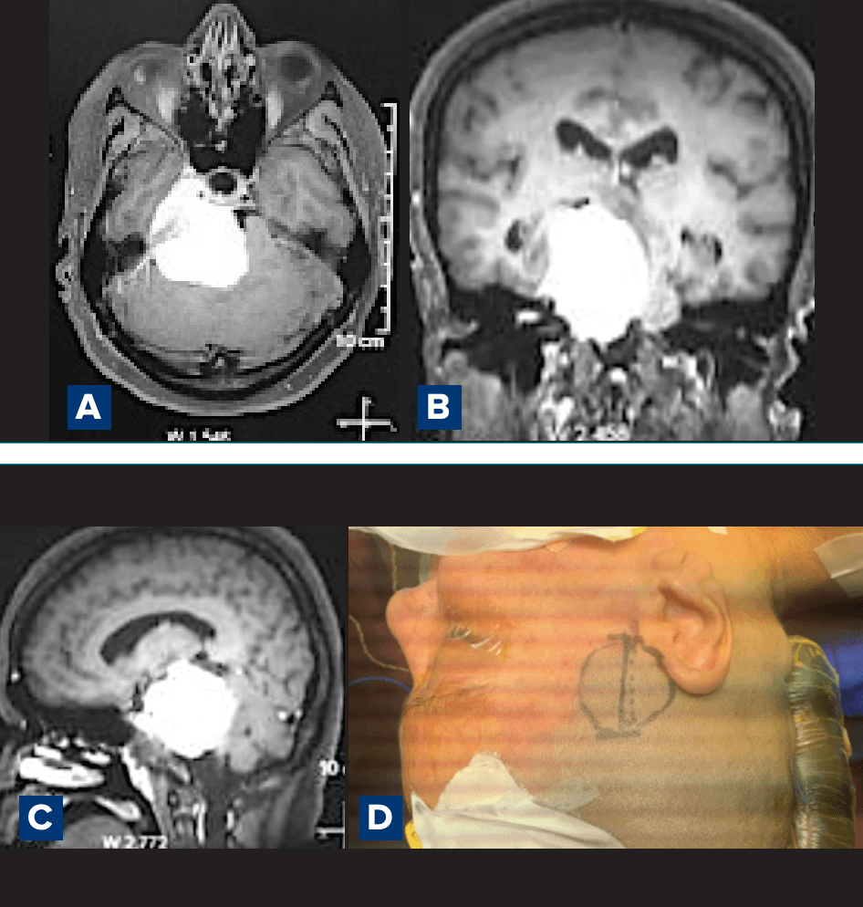
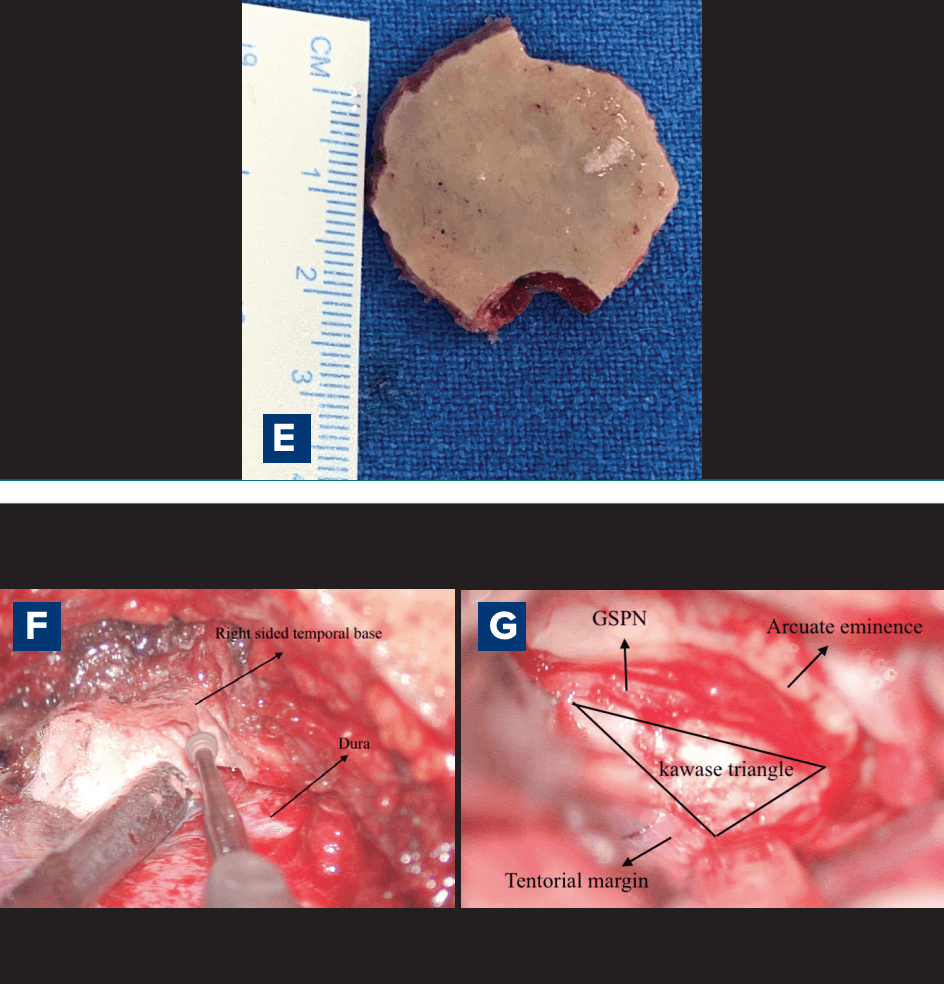
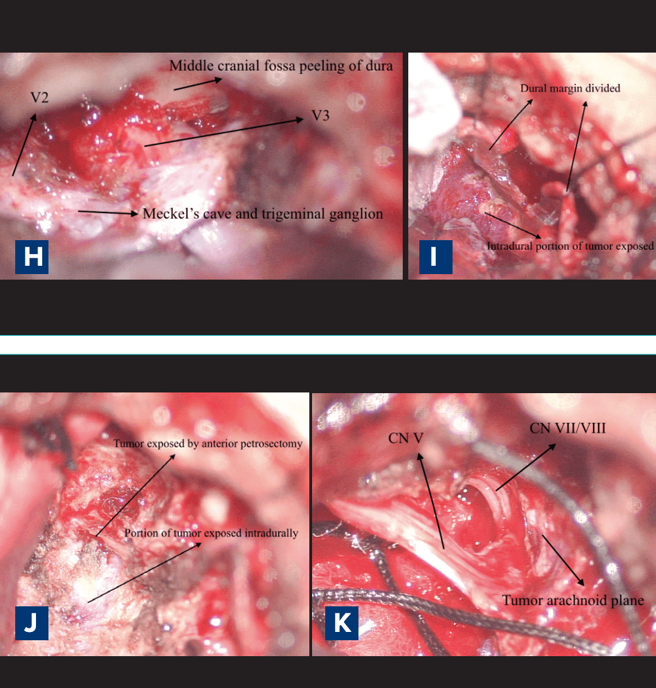
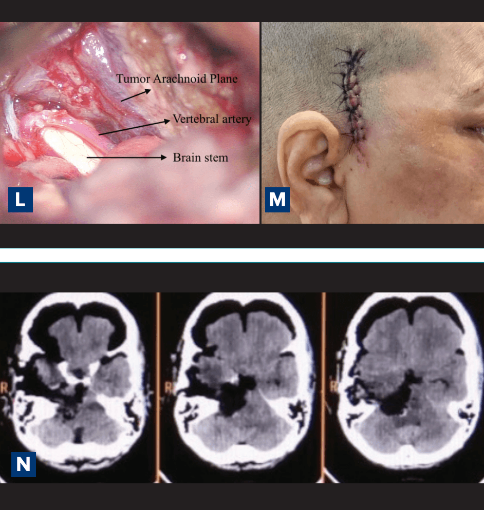
Figure 8: A–N. Contrast magnetic resonance imaging (MRI) of a right-sided petroclival meningioma primarily originating from the petrous apex and extending to the lower clivus; D. Patient placed on a horseshoe headrest. A 4 cm linear incision 1 cm anterosuperior to the tragus. The circular marking around the incision shows the size of the craniotomy; E. Craniotomy flap with size. A larger craniotomy than the incision size can be achieved by undermining the incision with the drill; F. Drilling of the temporal bone to reach the base of the middle cranial fossa. It is essential to make the temporal bone flat with the middle cranial fossa to have a better view during surgery. The reduced vision via keyhole approaches necessitates such steps. Peeling of the dura from the middle cranial fossa. The landmarks for surgery remain the same as in the conventional approach, but the window is smaller. A good understanding of anatomical relations is therefore important to guide direction. Neuro-navigation may assist in the direction of dissection but does not replace experience in anatomy; G. Margins of the Kawase triangle (region for anterior petrosectomy), the anterior portion of the arcuate eminence, and the greater superficial petrosal nerve are demonstrated; H–J. Post-petrosectomy and exposure of the tumour, the dura is opened and an intradural approach is taken. The figure shows the intradural portion of the tumour. The dura is divided and tented up with sutures, achieving a combined view of the intra- and extradural tumour. Further opening of the anterior petrosectomy with a 1 mm Kerrison punch helps provide a holistic view of the lesion. Occasionally, tumour decompression needs to be started on the visible lesion to create space for this exposure; K. Dissection of the tumour from the cisternal portion of the V nerve and VII/VIII nerve complex; L. Keyhole view towards the end of dissection with exposure of the vertebrobasilar complex and dissection from the brainstem; M. Showing the skin incision; N. Immediate post-operative computed tomography (CT) showing gross total tumour removal with a keyhole craniotomy.
Abbreviations: GSPN: Greater Superficial Petrosal Nerve; V2: Maxillary division of the Trigeminal Nerve; V3: Mandibular division of the Trigeminal Nerve; CN V: Cranial Nerve V; CN VII/VIII: Cranial Nerves VII and VIII.
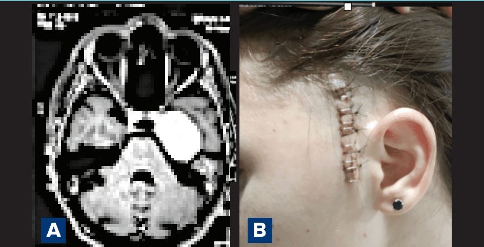
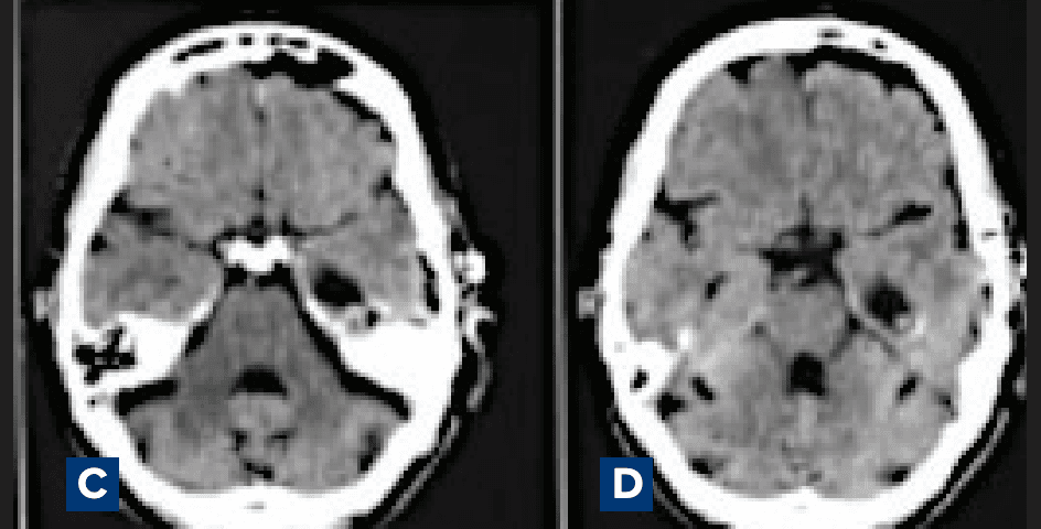
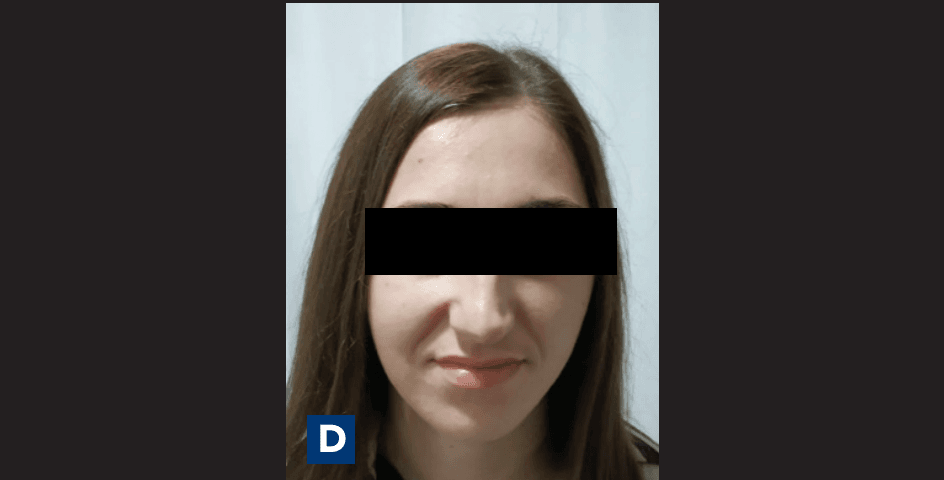
Figure 9A–E: Trigeminal schwannoma operated through a keyhole subtemporal approach: A. Contrast magnetic resonance imaging (MRI) in axial view showing the extent of the tumour; B. About 4 cm long linear incision was enough to remove such a formidable tumour. Such small incisions make the post-operative recovery faster, with minimal pain and excellent cosmesis; C–D. Post-operative computed tomography (CT) showing a good operative cavity; E. Happy patient in follow-up.
Morisako et al. described the details of a purely endoscopic subtemporal keyhole ATPA (eATPA) for petrous apex lesions.16 There have been numerous reports of keyhole approaches being described for petroclival meningiomas.17
Keyhole Retrosigmoid Approach
The keyhole retrosigmoid approach (KRSA) is similar to the standard conventional craniotomy, with which a neurosurgeon is familiar with. Any deep-seated lesion in the ventral posterior fossa, pons, or lateral cerebellum can be accessed with a keyhole retro- sigmoid approach. 18 The KRSA is also a valid choice for removing large vestibular schwannomas > 3 to 4 cm in size. Through this approach, the cerebellopontine angle can be effectively exposed. Skills to protect the facial nerve and extensive experience in microsurgical techniques can significantly improve the total resection rate and post-operative facial nerve function (Figures 10–12). 18,19
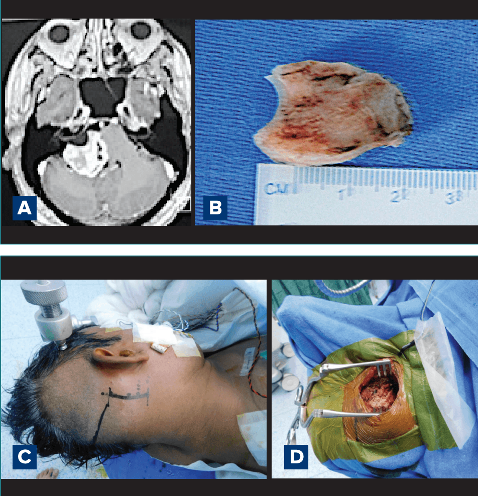
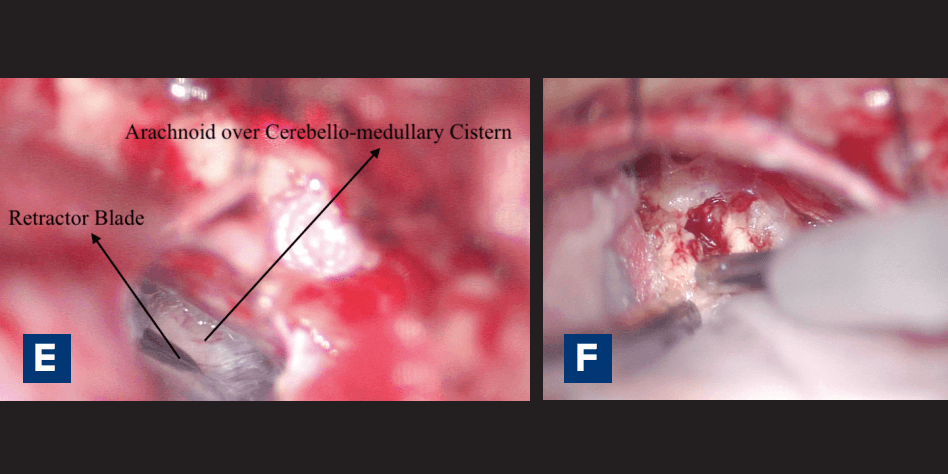
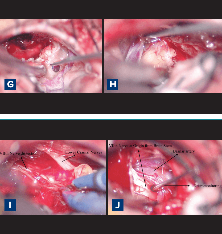
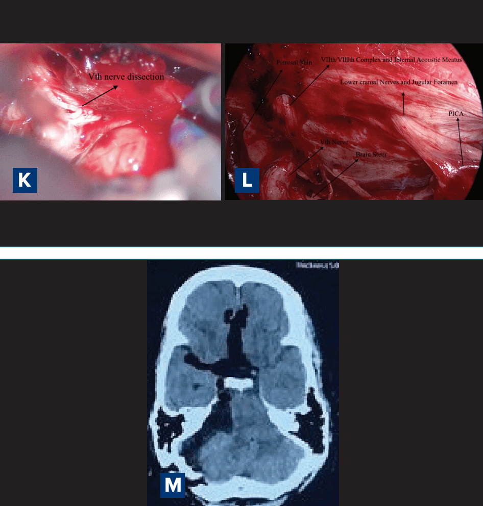
Figure 10: A Pre-operative contrast magnetic resonance imaging (MRI) of the patient showing a right-sided acoustic neuroma; B. Size of the craniotomy; C. Planning of the incision. The red line runs from the external acoustic meatus to the occipital protuberance and indicates the position of the transverse sinus. The incision is approximately 4 cm long and is lazy C-shaped; D. The completed bony exposure — the asterion is visualised, which marks the junction of the transverse and sigmoid sinuses. The burr hole should be placed here for making the craniotomy; E. Showing the first step after incising about 1 cm of dura at the lower edge — releasing the cerebrospinal fluid (CSF) from the lateral cerebellomedullary cistern. The dura is tented up with a suture and a retractor blade is inserted to retract the cerebellum and expose the arachnoid. A micro-scissors or needle dissector is then used to release the CSF and create space in the posterior fossa; F. Cavitron ultrasonic surgical aspirator (CUSA) decompression of the tumour after the overlying arachnoid has been peeled off the tumour capsule; G–H. Medial and superior dissection of the tumour from the surrounding structures; I–K. Intact trigeminal, facial, and lower cranial nerves after gross total tumour removal. Neuromonitoring is used at every step during tumour dissection to detect the facial nerve. The facial nerve is then traced along its course and dissected from the tumour; L. Endoscopic view of the tumour cavity after tumour resection showing intact nerves and complete tumour resection; M. Immediate post-operative contrast computed tomography (CT) of the patient showing complete tumour resection.
Abbreviation: PICA: Posterior Inferior Cerebellar Artery.
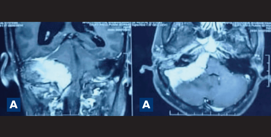
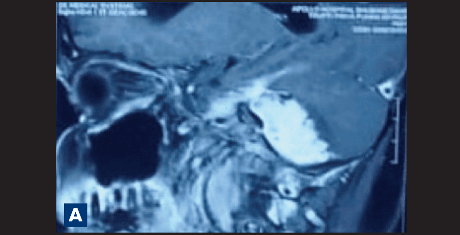
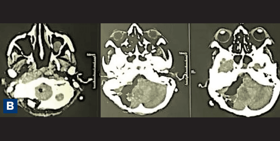
Figure 11: A. Pre-operative contrast magnetic resonance imaging (MRI) of the patient with a right-sided cerebellopontine angle meningioma; B. Immediate post-operative computed tomography (CT) of the same patient showing complete tumour resection. Note the size of the right-sided keyhole retromastoid craniotomy
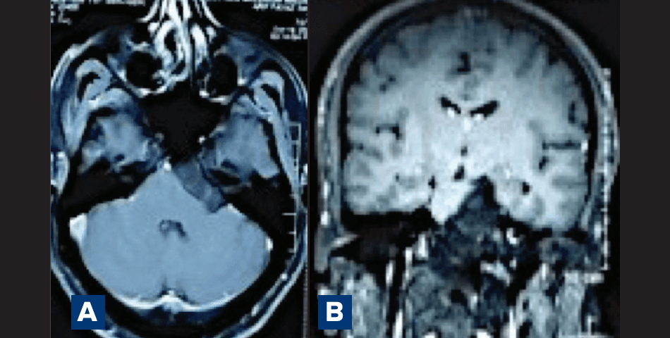
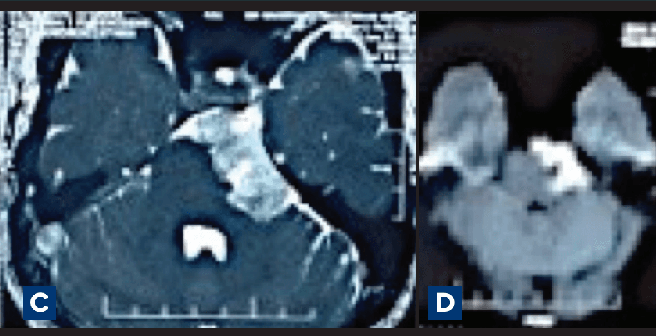
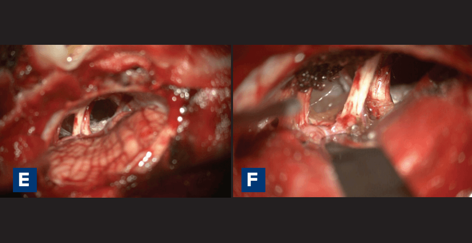
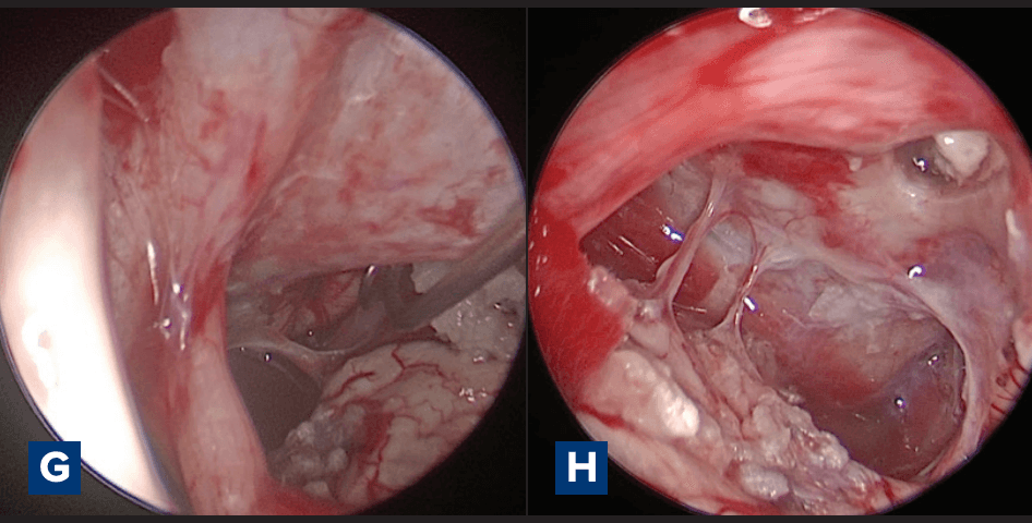
Figure 12 A–H: Keyhole approach for a left cerebellopontine (CP) angle epidermoid: A–C. Pre-operative magnetic resonance imaging (MRI) images showing the tumour in T1, T1 contrast, and T2-weighted sequences; D. Depicting diffusion-weighted imaging (DWI) with diffusion restriction, which is characteristic of an epidermoid; E. Microscopic image after keyhole retromastoid suboccipital (RMSO) craniotomy and durotomy showing the pearly white tumour located anterior to the VII–VIII nerve complex; F. Microscopic debulking of the tumour; G. Deployment of an angled endoscope and curved instruments is crucial to remove bits of tumour in the hidden areas; H. Endoscopic view of the resection cavity showing important structures after complete resection.
The patient positioning is of absolute paramount mportance in performing a retrosigmoid approach. The patient is being positioned laterally with the affected side up. The position of the head with respect to the torso depends upon the diagnosis for which surgery is being performed. In the most used scenario, if the KRSA is being used for any cerebellopontine angle lesion like acoustic schwannomas, then the head is flexed maximally (to open up the cisterna magna), tilted downwards towards the floor about 20–30 degrees, and rotated ipsilaterally (to view the brainstem) or contralaterally (to view the petrous bone).
The ipsilateral shoulder is taped gently downwards, avoiding excessive traction (to prevent inadvertent brachial plexus injury), to open the working space between the head and shoulder.
The incision is approximately 4 cm long and is a lazy C–shaped or a lazy S–shaped as depicted in the figure. The transverse–sigmoid sinus junction normally lies at the asterion, but we prefer to use image guidance always, as with all the keyhole craniotomies. A burr hole is fashioned here, and a 2–3 cm diameter craniotomy is made here for cerebellopontine and jugular foramen pathology, and slightly lower for petroclival region and Meckel’s cave pathology. The edges of the craniotomy should extend until a portion of the transverse and sigmoid sinuses is seen. A small dural incision of about 0.5 cm is then made at the lower part of the craniotomy, and an attempt is made to reach the cisterna magna and sharply incise the arachnoid here to patiently release the CSF through this small dural opening. Once the cerebellum gets lax with this manoeuvre, a dural incision is then given in an inverted Y-shaped manner to complete the dural opening.
Keyhole Posterior Interhemispheric Approach (Mini- Poppen’s Approach)
The minimally invasive techniques avoiding unnecessary tissue injuries were applied to refine traditional approaches for the removal of third ventricular tumours within a limited operative field. 20 These approaches allowing access to tumours of the pineal region are — keyhole lateral supracerebellar, midline/paramedian infratentorial supracerebellar, anterior interhemispheric transcallosal and occipital transtentorial. The posterior third ventricular tumours can be accessed through the anterior interhemispheric transcallosal approach. The position of the vein of Galen in the mid-sagittal cut magnetic resonance imaging (MRI) with respect to the tumour dictates the approach. The posterior third ventricular tumours push the vein downwards and posteriorly, while the pineal tumours push it upwards and forwards.
These deep-seated tumours are the best suited for these keyhole approaches. Choosing between the keyhole occipital transtentorial and midline/paramedian infratentorial supracerebellar approach depends upon several factors such as the long axis of the tumour, angle of the tentorium and venous anatomy. The midline/paramedian infratentorial supracerebellar is generally preferred when the long axis of the tumour projects to a point below the inion, while the keyhole occipital transtentorial is chosen when this long axis projects to a point above the inion.
A lateral 3/4 th oblique position is preferred for the keyhole occipital transtentorial approach, with the head end elevated and tilted 30° upwards (Figure 13). The incision (generally 4–6 cm in length) is linear and paramedian 1 cm lateral to the midline. The craniotomy is a long, thin one flush with the sagittal sinus. The dural flap is raised based on the sinus. The access to the falco-tentorial junction is achieved through meticulously approaching the interhemispheric cistern, dividing the arachnoid bands and draining the CSF, making the occipital lobe lax, without retraction (aided by gravity), and absolutely avoiding any injury to the lobe. Good anaesthesia is critical for this approach to avoid the occipital lobe from bulging out. Any bridging veins should be absolutely preserved and once the tentorial incisura is reached, an attempt is made to release the CSF, after which occipital lobe is further relaxed, and ample space is achieved even through this keyhole craniotomy. The straight sinus is identified and tentorium is divided. Any brisk bleeding encountered during the cutting of the tent is dealt with by cautery.
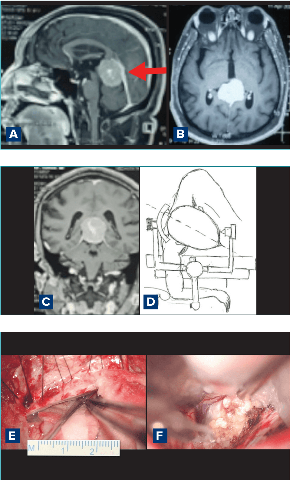
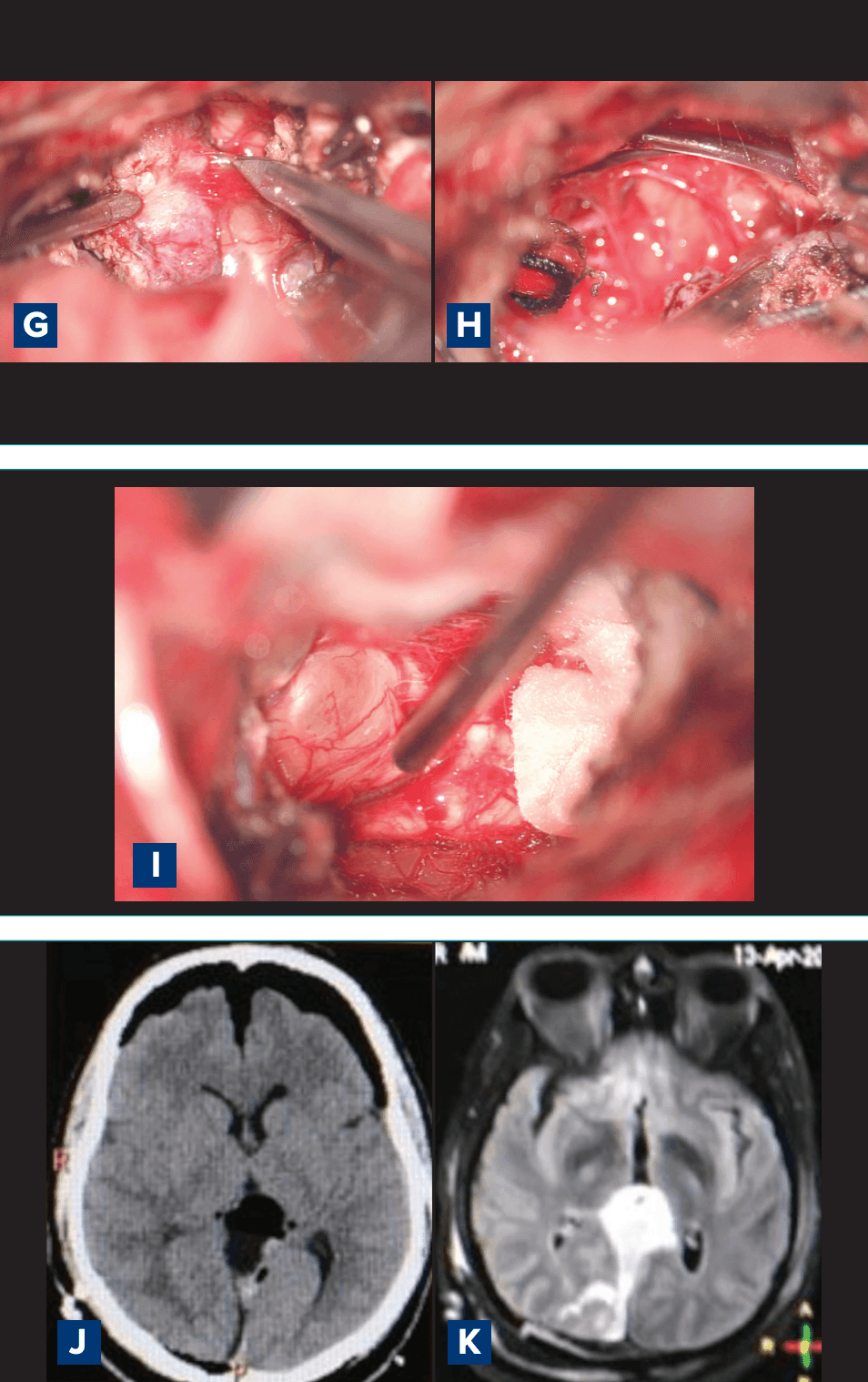
Figure 13A-K: Demonstrating keyhole approach for a falcotentorial meningioma operated via a keyhole occipital transtentorial approach: A–C. Pre-operative contrast magnetic resonance imaging (MRI) images showing the tumour extent in sagittal, axial, and coronal views; D. Pictorial diagram showing the positioning of the patient in a three-quarter prone position with the ipsilateral side down; E. Durotomy done based on the superior sagittal sinus. Gravity-aided retraction of the ipsilateral occipital lobe done to expose the tumour; F–G. Devascularisation and decompression of the tumour; H. Continued tumour decompression and dissection from the vein of Rosenthal and vein of Galen; I. Microscopic view of the resection cavity with the posterior wall of the third ventricle seen after total tumour resection; J. Immediate post-operative computed tomography (CT) showing a good post-operative cavity; K. Follow-up MRI showing gross total resection.
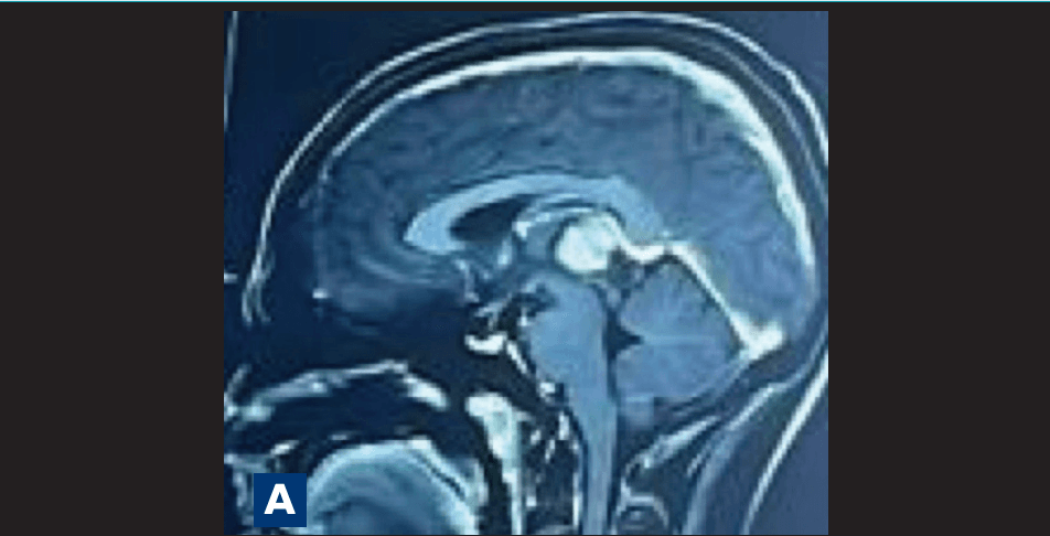
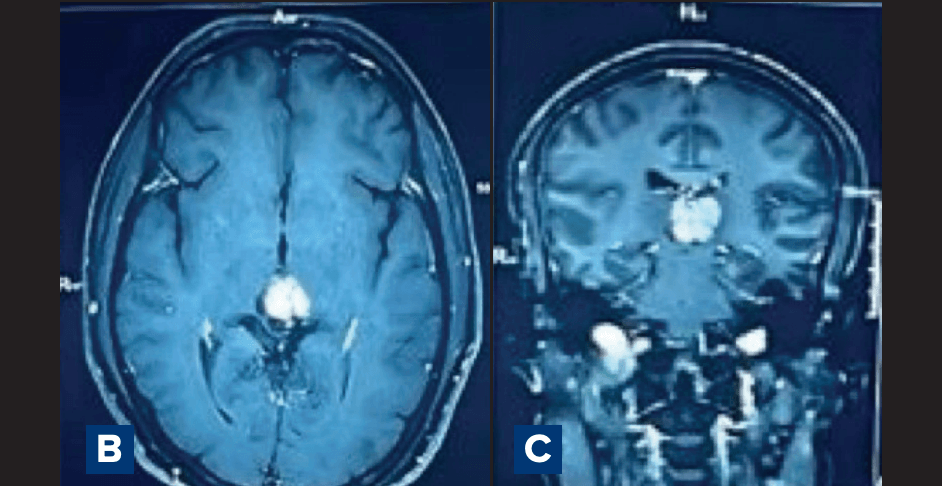
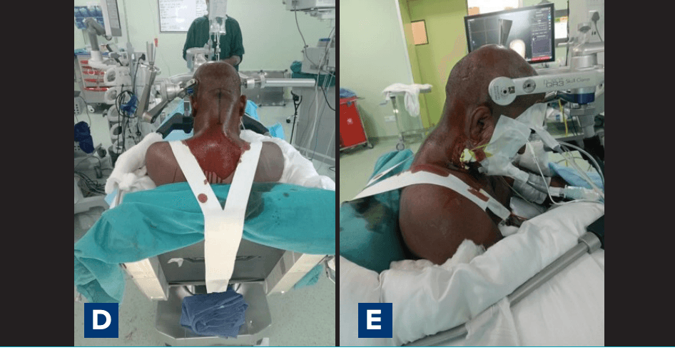
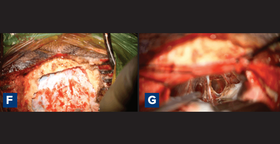
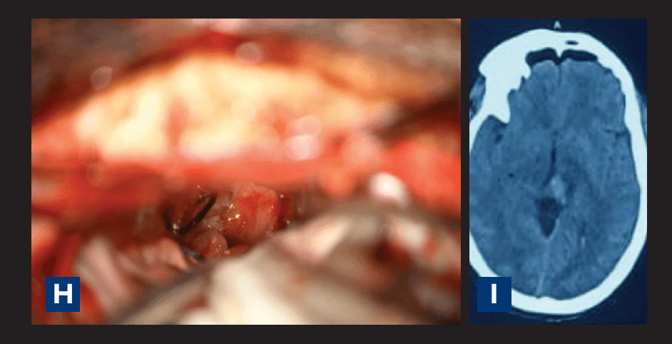
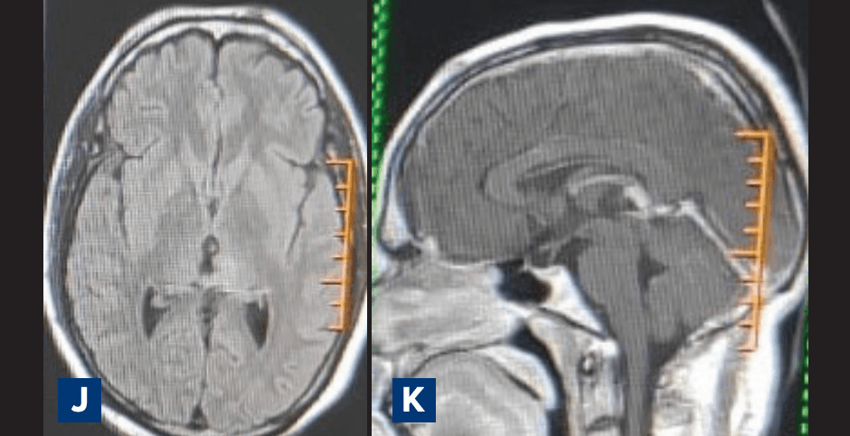
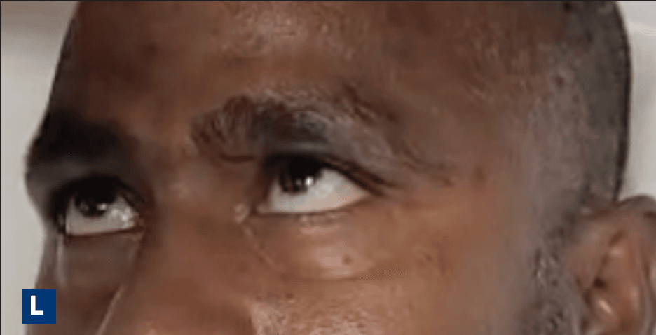
Figure 14A-L: Keyhole midline suboccipital approach for a pineal region tumour operated via the supracerebellar infratentorial (Krause) approach in the sitting position: A–C. Pre-operative contrast magnetic resonance imaging (MRI) images showing the tumour extent in sagittal, axial, and coronal views; D–E. Patient in the sitting position with the head fixed on a Mayfield 3-pin head holder. Adequate padding of all pressure points is essential; F. 2 × 3 cm midline suboccipital craniotomy is made, and it is essential to expose the lower margin of the transverse sinus; G. Gravity-aided retraction of the cerebellum is done inferiorly. A few bridging veins might have to be sacrificed. The precentral cerebellar vein can also be divided if required; H. Tumour seen after the dissection; I. Immediate post-operative computed tomography (CT) showing a satisfactory tumour cavity; J–K. Follow-up magnetic resonance imaging (MRI) of the patient after 6 months; L. Post-operative clinical photograph depicting preserved upgaze eye movements
Keyhole Supracerebellar Infratentorial Approach
The sitting position is preferred for this approach (Figure 14). The head is flexed maximally at the occipito-atlantal joint, keeping two finger space between the chin and manubrium. An imaginary line is extrapolated from the outer canthus to the external occipital protuberance. This line marks the surface marking of the tentorium. The head is flexed till this line is parallel to the ground. A pillow is adjusted behind the upper back. A 4–6 cm vertical skin incision is given in the midline and a craniotomy located just inferior to the transverse sinus is performed using a high-speed drill. The dura is opened in a U-shaped manner and reflected superiorly to expose the space between the cerebellum and tentorium. The draining veins should be preserved as much as possible to prevent venous infarction and post-operative neurological deficits
Conclusion
Keyhole approaches to skull base tumours represent a paradigm shift in neurosurgical practice, prioritising minimal invasiveness without compromising surgical efficacy. Over the past two decades, growing anatomical knowledge, advanced neuroimaging, neuro-navigation, and endoscopic integration have collectively enhanced the precision and safety of these approaches. Clinical studies have consistently demonstrated that keyhole strategies — such as the supraorbital, retrosigmoid, endonasal endoscopic, and combined mini-craniotomies — can achieve gross total or near-total resections in appropriately selected patients, with complication rates comparable to, or lower than, those of traditional craniotomies.
However, keyhole surgery requires rigorous case selection and a steep learning curve. The limited working corridor demands refined microsurgical skills, detailed anatomical understanding, and experience with endoscopic or microscopic assistance. In conclusion, keyhole approaches are a powerful, evidence-based addition to the skull base surgeon’s repertoire. When employed judiciously, they have the potential to replace the time-consuming morbid skull base approaches, can improve outcomes, minimise morbidity, and meet the increasing demand for patient-centred, minimally invasive neurosurgical care.
Sumit Sinha. Surgical Minimalism in Neurosurgery: The “Keyhole” Concept. MMJ. 2026, June. Vol 3 (2).
DOI: XXXX_XXXX_XXXX_XXXX
References
- Burks JD, Conner AK, Bonney PA, et al. Management of Intracranial Meningiomas Using Keyhole Techniques. Cureus. 2016;8(4):e588.
- Lan Q, Sughrue M, Hopf NJ, et al. International expert consensus statement about methods and indications for keyhole microneurosurgery from International Society on Minimally Invasive Neurosurgery. Neurosurg Rev. 2021;44(1):1–17.
- Lan Q, Sughrue ME, Briggs RG. Minimally invasive keyhole techniques for resection of giant intracranial tumors. Chin Neurosurg J. 2022;8(1):19.
- Fischer G, Stadie A, Reisch R, et al. The keyhole concept in aneurysm surgery: results of the past 20 years. Neurosurgery. 2011;68(1 Suppl Operative):45–51.
- Perneczky A, Reisch R. Volume 1: Concept and Surgical Technique. Keyhole Approaches in Neurosurgery. Vienna: Springer; 2009.
- Teo Charles, Sughrue ME. Principles and Practice of Keyhole Brain Surgery. Stuttgart: Thieme; 2015.
- Reisch R, Stadie A, Kockro RA, et al. The keyhole concept in neurosurgery. World Neurosurg. 2013;79(2 Suppl):S17.e9-S17.e13.
- Figueiredo EG, Deshmukh P, Nakaji P, et al. The minipterional craniotomy: technical description and anatomic assessment. Neurosurgery. 2007;61(5 Suppl 2):256–65.
- Ormond DR, Hadjipanayis CG. The Supraorbital Keyhole Craniotomy through an Eyebrow Incision: Its Origins and Evolution. Minim Invasive Surg. 2013;2013:296469.
- Valerio J, Fernandez Gomez MP, Ayala Arcipreste A, et al. Exploring the Potential Use of Virtual Reality with a Supraorbital Keyhole Craniotomy for Anterior Skull Base Meningiomas: Two Case Reports. J Pers Med. 2024;14(11):1074.
- Khan DZ, Muskens IS, Mekary RA, et al. The endoscope-assisted supraorbital "keyhole" approach for anterior skull base meningiomas: an updated meta-analysis. Acta Neurochir (Wien). 2021;163(3):661–76.
- Cai M, Ye Z, Ling C, et al. Trans-eyebrow supraorbital keyhole approach in suprasellar and third ventricular craniopharyngioma surgery: the experience of 27 cases and a literature review. J Neurooncol. 2019;141(2):363–71.
- Igressa A, Pechlivanis I, Weber F, et al. Endoscope-assisted keyhole surgery via an eyebrow incision for removal of large meningiomas of the anterior and middle cranial fossa. Clin Neurol Neurosurg. 2015;129:27–33.
- Bander ED, Pandey A, Yan J, et al. Olfactory groove meningiomas: supraorbital keyhole versus orbitofrontal, frontotemporal, or bifrontal approaches. J Neurosurg. 2023;140(6):1568–75.
- Lin YJ, Chen KT, Lee CC, et al. Anterior Skull Base Tumor Resection by Transciliary Supraorbital Keyhole Craniotomy: A Single Institutional Experience. World Neurosurg. 2018;111:e863-e870.
- Morisako H, Sasaki T, Ikegami M, et al. Purely endoscopic subtemporal keyhole anterior transpetrosal approach to access the petrous apex region: surgical techniques and early results. J Neurosurg. 2024;141(3):752–61.
- Zhu W, Mao Y, Zhou LF, et al. Keyhole approach surgery for petroclival meningioma. Chin Med J (Engl). 2006;119(16):1339–42.
- Daming C, Yiwen S, Bin Z, et al. Large vestibular schwannoma resection through the suboccipital retrosigmoid keyhole approach. J Craniofac Surg. 2014;25(2):463–68.
- Hoshide R, Faulkner H, Teo M, et al. Keyhole retrosigmoid approach for large vestibular schwannomas: strategies to improve outcomes. Neurosurg Focus. 2018;44(3):E2.
- Lan Q, Dong J, Huang Q. Minimally invasive keyhole approaches for removal of tumors of the third ventricle. Chin Med J (Engl). 2006;119(17):1444–50.