 WhatsApp Us (24/7)
 +91 926 888 0303 (24/7)


Abstract:
In an era of rapidly advancing diagnostic technologies, the value of bedside clinical examination is gradually diminishing despite its enduring relevance in everyday medical practice. Careful observation and elicitation of physical signs remain fundamental to early diagnosis, prompt decision-making, and cost-effective patient care, particularly in resource-constrained settings. This review, “Clinical Signs – The Forgotten Art! (Part II),” revisits a selected collection of high-yield clinical signs that continue to hold substantial diagnostic importance across multiple specialities, including internal medicine, surgery, neurology, and dermatology. Emphasis has been placed on signs that are simple to elicit, clinically reliable, and frequently encountered in undergraduate and postgraduate medical training as well as routine clinical practice. For each clinical sign, the review discusses its historical origin, methodology of elicitation, pathophysiological basis, associated clinical conditions, and contemporary diagnostic relevance. In addition, the article highlights how bedside examination can complement modern investigations by guiding differential diagnosis, prioritising investigations, and enhancing physician– patient interaction. By re-emphasising these classical examination skills, this review aims to encourage clinicians and trainees to preserve the art of bedside medicine while integrating it effectively with current evidence-based diagnostic approaches and technological advancements in healthcare.
Key words: Clinical Signs, Bedside Diagnosis, Physical Examination, Clinical Pearls, Postgraduate Medicine.
Introduction
Even with significant advances in medical technology, the first clinical evaluation still depends heavily on bedside examination. The capacity to derive important diagnostic insights from basic physical signs remains a core competency of an effective clinician. However, with increasing dependence on imaging and laboratory investigations, many such signs are no longer routinely elicited, particularly in fast-paced clinical environments.1-3
Unlike classical signs that are often descriptive and occasionally of limited sensitivity, there exists a subset of clinical signs that are highly practical, reproducible, and immediately informative. These signs frequently provide early diagnostic direction, help differentiate between competing clinical possibilities, and, in certain situations, may even obviate the need for urgent investigations.
For postgraduate trainees, mastery of these signs is particularly important. They form the backbone of clinical examinations such as objective structured clinical examinations (OSCEs) and viva voce, and more importantly, they cultivate a disciplined approach to patient evaluation. Recognising a ‘hung-up reflex’, eliciting a ‘Gower’s sign’, or identifying ‘Hoover’s sign’ in suspected functional weakness are examples of how bedside skills can directly influence clinical reasoning.
This review, as a continuation of the earlier discussion on forgotten clinical signs, shifts focus toward twelve high-yield, clinically actionable signs that every postgraduate student should be familiar with. These signs have been selected not only for their diagnostic value but also for their enduring relevance in everyday clinical practice.
Twelve High-Yield Clinical Signs Every Postgraduate
Must Know In modern clinical practice, rapid access to investigations has reduced reliance on bedside examination. However, certain clinical signs remain highly discriminative, elegant, and diagnostically powerful. This curated list highlights twelve such signs across multiple specialities that every postgraduate should confidently elicit and interpret.
1. Hung-up reflex (Woltman’s sign) (Hypothyroidism)
First described by: Henry Woltman.
Finding: Delayed relaxation phase of deep tendon reflex, especially ankle jerk (Figure 1).
Significance: Suggestive of hypothyroidism.
2. Facial (Malar) rash in systemic lupus erythematosus (SLE) (Dermatology/Rheumatology)
Explained in classical dermatology by Ferdinand von Hebra.
Finding: Appearance of a butterfly rash over the cheeks and nose, sparing the nasolabial folds (Figure 2).
Significance: Suggestive of SLE.
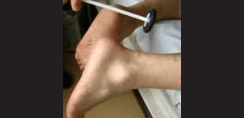
Figure 1: Delayed relaxation reflex.

Figure 2: Malar rash in systemic lupus erythematosus (SLE).
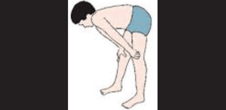
Figure 3: Using hands to push on the legs to stand.
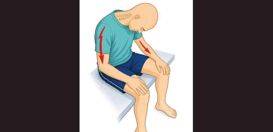
Figure 4: Electric shock sensation on neck.
3. Gower’s sign (Neurology)
First described by: William Richard Gowers.
Finding: The patient usually uses their hands to “climb up” the thighs while rising (Figure 3).
Significance: It is suggestive of proximal muscle weakness (e.g., Duchenne muscular dystrophy).5
4. Lhermitte’s sign (Neurology)
First described by: Jean Lhermitte.
Finding: An electric shock sensation appears on neck flexion (Figure 4).
Significance: Usually seen in multiple sclerosis and cervical cord lesions.5
5. Cervical venous hum in severe anaemia
Finding: Continuous humming sound over the jugular veins, which is abolished by compression (Figure 5).
Significance: Suggestive of severe anaemia or a hyperdynamic circulation.
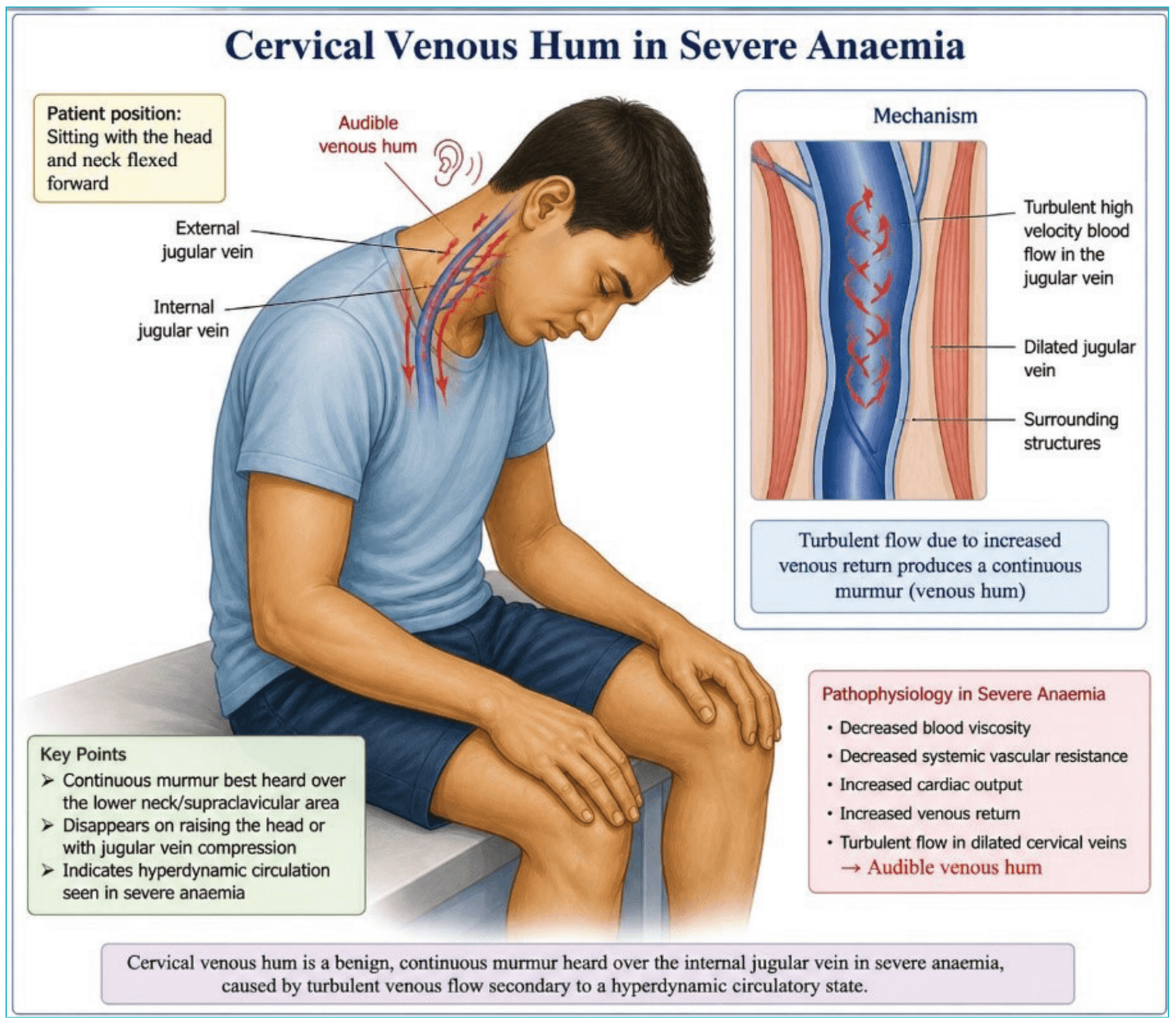
Figure 5: Cervical venous hum in severe anaemia.
6. Hoover’s Sign (Functional vs organic weakness) (Neurology)
First described by: Charles Franklin Hoover (1908).
Method:
- The patient lies supine.
- The examiner places a hand under the heel of the "normal" leg.
- The patient is asked to lift the "weak" leg against resistance.
Findings:
- Organic weakness: Downward pressure is felt under the opposite heel.
- Functional (Non-organic) weakness: No pressure felt.
Principle: Based on crossed extensor reflex and synergistic muscle activation (Figure 6).
Clinical Significance: Differentiates between:
- True neurological weakness
- Functional (conversion disorder) weakness
Clinical Pearl:
“Absence of involuntary extension in the contralateral limb suggests non-organic weakness.”
7. Spider naevi (Hepatology/Pregnancy)
Finding: Central arteriole with radiating vessels (Figure 7).
Significance: Seen in chronic liver disease and pregnancy (hyperestrogenism).3
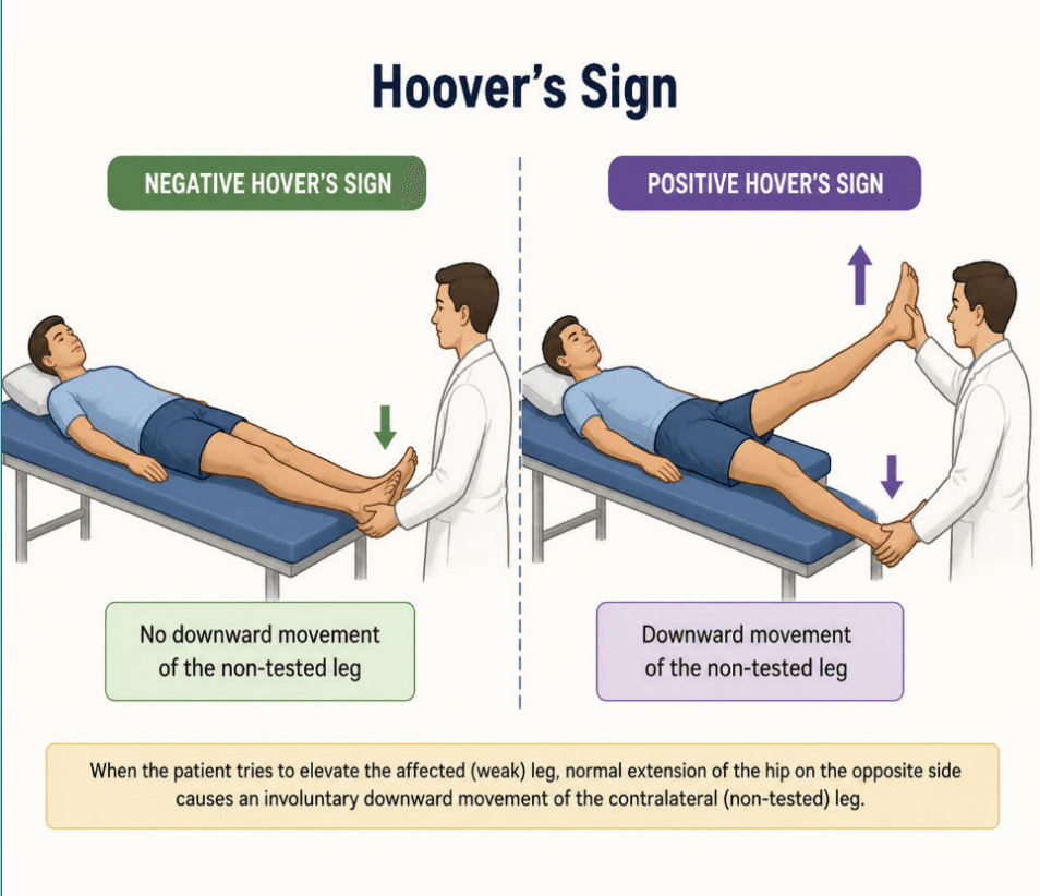
Figure 6: Hoover’s sign.
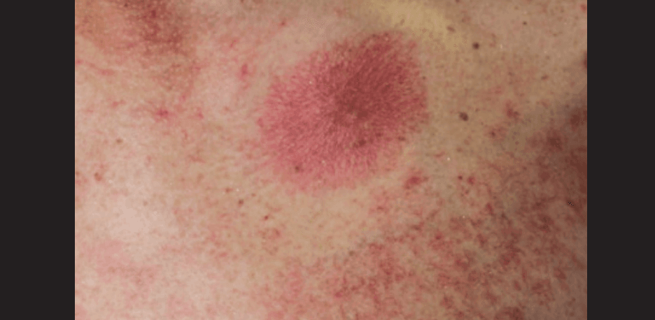
Figure 7: Spider naevi.
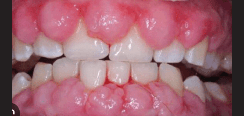
Figure 8: Gingival hyperplasia.
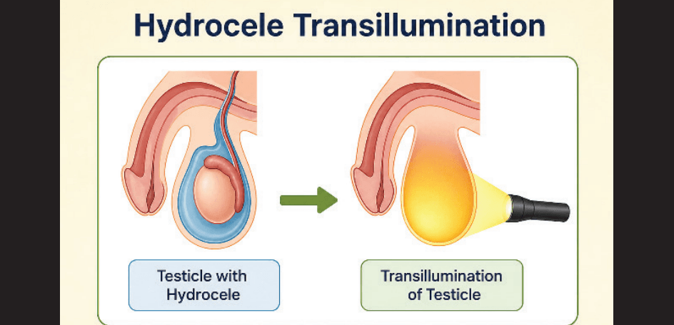
Figure 9: Hydrocele transillumination
8. Gingival hyperplasia
Finding: Overgrowth of gums (Figure 8).
Causes:
- Phenytoin
- Cyclosporine
- Leukaemia
- Pregnancy
Significance: Clue to a drug effect or systemic disease.
9. Transillumination test of the testis (Surgery/Urology)
Finding:
- Hydrocele → Transilluminates
- Tumour → Does not transilluminate
Significance: Differentiates cystic from solid scrotal swelling (Figure 9)
10. Courvoisier’s law (Palpable gallbladder) (Surgery)
First described by: Ludwig Courvoisier.
Finding: Palpable gallbladder with painless jaundice (Figure 10).
Significance:Suggestive of malignant obstruction (not gallstones)
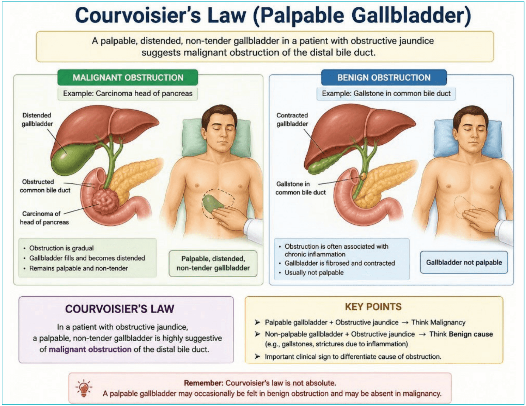
Figure 10: Palpable gallbladder with painless jaundice.
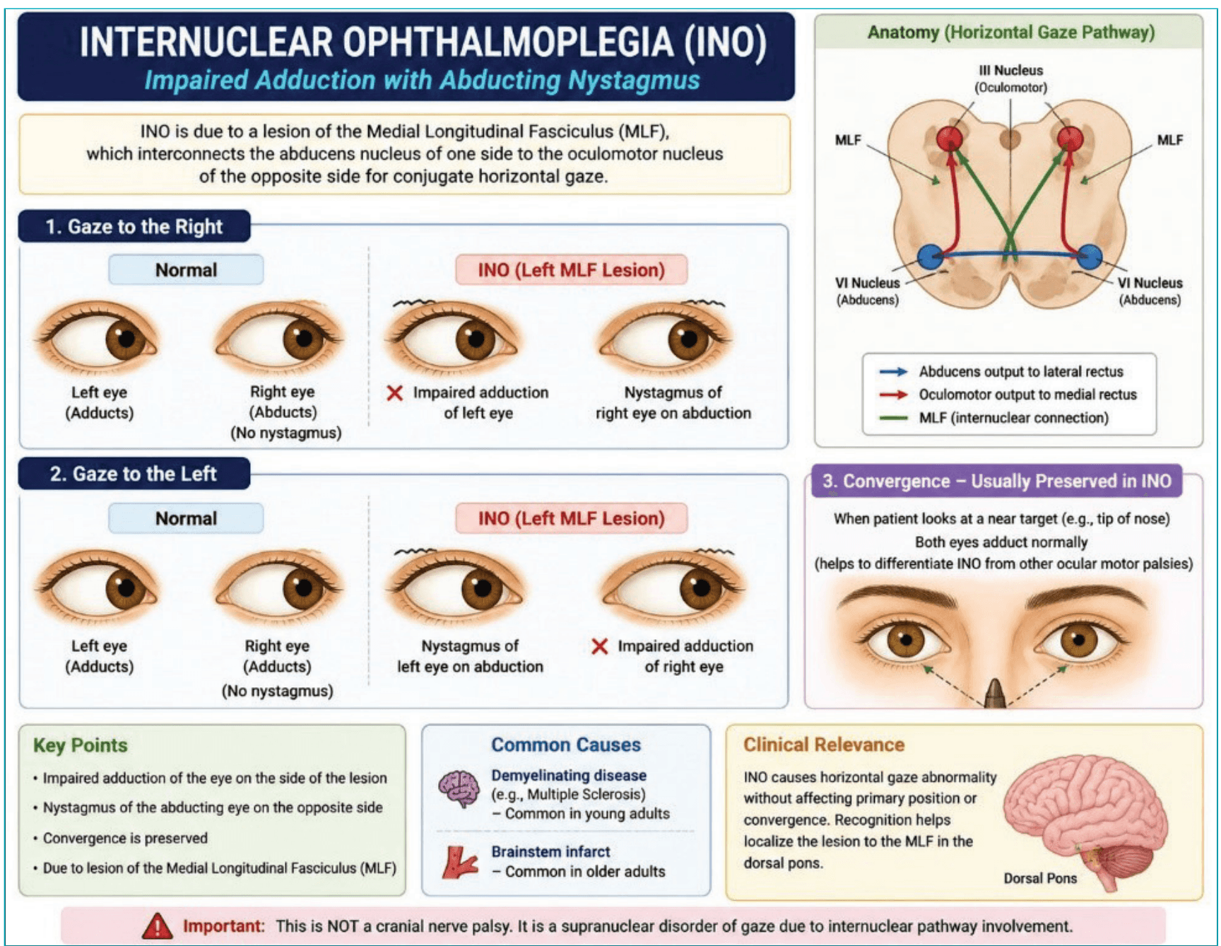
Figure 12: Internuclear ophthalmoplegia.
11. Argyll Robertson pupil (Neurosyphilis)
First described by: Douglas Argyll Robertson.
Finding:Accommodation present, light reflex absent (Figure 11).
Significance: Classic finding of neurosyphilis.
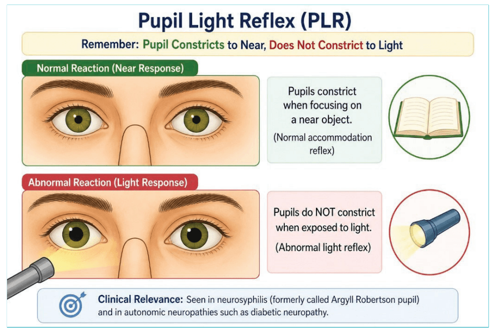
Figure 11: Argyll Robertson pupil.
12. Internuclear ophthalmoplegia (INO) (Neurology)
Described by: David Cogan.
Finding:
- Failure of adduction of one eye.
- Nystagmus of the abducting eye (Figure 12).
Significance: Seen in multiple sclerosis. 9 Figure 11: Argyll Robertson pupil.
Conclusion
This revised list emphasises clinically powerful, examination-relevant, and conceptually rich signs. Among them, Hoover’s sign stands out as a brilliant example of how bedside examination can distinguish organic disease from functional disorder without any investigation.
“The best clinicians are not those who order the most tests, but those who observe the most carefully.”
Narinder Pal Singh. Clinical Signs – The Forgotten Art! (Part II). MMJ. 2026, June. Vol 3 (2).
DOI: XXXX-XXXX
References
- Bickley LS. Bates’ Guide to Physical Examination and History Taking. 13th Edition. Philadelphia: Wolters Kluwer; 2020.
- Talley NJ, O’Connor S. Clinical Examination: A Systematic Guide to Physical Diagnosis. 8th Edition. Sydney: Elsevier; 2019.
- Douglas G, Nicol F, Robertson C. Macleod’s Clinical Examination. 14th Edition. Edinburgh: Elsevier; 2018.
- Corona-Vázquez CP, Orozco-Puga P, Macias-Ortiz FG, et al. Hung-up reflex: the history of a clinical sign and differential diagnosis. Acta Neurol Belg. 2025;125(6):1521–4.
- Gowers WR. A Manual of Diseases of the Nervous System. London: Churchill; 1886.
- Shiota T, Sakamoto T, Amano K, et al. [Venous hum and innominate vein flow velocity in chronic anemia: a pulsed Doppler echocardiographic study]. J Cardiol. 1989;19(3):885–92.
- Agrawal AA. Gingival enlargements: Differential diagnosis and review of literature. World J Clin Cases. 2015;3(9):779–88.
- Dichter SL, Khan Suheb MZ, Shubert GS. Argyll Robertson Pupil. [Updated 2024 Jan 31]. In: StatPearls [Internet]. Treasure Island (FL): StatPearls Publishing; 2026 Jan. Available at: https://www.ncbi.nlm.nih.gov/books/NBK537179/. Accessed on: 20th April 2026.
- Feroze KB, Wang J. Internuclear Ophthalmoplegia. [Updated 2023 Jun 26]. In: StatPearls [Internet]. Treasure Island (FL): StatPearls Publishing; 2026 Jan. Available at: https://www.ncbi.nlm.nih.gov/books/NBK441970/. Accessed on: 20th April 2026.