 WhatsApp Us (24/7)
 +91 926 888 0303 (24/7)


Abstract:
Spontaneous cerebrospinal fluid (CSF) rhinorrhoea is an increasingly recognised clinical entity defined by the leakage of CSF into the nasal cavity in the absence of trauma, surgery, or congenital skull base defects. This condition is strongly associated with idiopathic intracranial hypertension (IIH), particularly in middleaged, obese women, where chronically elevated or pulsatile intracranial pressure leads to dural thinning and progressive skull base remodelling. We describe the radiological evaluation of a 34-year-old obese female who presented with a two-week history of intermittent, clear, watery nasal discharge from the right nostril, which exacerbated upon bending forward and was associated with a salty taste. This case was selected to underscore the pivotal role of magnetic resonance imaging (MRI) surrogate markers — such as empty sella, optic nerve sheath dilatation, and posterior globe flattening — in the diagnosis of underlying IIH. While highresolution computed tomography (CT) is excellent for localising osseous defects, MRI and heavily T2-weighted sequences like constructive interference of steady state (CISS) are superior for identifying the markers of raised intracranial pressure. Recognising these features is critical for clinical management, as failure to address the underlying intracranial hypertension significantly increases the risk of postoperative recurrence. Consequently, an integrated multidisciplinary approach involving both surgical repair and medical management of intracranial pressure is required for optimal patient outcomes. This case report illustrates how advanced cross-sectional imaging provides the necessary diagnostic information to guide these complex therapeutic decisions.
Key words: Spontaneous Cerebrospinal Fluid Rhinorrhoea, Idiopathic Intracranial Hypertension, Magnetic Resonance Imaging Surrogate Markers, Empty Sella, Skull Base Defect, Magnetic Resonance Cisternography.
Introduction
Spontaneous cerebrospinal fluid (CSF) rhinorrhoea is defined as the leakage of CSF into the sinonasal cavity without an identifiable inciting event, such as trauma, surgical intervention, neoplasm, or congenital malformation. Epidemiologically, this condition demonstrates a clear predilection for middle-aged, obese women, a demographic profile that mirrors that of idiopathic intracranial hypertension (IIH).1
The pathophysiological basis of these spontaneous leaks is increasingly attributed to a chronic state of persistently elevated or pulsatile intracranial pressure. This physiological strain results in progressive dural thinning and gradual bone remodelling of the skull base.2 When this process affects highly pneumatised regions, such as the cribriform plate or the sphenoid sinus, it can lead to the formation of focal osseodural defects and subsequent CSF fistulas.
Furthermore, the presence of arachnoid granulations adjacent to the ethmoid and sphenoid sinuses is believed to facilitate focal erosion, thereby predisposing these sites to dehiscence.
Case Report
A 34-year-old obese female presented for neuroimaging evaluation with a two-week history of intermittent, clear, watery discharge from the right nostril. The patient noted that the rhinorrhoea was predominantly elicited by bending forward and was consistently associated with a salty taste.
The clinical history was negative for head trauma, previous sinonasal or neurosurgical procedures, chronic sinusitis, or prior radiation therapy. While the patient reported a history of intermittent headaches, she denied fever, visual disturbances, or symptoms of meningeal irritation. A comprehensive neurological examination was unremarkable. Given the high clinical suspicion of a spontaneous CSF fistula, a diagnostic evaluation was initiated to localise the site of the leak and evaluate for radiological hallmarks of intracranial hypertension using magnetic resonance imaging (MRI) (Figure 1A–D) and computed tomography (CT) (Figure 2A–C).3
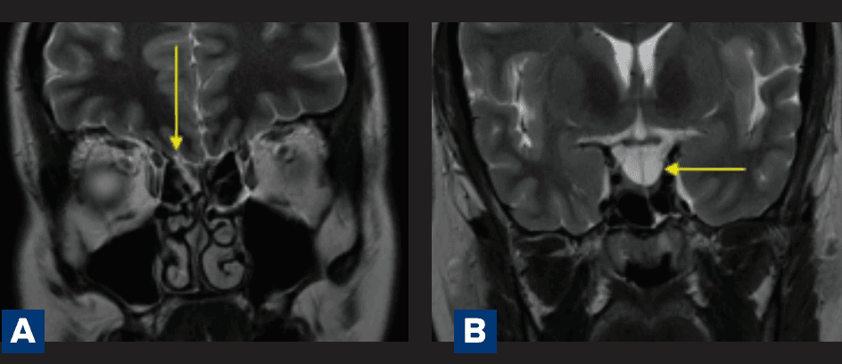
Figure 1 A-B: A. Magnetic resonance imaging (MRI) demonstrates a cerebrospinal fluid (CSF)-intensity linear tract extending inferior to the right cribriform plate, consistent with a CSF fistula (yellow arrow); B. MRI shows a partial empty sella (yellow arrow) and prominent CSF spaces within Meckel’s caves, consistent with idiopathic intracranial hypertension (IIH);
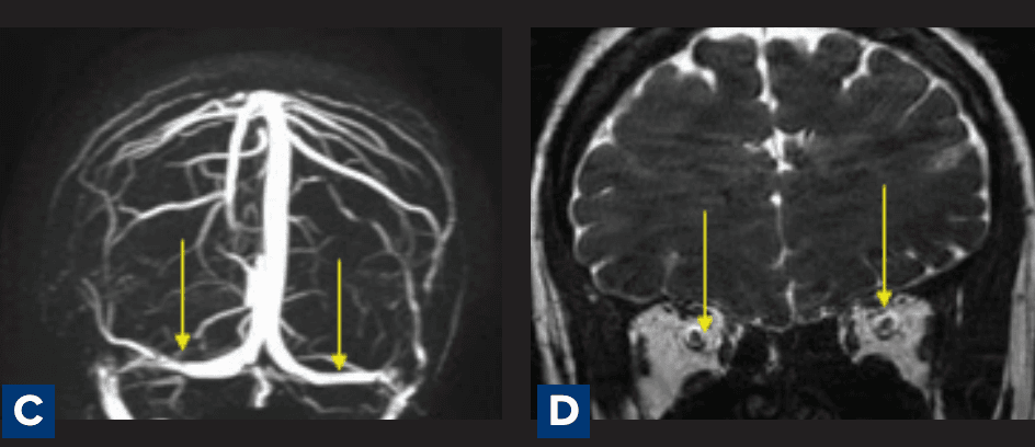
Figure 1 C-D: C. Magnetic resonance venography demonstrating stenosis along the lateral aspects of the bilateral transverse sinuses (yellow arrows), without evidence of venous sinus thrombosis. ; D. Prominence of perioptic CSF spaces with vertical tortuosity of the optic nerves
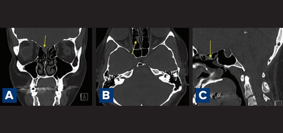
Figure 2: A–C: Computed tomography (CT) images demonstrating asymmetrical, slight widening of the right olfactory fossa with a focal defect involving the right cribriform plate, concerning for a cerebrospinal fluid (CSF) fistula (yellow arrows): A. Coronal CT image demonstrating subtle widening of the right olfactory fossa with focal thinning of the right cribriform plate (yellow arrow), suspicious for a CSF fistula; B. Coronal high-resolution CT image showing focal dehiscence involving the right cribriform plate with adjacent widening of the olfactory fossa (yellow arrow), suggestive of the leak site; C. Coronal CT image demonstrating a focal defect along the right cribriform plate with asymmetry of the right olfactory fossa (yellow arrow), concerning for spontaneous CSF fistula.
Discussion
The aetiology of CSF rhinorrhoea is fundamentally categorised into traumatic and non-traumatic origins. While post-traumatic fractures remain the most frequent cause, non-traumatic aetiologies are relatively rare, comprising approximately 3% of all cases.4 Within this non-traumatic subset, spontaneous leaks are the most prevalent and are now recognised as a primary radiological manifestation of IIH.
In patients presenting with spontaneous leaks, the prevalence of MRI surrogate markers for IIH ranges from 60% to 80%. These markers include the empty sella sign, optic nerve sheath dilatation, posterior globe flattening, and transverse sinus stenosis.5 It is vital for the clinician to recognise that the fistula may act as a natural pressure release valve; consequently, the standard clinical symptoms of IIH may be absent until the defect is surgically closed.
Effective therapeutic management must address both the anatomical breach and the underlying physiological driver. While endoscopic surgical repair is the definitive treatment for the osseodural defect, the high risk of recurrence necessitates medical intervention to lower intracranial pressure. This typically involves aggressive weight reduction and the administration of acetazolamide to decrease CSF production. In the absence of a combined treatment approach, the repaired site remains vulnerable to the same pulsatile forces that caused the initial erosion.
Conclusion
The radiological evaluation of spontaneous CSF rhinorrhoea requires a dual focus: the precise localisation of the osseodural fistula and the systematic assessment for underlying IIH. Identifying surrogate markers such as the empty sella and considering anatomical variations like arachnoid pits and extensive sinus pneumatisation are vital for characterising the aetiology of the leak. 6 Recognising these features is essential for long-term management and the prevention of postoperative recurrence. Ultimately, achieving optimal patient outcomes requires a coordinated multidisciplinary approach that integrates high-resolution imaging, targeted surgical intervention, and medical management of intracranial pressure.
Mohit Tamakuwala, Mitusha Verma, Deepak Patkar. Beyond Direct Leak Visualisation: Magnetic Resonance Imaging as a Surrogate Marker in Spontaneous Cerebrospinal Fluid Rhinorrhoea. MMJ. 2026, June. Vol 3 (2).
DOI: XXXX-XXXX
References
- Hiremath SB, Gautam AA, Sasindran V, et al. Cerebrospinal fluid rhinorrhea and otorrhea: a multimodality imaging approach. Diagn Interv Imaging. 2019;100(1):3–15.
- Schuknecht B, Simmen D, Briner HR, et al. Nontraumatic skull base defects with spontaneous CSF rhinorrhoea and arachnoid herniation: imaging findings and correlation with endoscopic sinus surgery in 27 patients. AJNR Am J Neuroradiol. 2008;29(3):542–9.
- Vemuri NV, Karanam LS, Manchikanti V, et al. Imaging review of cerebrospinal fluid leaks. Indian J Radiol Imaging. 2017;27(04):441–6.
- Goodier M, Lubbe D, Andronikou S, et al. Spontaneous lateral sphenoid cerebrospinal fluid fistula: MRI diagnosis. S Afr J Radiol. 2012;16(1):12–3.
- Matsubara T, Akutsu H, Tanaka S, et al. A case of spontaneous cerebrospinal fluid rhinorrhea: Accurate detection of the leak point by magnetic resonance cisternography. Surg Neurol Int. 2014;5:54.
- Almontasheri A, Al-Qahtani B, Aldajani N. Arachnoid pit and extensive sinus pneumatization as the cause of spontaneous lateral intrasphenoidal encephalocele. J Clin Imaging Sci. 2012;2:1.