 WhatsApp Us (24/7)
 +91 926 888 0303 (24/7)


Abstract:
Metallosis is a rare but serious long-term complication following total knee arthroplasty (TKA), most commonly resulting from polyethylene insert wear that leads to unintended metal-on-metal articulation and subsequent metallic debris deposition. We present the case of a 70-year-old female who developed progressive pain, stiffness, and instability in her right knee, 19 years after undergoing primary TKA. Radiographic evaluation revealed loosening of both femoral and tibial components, while laboratory investigations ruled out infection. Intraoperatively, extensive metallosis was identified with diffuse, black-stained synovium, metallic debris accumulation, and complete wear of the medial polyethylene insert exposing the tibial tray. Both prosthetic components were found to be loose, accompanied by collateral ligament laxity. Thorough synovectomy and debridement were performed, followed by revision TKA using a constrained condylar knee prosthesis (Zimmer Rotating Hinge Knee [RHK]). Metallosis-associated inflammatory reaction can result in osteolysis, soft tissue destruction, and eventual implant failure, making early diagnosis and timely intervention essential despite the non-specific clinical presentation. Revision arthroplasty with appropriately constrained implants can achieve satisfactory outcomes in cases associated with instability and severe component loosening.
Key words: Metallosis, Total Knee Arthroplasty, Aseptic Loosening, Polyethylene Wear, Revision Total Knee Arthroplasty, Rotating Hinge Knee.
Introduction
Metallosis is an uncommon but recognised complication following total knee arthroplasty (TKA), characterised by the deposition of metallic debris in peri-prosthetic tissues due to abnormal metal-on-metal articulation.1 This phenomenon is most commonly associated with polyethylene wear or failure, leading to exposure of metallic components and subsequent abrasive interaction between prosthetic surfaces. The resulting metallic debris incites a chronic inflammatory response, termed metal-induced synovitis, which may progress to osteolysis, soft tissue destruction, and eventual implant loosening.2,3
Although metallosis is more frequently reported in total hip arthroplasty, its occurrence in TKA remains relatively rare, with limited cases described in the literature. The diagnosis is often delayed due to its insidious onset and nonspecific clinical presentation, including pain, swelling, and reduced joint function. Radiographic findings may suggest implant loosening or osteolysis, but a definitive diagnosis is typically established intraoperatively.
In this report, we present a rare case of metallosisinduced aseptic loosening in a long-standing primary TKA, highlighting its clinical presentation, intraoperative findings, and management with revision arthroplasty.
Case Report
A 70-year-old female patient presented with complaints of progressively increasing pain, stiffness, and difficulty in ambulation involving the right knee for one year. She had undergone a primary right TKA in January 2006 and had remained asymptomatic for several years postoperatively.
On clinical examination, the patient exhibited a painful range of motion with reduced flexion, joint line tenderness, and signs suggestive of instability. There was no history of trauma, fever, or systemic symptoms suggestive of infection.
Radiographic evaluation revealed features consistent with loosening of both femoral and tibial components, including radiolucent lines and possible osteolysis. Laboratory investigations, including inflammatory markers, were within normal limits, thereby reducing the likelihood of peri-prosthetic joint infection.
Based on clinical and radiological findings, a diagnosis of aseptic loosening of the TKA implant was made, and the patient was planned for revision arthroplasty.
Intraoperative findings
Intraoperatively, there was extensive black discolouration of synovial and peri-prosthetic tissues, characteristic of metallosis. The synovium appeared markedly thickened and inflamed, with visible metallic debris deposits. There was complete wear of the medial half of the polyethylene insert, exposing the underlying metal tibial tray, resulting in direct metal-on-metal articulation.
Both femoral and tibial components were found to be loose. Additionally, there was associated collateral ligament laxity, contributing to joint instability (Figures 1 and 2).
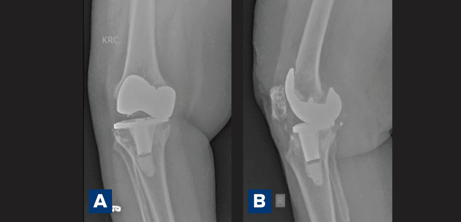
Figure 1A and B: Preoperative radiograph showing laxity and loosening of the implant.
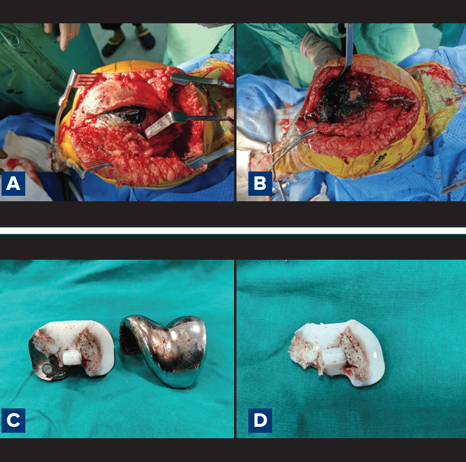
Figure 2A–D: Intraoperative finding of metallotic synovium, polyethylene wear exposing the metal surface.
A thorough synovectomy and debridement of metallotic tissue were performed. The failed components were removed, and revision was carried out using a constrained condylar knee prosthesis (Zimmer Rotating Hinge Knee [RHK]), to address the ligamentous instability (Figures 3 and 4).
Postoperative recovery was uneventful, and the patient demonstrated improved pain relief and functional outcome on follow-up.
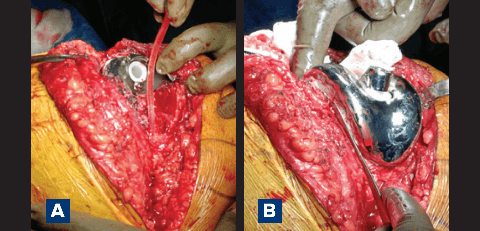
Figure 3A and B : Revision using a constrained condylar knee prosthesis (Zimmer Rotating Hinge Knee [RHK]).
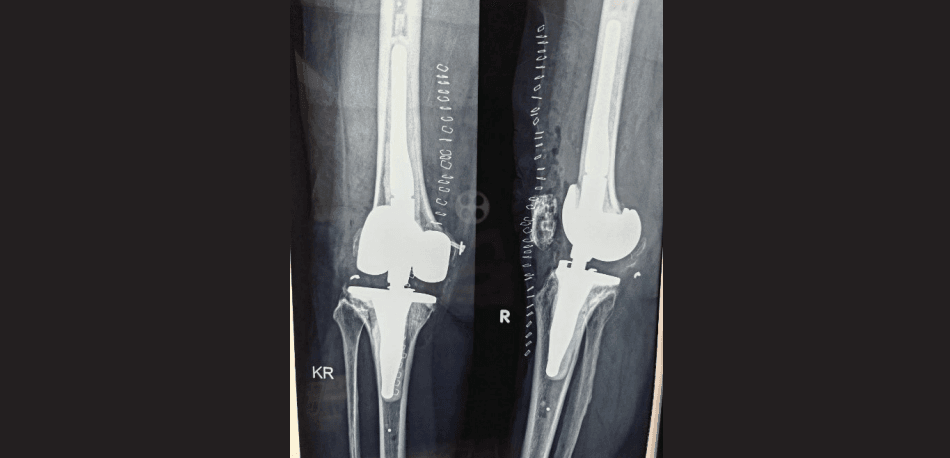
Figure 4: Postoperative radiograph.
Discussion
Metallosis is a rare complication of TKA due to metal-onmetal contact secondary to polyethylene wear.5 It is caused by the infiltration and accumulation of metallic debris into the peri-prosthetic structures, deriving from friction between metallic prosthetic components.6 The metallic debris induce a massive release of cytokines from inflammatory cells, causing osteolysis and loosening of the prosthesis.7–9 Over time, extensive soft tissue involvement may also compromise ligamentous structures, further exacerbating joint instability.
Several risk factors have been implicated in the development of metallosis in TKA, including:10
- Polyethylene wear or fracture
- Malalignment of components
- Instability leading to abnormal loading
- Use of thinner polyethylene inserts
- Long implant survival duration
Clinically, patients typically present with pain, swelling, reduced range of motion, and sometimes instability. However, these symptoms are non-specific and overlap with other causes of TKA failure, making early diagnosis challenging.
Radiographically, findings may include:
- Radiolucent lines indicating loosening
- Osteolytic defects
- Component malposition
- "Metal-line sign" (a thin rim of linear density outlining the joint capsule) and the "Cloud sign" (amorphous cloudy densities within the soft tissues) in X-ray image.10
Advanced imaging modalities such as computed tomography (CT) or magnetic resonance imaging (MRI) may help detect soft tissue involvement, but definitive diagnosis is often made intraoperatively, as seen in our case.
Metallosis is more common in hip than in knee arthroplasty.10 In the present case, prolonged implant survival (approximately 19 years) likely contributed to progressive polyethylene wear, eventually leading to exposure of the tibial tray and subsequent metallosis. It highlights the importance of lifelong follow-up for TKA patients to detect polyethylene wear before it reaches the metal-on-metal stage.
The choice of a constrained or hinged implant is often a mechanical necessity. The chronic inflammatory environment associated with metallosis can compromise the collateral ligaments, making standard semi-constrained liners insufficient.4 Furthermore, the extensive osteolysis caused by cytokine-mediated bone resorption often leaves significant voids that require modular revision systems.3 Early diagnosis and aggressive synovectomy are critical to prevent "pseudotumour" formation and systemic toxicity from metal ion release.11
Conclusion
Metallosis following TKA is an uncommon but potentially serious complication, most often resulting from polyethylene wear leading to unintended metal-on-metal articulation. Early recognition is crucial, as progressive metal debris–induced inflammation can cause osteolysis, implant loosening, and extensive soft-tissue damage. Timely revision surgery with appropriate implant selection can lead to favourable clinical outcomes.
Debasish Sarkar, Pradeep Bhosale. A Case Report of Metallosis-Related Aseptic Loosening of Primary Total Knee Arthroplasty Implant Managed with Revision Total Knee Arthroplasty. MMJ. 2026, June. Vol 3 (2).
DOI: XXXX-XXXX
References
- Koonen L, Duit-van den Belt L, Veenstra K, et al. Late Onset Metallosis after Revision Total Knee Arthroplasty with Metal-Backed Patellar Component In Situ. Arthroplast Today. 2020;6(2):262–6.
- Romesburg JW, Wasserman PL, Schoppe CH. Metallosis and Metal-Induced Synovitis Following Total Knee Arthroplasty: Review of Radiographic and CT Findings. J Radiol Case Rep. 2010;4(9):7–17.
- Zhong Q, Pan X, Chen Y, et al. Prosthetic Metals: Release, Metabolism and Toxicity. Int J Nanomedicine. 2024;19:5245–67.
- Gopi J, Harafan A, Roshan SH, et al. Severe Metallosis-Induced Osteolysis and Polyethylene Liner Dissociation as a Cause of Total Knee Replacement Failure. J Orthop Case Rep. 2026;16(1):208–11.
- Rader CP, Löhr J, Wittmann R, et al. Results of total knee arthroplasty with a metal-backed patellar component: a 6-year follow-up study. J Arthroplasty. 1996;11(8):923–30.
- Salem KH, Lindner N, Tingart M, et al. Severe metallosis-related osteolysis as a cause of failure after total knee replacement. J Clin Orthop Trauma. 2020;11(1):165–70.
- Schiavone Panni A, Vasso M, Cerciello S, et al. Metallosis following knee arthroplasty: a histological and immunohistochemical study. Int J Immunopathol Pharmacol. 2011;24(3):711–19.
- Burge AJ, Gold SL, Lurie B, et al. MR Imaging of Adverse Local Tissue Reactions around Rejuvenate Modular Dual-Taper Stems. Radiology. 2015;277(1):142–50.
- Dapunt U, Giese T, Lasitschka F, et al. On the inflammatory response in metal-on-metal implants. J Transl Med. 2014;12:74.
- Oliveira CA, Candelária IS, Oliveira PB, et al. Metallosis: A diagnosis not only in patients with metal-on-metal prostheses. Eur J Radiol Open. 2014;2:3–6.
- Bogdonoff YM, Amirouche F. Addressing metallosis in knee arthroplasty: From diagnostic challenges to innovative treatments. World J Orthop. 2024;15(5):386–9.