 WhatsApp Us (24/7)
 +91 926 888 0303 (24/7)


Abstract:
Presence of synchronous primary tumours and oral potentially malignant lesions in the oral cavity over the bilateral buccal mucosa warrants surgical treatment and creates synchronous defects. Functional rehabilitation of such defects is challenging and requires free tissue transfer to reconstruct the bilateral buccal mucosa defects created from simultaneous resection of such lesions. Advances in perforator flap concepts now allow the harvesting of multiple skin islands from a single donor area without sacrificing the donor vessels. The vascular anatomy of the free anterolateral thigh (FALT) flap is variable, with several variations seen due to the presence of multiple musculocutaneous perforators, many of which originate from vessels other than the lateral circumflex artery, allowing for two separate skin paddles to be harvested from a single donor site. This case report presents a modified FALT flap harvesting technique and its application in reconstructing such extensive defects with the use of an intraoral anastomosis technique.
Key words: Anterolateral Thigh Flap, Bilateral Mucosal Defect, Intraoral Anastomosis.
Introduction
Free flaps have become a cornerstone in reconstructing oral malignancy defects, offering a broad spectrum of options, including chimeric and multiple free flaps. These advancements enable the reconstruction of complex defects with excellent aesthetic and functional outcomes.
However, certain practical challenges remain. The presence of synchronous primary tumours in the oral cavity is seen in 1%–11% of patients due to field cancerisation, and surgical treatment of these lesions creates synchronous defects with intervening normal tissue, which complicates the use of a single flap. 1,2 Bilateral buccal mucosa defects often necessitate two separate flaps. Preoperative planning for such defects can be particularly challenging, especially with reduced mouth opening seen in this group of patients due to the disease or pre-existing oral submucous fibrosis. Utilising two free flaps increases donor site morbidity, potentially requiring skin grafting. This also increases operative time and involves challenges in preoperative counselling. Another challenge arises with potentially malignant lesions, such as erythroleukoplakia, where treatment warrants the achievement of clear margins but does not necessitate neck dissection. 3 Using a free flap in these cases requires neck exploration and tunnelling for the vascular pedicle, adding risks and complications. This case study explores these scenarios and highlights how expanding the limits of microvascular reconstruction can address them effectively.
Case Report
A male patient in his 50s, diagnosed preoperatively via incisional biopsy with right buccal mucosa squamous cell carcinoma underwent planned bite margin resection, including marginal mandibulectomy and upper alveolectomy. Preoperative mouth opening was less than 1 cm. During examination under anaesthesia, a superficial lesion was identified on the left buccal mucosa. An additional wide local excision of the left buccal mucosa was performed with adequate margins.
A frozen section confirmed moderate dysplasia, eliminating the need for a formal neck dissection. The left-sided defect measured 6 × 4 cm, leaving only skin and subcutaneous tissues (Figure 1).
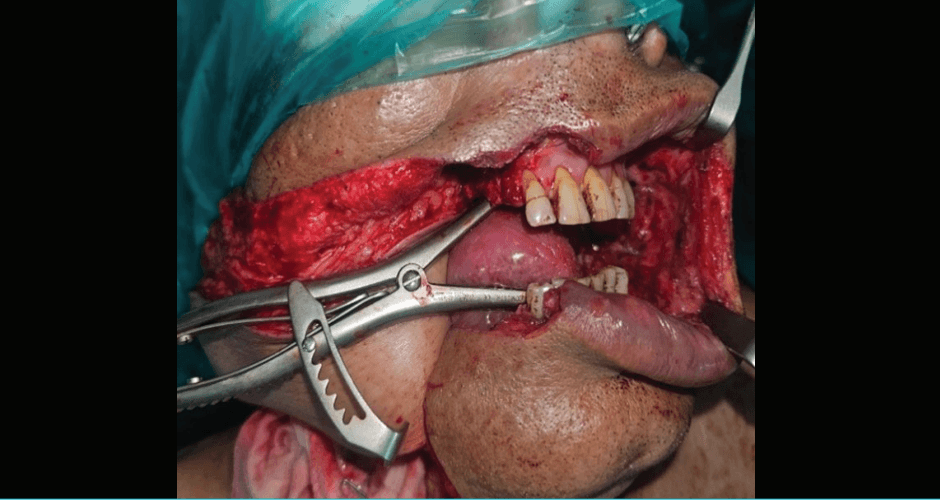
Figure 1: Post-extirpation defect over the left buccal mucosa following excision of the mucosal lesion.
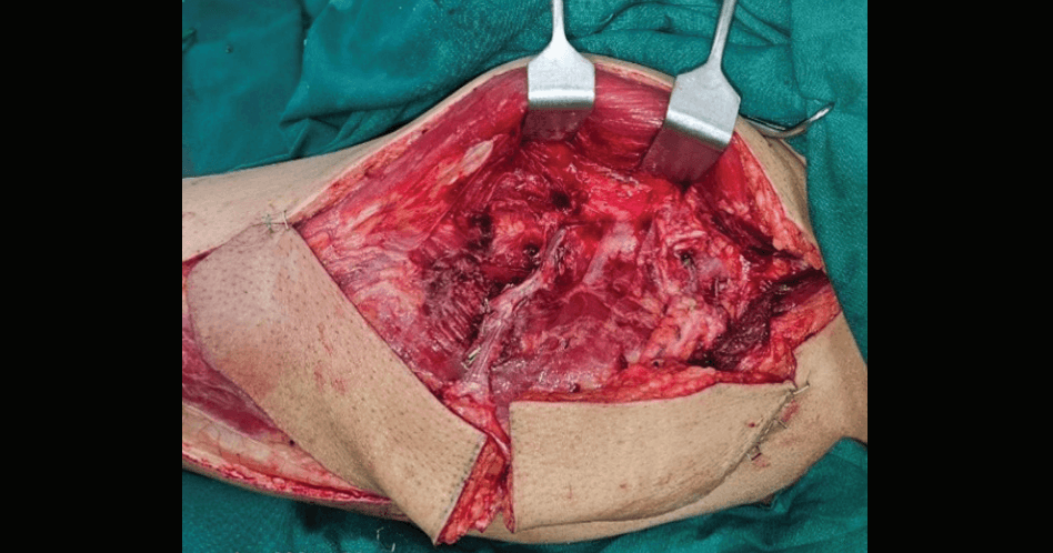
Figure 2: Harvest of free anterolateral thigh flap (FALT) with two cutaneous perforators — one from the oblique branch and one from the descending branch of the lateral circumflex femoral artery (LCFA) — subsequently separated and used individually.
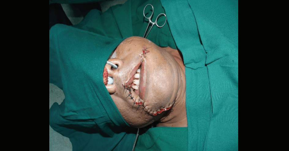
Figure 3: Inset of a smaller skin paddle of the free anterolateral thigh flap (FALT) from the distal perforator — descending branch of the lateral circumflex femoral artery (LCFA) — with intraoral anastomosis.
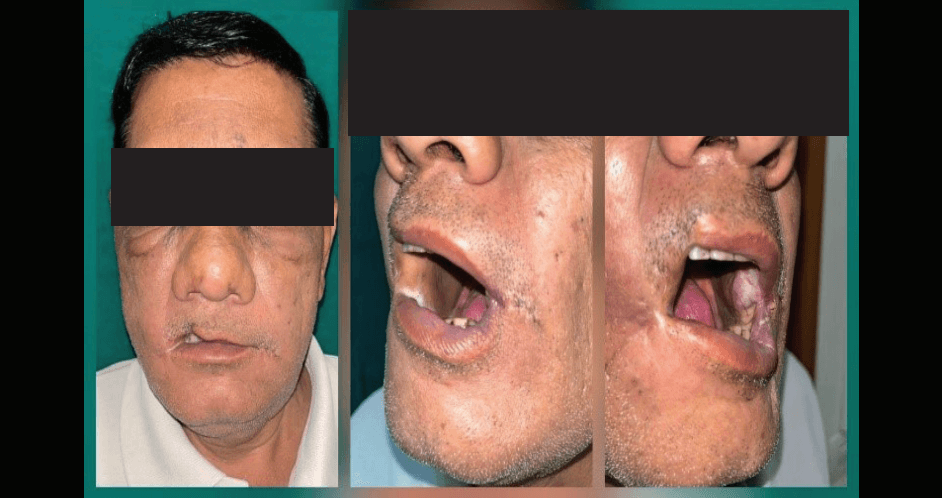
Figure 4: Postoperative appearance of the patient demonstrating the absence of neck incision on the left side.
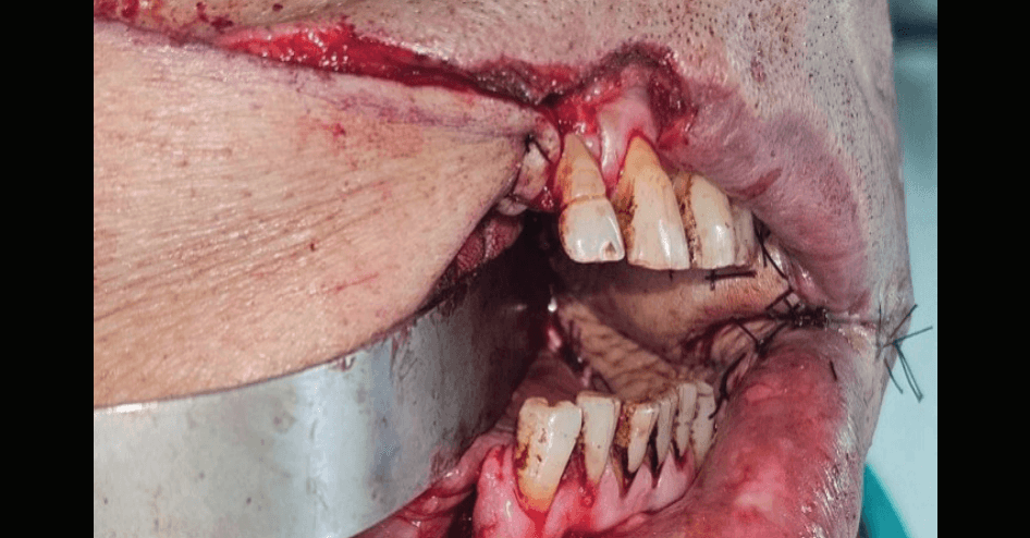
Figure 5: Postoperative result at 6 months follow-up.
As per the initial plan, the left anterolateral thigh (ALT) flap territory was explored via a non-committal incision. Two cutaneous perforators were identified — one from the oblique branch and one from the descending branch of the lateral circumflex femoral artery (LCFA) (Figure 2). Two separate skin islands were harvested on each of these perforators from the same donor area.
The larger island, based on the oblique branch, was used to reconstruct the right-sided bite defect and was anastomosed to vessels in the right neck. A smaller paddle, measuring 6 × 5 cm, was harvested from the distal perforator. The pedicle was divided at its junction with the descending branch of the LCFA, resulting in a pedicle length of only 6 cm.
The distal continuation of the facial artery and vein within the left-sided intraoral defect was identified and used for microvascular anastomosis (Figures 3 and 4). The donor site was closed primarily. The postoperative period was uneventful, with timely initiation of oral intake and smooth suture removal. At the 6-month follow-up, the patient showed excellent cosmetic outcomes and significant improvement in mouth opening (Figure 5). No adjuvant therapy was required as final clinical staging for the left buccal mucosa was T2N0 as per the American Joint Committee on Cancer (AJCC) 8th Edition.
Discussion
Bilateral buccal mucosa defects following resection of synchronous oral lesions may require either two flaps or a chimeric configuration. Advances in perforator flap concepts now allow the harvesting of multiple skin islands from a single donor area without sacrificing the donor vessels. 4 For example, the LCFA axis supports harvesting perforators from various branches: transverse (tensor fascia lata flap), oblique or descending (ALT flap), or rectus muscle branch (anteromedial thigh [AMT] flap).
Harvesting multiple flaps from a single donor site minimises morbidity and optimises outcomes while allowing primary closure. Potentially malignant lesions and conditions, such as leukoplakia, erythroplakia, oral submucous fibrosis, etc., have often been reconstructed using suboptimal methods like primary closure, skin grafting, or nasolabial flaps, which can lead to functional limitations such as trismus and cosmetic deformities. Full-thickness flaps are essential to prevent such complications, as local nasolabial flaps often fail to provide sufficient width to fill these defects.5
Microvascular free flaps, such as the radial forearm flap, medial sural artery flap, or distal ALT flap, offer the appropriate thickness and adaptability required. Intraoral microvascular anastomosis eliminates the need for unnecessary neck dissection and tunnelling, simplifying the procedure and reducing complications.6
The use of smaller, thinner microvascular free flaps with intraoral vascular anastomosis for premalignant lesions is gaining acceptance. The field of microsurgery is pushing boundaries, addressing challenges of depth, precision, and spatial limitations through perforator-based super microsurgery.
Conclusion
- Single donor site dual flap harvesting: Rebuilding bilateral oral defects without increasing donor site morbidity or needing multiple donor sites is possible by using two skin paddles from the ALT flap territory, which are based on different perforators.
- Intraoral microvascular anastomosis: When nodal dissection is not required, microvascular anastomosis within the oral cavity reduces surgical complexity and related risks by doing away with the need for neck dissection and pedicle tunnelling.
- Better results in premalignant lesions: Full-thickness microvascular free flaps, as opposed to more conventional techniques like skin grafts or nasolabial flaps, offer better functional and cosmetic results, particularly in premalignant conditions with field cancerisation or poor mouth opening.
- Problems with synchronous oral lesions: When defects are separated by healthy mucosa, field cancerisation can lead to synchronous lesions that need intricate reconstructions.
Bharat Saxena, Raj Lakshmi Pandey, Devendra Chaukar. Reconstruction of Synchronous Bilateral Buccal Mucosa Defects with a Single Free Anterolateral Thigh Flap with Intraoral Anastomosis. MMJ. 2026, June. Vol 3 (2).
DOI: XXXX_XXXX_XXXX_XXXX
References
- Gluckman JL. Synchronous Multiple Primary Lesions of the Upper Aerodigestive System. Arch Otolaryngol. 1979;105(10):597–
- Mariela Peralta-Mamani, Ángel Terrero-Pérez, Rosana Mara Adami Tucunduva, et al. Occurrence of field cancerization in clinically normal oral mucosa: A systematic review and meta-analysis. Arch Oral Biol. 2022;143:105544.
- Warnakulasuriya S. Oral potentially malignant disorders: A comprehensive review on clinical aspects and management. Oral Oncol. 2020;102:104550.
- Huang JJ, Wallace C, Lin JY, et al. Two small flaps from one anterolateral thigh donor site for bilateral buccal mucosa reconstruction after release of submucous fibrosis and/or contracture. J Plast Reconstr Aesthet Surg. 2010;63(3):440–
- Lin PY, Chen CC, Kuo YR, et al. Simultaneous reconstruction of head and neck defects following tumor resection and trismus release with a single anterolateral thigh donor site utilizing a lateral approach to flap harvest. Microsurgery. 2012;32(4):289–
- Brandtner C, Bürger H, Hachleitner J, et al. The intraoral anastomosing technique in reconstructive surgery of the face – a consecutive case series of 70 patients. J Craniomaxillofac Surg. 2015;43(9):1763–8