 WhatsApp Us (24/7)
 +91 926 888 0303 (24/7)


Abstract:
Complete atrioventricular (AV) block during one-lung ventilation (OLV) is a rare but life-threatening complication. We report the case of a 71-year-old male with pre-existing right bundle branch block (RBBB) undergoing left lower lobectomy who developed atropine-refractory complete heart block after two hours of OLV. Following a transient episode of asystole, return of spontaneous circulation (ROSC) was achieved with cardiopulmonary resuscitation (CPR) and adrenaline administration; however, persistent bradycardia required transcutaneous pacing (TCP) (70/min, 30 mA) and an isoprenaline infusion. The patient subsequently required transvenous pacing in the Intensive Care Unit (ICU). This case underscores the importance of preparedness for conduction abnormalities during high-risk thoracic surgeries, particularly with pre-existing RBBB, and highlights the role of TCP in refractory AV block.
Key words: Complete Atrioventricular Block, One-lung Ventilation, Thoracic Surgery, Transcutaneous Pacing, Right Bundle Branch Block.
Introduction
Complete atrioventricular (AV) block during surgery is a rare yet life-threatening condition that can pose significant challenges, particularly in patients with pre-existing cardiac conditions. Conduction disturbances during anaesthesia may arise due to underlying structural conduction abnormalities, autonomic imbalance, electrolyte disturbances, myocardial ischaemia, or direct surgical manipulation near the cardiac conduction pathways. Patients with pre-existing bundle branch block, especially right bundle branch block (RBBB), may be particularly vulnerable to progression towards highergrade AV block under perioperative stress conditions.
Thoracic surgeries requiring one-lung ventilation (OLV) introduce additional physiological challenges that may predispose patients to cardiovascular instability. OLV results in altered pulmonary vascular resistance, ventilation-perfusion mismatch, hypoxaemia, and increased right ventricular afterload, all of which can adversely affect myocardial oxygen supply-demand balance. Furthermore, mediastinal manipulation and traction during thoracic procedures can enhance vagal stimulation, precipitating severe bradyarrhythmia and conduction abnormalities in susceptible individuals. Although transient bundle branch blocks and arrhythmias during surgery have been described in the literature, complete AV block occurring specifically during OLV remains exceedingly rare. It can lead to significant haemodynamic instability and is often refractory to vagolytic medications. 1
Prompt recognition and immediate intervention are critical because atropine-resistant AV block may rapidly progress to profound bradycardia, circulatory collapse, and asystole. In such situations, transcutaneous pacing (TCP) serves as an important bridge until definitive transvenous or permanent pacing can be established. Awareness of this rare but life-threatening complication is therefore essential for anaesthesiologists managing high-risk thoracic surgical patients.
A meticulous plan of action to manage such a crisis intraoperatively is vital for optimal patient care and outcomes. Very few studies are available to date on this condition, and none of them describes its occurrence during OLV. Here, we present the case of a 71-year-old male posted for left lower lobectomy with a history of lower anterior resection and hepatic metastasectomy. The patient developed complete heart block after two hours of OLV, which was managed promptly with TCP.
Case Report
A 71-year-old male with a history of lower anterior resection for carcinoma of the sigmoid colon 6 years earlier and hepatic metastasectomy 5 years earlier presented with left lung metastasis. He was scheduled for left lower lobectomy. The patient was a known case of hypertension, which was optimised on tablet amlodipine 5 mg once daily (OD), and hypothyroidism for which he was on tablet thyronorm 50 mcg OD. On preoperative assessment, his electrocardiogram (ECG) revealed RBBB, for which a cardiology consultation was taken for perioperative risk stratification. A two-dimensional echocardiogram (2D echo) was done, which was normal with left ventricular ejection fraction (LVEF) 55%–60%, and the patient was stratified under moderate risk of major adverse cardiovascular events (MACE). The patient had received neoadjuvant chemotherapy with two cycles of capecitabine plus oxaliplatin (CAPOX) and folinic acid, fluorouracil, and irinotecan (FOLFIRI), with the last dose administered 6 weeks prior to evaluation. Preand post-ECG revealed normal findings, with an ejection fraction within the range of 55%–60%, no regional wall motion abnormalities (RWMA), and normal chamber dimensions.
On the day of surgery, the patient was transferred to the operating theatre after confirming nil per os (NPO) status. All standard American Society of Anesthesiologists (ASA) monitors, including ECG, pulse oximetry, and non-invasive blood pressure monitoring, were connected. The patient had a baseline heart rate of 45/min. An 18 G cannula was secured. An epidural was placed at T6–T7 level in the lateral position. Patient was preoxygenated using transnasal humidified rapid-insufflation ventilatory exchange (THRIVE) with settings of 100% fraction of inspired oxygen (FiO2) and flow rate of 30 L/min till end-tidal oxygen concentration (EtO2) of 90%. After adequate preoxygenation, intravenous induction was started using injection fentanyl (2 mcg/kg), injection propofol titrated to loss of verbal response and injection rocuronium (0.6 mg/kg) and the patient’s trachea was intubated using a right double-lumen tube (DLT) of 39 Fr under videolaryngoscope guidance. The position of the tube was confirmed with a fibreoptic bronchoscope, and the circuit was connected to the ventilator. Additionally, a 20 G arterial cannula was secured in the right upper limb to allow beat-to-beat haemodynamic monitoring and frequent blood gas analysis during OLV.
The patient was then positioned in the right lateral decubitus position, and all the pressure points were adequately padded to prevent pressure injury. OLV was initiated after the commencement of surgery. After 30 minutes of initiating OLV, the patient started desaturating gradually, which was managed by increasing FiO 2 and positive end-expiratory pressure (PEEP) to the ventilated lung. There was a requirement of intermittent continuous positive airway pressure (CPAP) to the non-ventilated lung as well to maintain SpO2 > 90%. The patient had intermittent arrhythmias and occasional ventricular premature complex (VPC) (< 5/rhythm) secondary to pericardial stimulation during video-assisted thoracic surgery (VATS), though he was haemodynamically stable throughout.
There was a sudden decrease in heart rate (minimum 32/min) as soon as the lobectomy specimen was removed, and he developed hypotension, for which injection atropine 1 mg was administered; however, there (CPR) was no response, and the patient developed asystole. Cardiopulmonary resuscitation was started according to the standard advanced cardiovascular life support (ACLS) guidelines in the lateral position with 1 mg of adrenaline administered every 3 minutes for a total of two cycles, following which the patient was positioned supine. Return of spontaneous circulation (ROSC) was achieved, but the patient was still having bradycardia and rhythm was depicting Mobitz type 1 (Wenckebach phenomenon) heart block, which got converted to complete heart block. Bradycardia was nonresponsive to injection atropine.
Immediate transcutaneous pads were applied, and pacing was initiated at a rate of 70/min and an output of 30 mA, along with isoprenaline at a rate of 4–10 µg/min for pharmacological pacing. The patient’s heart rate responded to pacing, and he was haemodynamically stable thereafter. Arterial blood gas, electrolytes and cardiac enzymes showed no remarkable findings. A chest tube was inserted, and the DLT was changed to single-lumen tube (SLT), and the patient was shifted to the Intensive Care Unit (ICU) for further management.
A cardiology consultation was sought, and the patient underwent transvenous pacing in the ICU under radiological He was weaned off the ventilator on postoperative day (POD) 1 and subsequently extubated without any evidence of hypoxic brain injury. All the laboratory investigations and vital parameters were within normal limits. The patient was shifted to the ward on POD 2 and was planned for permanent pacing by the cardiology team.
An implantable cardioverter-defibrillator (ICD) with cardiac resynchronisation therapy (CRT) (Figure 2) was inserted on POD 4, and the patient was discharged with the pacemaker in situ on POD 7.
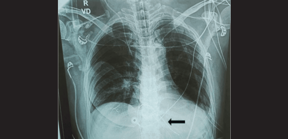
Figure 1: Chest X-ray of the patient showing the transvenous pacing wire (black arrow) after placement inside the ventricle.
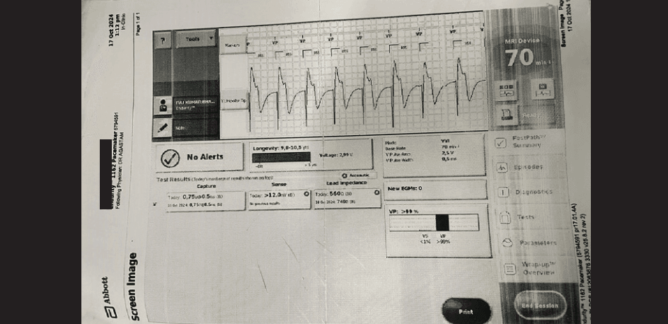
Figure 2: Mode of pacemaker.
Discussion
Intraoperative transient bundle branch blocks (BBBs) are mostly paroxysmal, with tachycardia-associated, ratedependent left bundle branch block (LBBB) being the more common type.2 The bradycardia-dependent blocks caused by phase-4 block are rare and usually associated with structural heart disease.3 Acquired complete AV block involves blockade of the His-Purkinje system distal to the AV node. In this type of heart block, the ECG typically demonstrates a wide QRS complex and a relatively slow heart rate.4 However, in our case, the patient showed a normal QRS and severe bradycardia that was refractory to atropine administration. This rare and atypical AV block is referred to as paroxysmal AV block.5
The precise mechanism of paroxysmal AV block remains unclear but is thought to be related to the hyperreactivity of the AV node to vagotonic stimulation. Sudden severe bradycardia in the periprocedural setting is often caused by physical manipulations that increase vagal tone and is potentiated by the combination of vagotonic anaesthetics and the sympatholysis that accompanies almost all anaesthetics. The left lateral decubitus position in our case may have further contributed to the increase in vagal tone.
The transition to OLV leads to a reduction in ventilated lung volume, causing hypoxic pulmonary vasoconstriction and increased pulmonary vascular resistance. These changes can elevate right ventricular afterload and potentially compromise coronary perfusion, especially in patients with pre-existing cardiac conditions. The resultant myocardial ischaemia can disrupt the cardiac conduction system, leading to heart blocks. Additionally, surgical manipulation near the heart or major vessels can directly affect the conduction pathways, increasing the risk of intraoperative AV block.
A case reported by Morita et al. 6 illustrates the management of intraoperative complete AV block during laparoscopic surgery. An 82-year-old patient developed a third-degree AV block unresponsive to atropine and TCP. The team successfully employed a transvenous temporary pacemaker, stabilising the patient's haemodynamic status and allowing the surgery to proceed without further complications. 6
Medications such as digitalis and antiarrhythmic drugs, coronary artery disease, and ageing-related degenerative changes in the conduction system are common causes of paroxysmal AV block among various causes, including surgery, electrolyte imbalance, endocarditis, tumour, Chagas’ disease, rheumatic heart disease, calcified aortic valve stenosis, myxoedema, and inflammatory and infiltrative heart diseases. ACLS in the lateral position is technically difficult. Manual compressions can be performed over the lower sternum or the mid-axillary line, although repositioning the patient to the supine position remains optimal. In our case, initial lateral CPR achieved adequate circulation until repositioning.
The possible mechanism in this case may be attributed to pre-existing degenerative changes in the conduction system. This is inferred from the presence of a firstdegree AV block in an elderly patient, which may have been precipitated by vagotonic stimuli such as surgical manipulation of the cardiac fibres while dissecting.
Educational Take-Home Points
- Patients with pre-existing RBBB undergoing thoracic surgery are at risk of progression to high-grade AV block, especially during OLV and mediastinal manipulation.
- Preparedness with TCP pads and immediate pacing capability is essential.
- ACLS in the lateral decubitus position is feasible; however, the patient should be repositioned to the supine position as soon as possible.
- Meticulous review of anaesthetic and epidural medications is warranted, as high neuraxial block or local anaesthetic spread may exacerbate bradycardia.
Acknowledgements
We thank Dr. Sunil Kumar, Professor, Department of Surgical Oncology, Dr. B.R.A. Institute-Rotary Cancer Hospital, AIIMS.
Conclusion
This case highlights the importance of vigilance for conduction disturbances during OLV, especially in patients with baseline conduction defects. Prompt initiation of CPR, timely pacing, and interdisciplinary collaboration between anaesthesiology and cardiology teams can ensure favourable outcomes in such rare but critical events.
Priya Singla, Abhilasha Mishra, Sachidanand Jee Bharti. Management of Conversion of Atrioventricular Block to Complete Heart Block During One-Lung Ventilation in Thoracic Surgery: A Case Report. MMJ. 2026, June. Vol 3 (2).
DOI: XXXX-XXXX
References
- Epstein AE, DiMarco JP, Ellenbogen KA, et al. ACC/AHA/HRS 2008 Guidelines for Device-Based Therapy of Cardiac Rhythm Abnormalities: A report of the American College of Cardiology/American Heart Association Task Force on Practice Guidelines (Writing Committee to Revise the ACC/AHA/NASPE 2002 Guideline Update for Implantation of Cardiac Pacemakers and Antiarrhythmia Devices): developed in collaboration with the American Association for Thoracic Surgery and Society of Thoracic Surgeons. J Am Coll Cardiol. 2008;51(21):e350–408.
- Nasa P, Juneja D, Jain R, et al. Transient bundle branch block during the perioperative period—A meta-summary of case reports. Indian J Anaesth. 2023;67(12):1123–5.
- Frisch DR, Zimetbaum PJ, Josephson ME, et al. Alternating left bundle branch block and right bundle branch block during tachycardia: what is the mechanism? Heart Rhythm. 2007;4(5):679–80.
- Zipes DP. Special arrhythmias: diagnosis and treatment. In: Braunwald E, ed. Heart Disease. 5th ed. Philadelphia: W.B. Saunders Company; 1997. p. 687–93.
- Shohat-Zabarski R, Iakobishvili Z, Kusniec J, et al. Paroxysmal atrioventricular block: clinical experience with 20 patients. Int J Cardiol. 2004;97(3):399–405.
- Morita T, Kishikawa H, Sakamoto A. Management of third-degree atrioventricular block with intraoperative transvenous temporary pacemaker. J Anesth Clin Res. 2020;11(8):963.