 WhatsApp Us (24/7)
 +91 926 888 0303 (24/7)


Abstract:
Spontaneous porto-systemic shunts (SPSS) are common vascular adaptations in patients with portal hypertension and cirrhosis, typically occurring through recognised collateral pathways such as splenorenal, gastrorenal, or paraumbilical routes; however, atypical thoracic venous shunts are extremely rare and may have important implications during liver transplantation. We report a rare case of a transdiaphragmatic SPSS arising from the intrahepatic left portal vein and draining into the pericardiacophrenic venous system in a patient with hepatitis B–related decompensated cirrhosis who underwent living donor liver transplantation. Preoperative triphasic computed tomography (CT) demonstrated a large tortuous collateral vessel originating from the intrahepatic portion of the left portal vein, extending along the left coronary ligament, traversing the left hemidiaphragm, and draining into the pericardial venous system. During recipient hepatectomy, the shunt was carefully dissected from the left lobe of the liver and the left hemidiaphragm, to avoid a sudden increase in portal pressure during dissection, it was preserved during the early stages of surgery and ligated only after completion of the major dissection, immediately before division of the middle and left hepatic vein confluence. This case highlights a rare transdiaphragmatic porto-systemic collateral with an intrahepatic portal origin draining into the pericardiacophrenic venous system and emphasises the importance of preoperative identification of unusual vascular variants for safe surgical planning and maintenance of portal haemodynamic stability during liver transplantation.
Key words: Spontaneous Porto-Systemic Shunt, Transdiaphragmatic Shunt, Pericardiacophrenic Vein, Portal Hypertension, Cirrhosis, Hepatitis B, Living Donor Liver Transplantation.
Introduction
Spontaneous porto-systemic shunts (SPSS) are a wellrecognised vascular adaptation in patients with portal hypertension and cirrhosis. These collateral pathways develop due to a combination of reopening of embryological venous channels and angiogenesis driven by sustained portal hypertension. The prevalence of SPSS in cirrhotic patients has been reported to be approximately 60%, with large shunts (> 8–10 mm) present in nearly half of these cases. 1
The most commonly described SPSS include splenorenal, gastrorenal, paraumbilical, and mesocaval shunts. These pathways decompress the portal venous system but may also lead to complications such as hepatic encephalopathy, portal steal phenomenon, and altered portal haemodynamics following liver transplantation.2
While typical collateral pathways are well documented, unusual routes of spontaneous shunts are rarely encountered. Collaterals extending through diaphragmatic venous channels into mediastinal or pericardial venous systems are exceedingly uncommon and sparsely reported in the literature. Identification of such variants is clinically relevant, particularly in patients undergoing liver transplantation, where unrecognised collateral vessels may lead to significant intraoperative bleeding or altered portal haemodynamics.
Here, we present a rare case of a large SPSS arising from the left intrahepatic portal vein and draining into the pericardial venous system, identified preoperatively on triphasic computed tomography (CT) and confirmed intraoperatively during living donor liver transplantation (LDLT).
Case Report
A 51-year-old male with hepatitis B–related chronic liver disease presented with progressive abdominal distension, jaundice, and generalised weakness. His clinical course had been complicated by recurrent ascites and episodes of upper gastrointestinal bleeding requiring endoscopic variceal ligation. He also had evidence of renal dysfunction consistent with chronic kidney disease. The patient was evaluated for liver transplantation due to decompensated cirrhosis with portal hypertension.
On presentation to our centre, he had features of advanced liver disease including ascites, splenomegaly, and sarcopenia. Upper gastrointestinal endoscopy demonstrated residual oesophageal varices and portal hypertensive gastropathy. Pre-transplant evaluation revealed a Model for End-Stage Liver Disease (MELD) score of 29 and Child–Pugh class C disease, consistent with advanced decompensated cirrhosis. The patient was planned for LDLT, with his son as the donor.
As part of routine preoperative vascular evaluation, a triphasic CT scan of the abdomen was performed. Portal venous phase images demonstrated an unusual vascular channel arising from the intrahepatic segment of the left portal vein. The vessel formed a large tortuous SPSS that extended superiorly along the left coronary ligament, traversed the left hemidiaphragm, and drained into the pericardial venous plexus (Figure 1A and B). The course of the vessel suggested communication with mediastinal/ pericardiacophrenic venous channels. No evidence of portal vein thrombosis was noted.
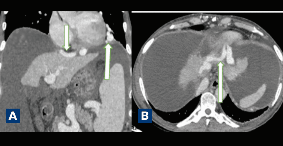
Figure 1: Triphasic computed tomography (CT) scan of the abdomen (portal venous phase): A . Coronal reconstruction demonstrating a large tortuous spontaneous porto-systemic shunt arising from the intrahepatic portion of the left portal vein, coursing superiorly along the left coronary ligament, traversing the left hemidiaphragm, and extending into the pericardial venous system; B. Axial image in the portal venous phase depicting the same collateral vessel extending cranially from the left portal vein towards the pericardial venous plexus, consistent with a transdiaphragmatic porto-systemic shunt.
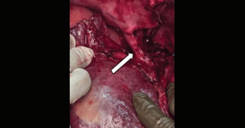
Figure 2: Intraoperative photograph demonstrating the spontaneous porto-systemic shunt following meticulous dissection and separation from the left lobe of the liver and the left hemidiaphragm during recipient hepatectomy.
The patient subsequently underwent LDLT. During recipient hepatectomy, careful exploration of the suprahepatic and left diaphragmatic region revealed a large tortuous collateral vessel corresponding to the shunt identified on CT imaging. Meticulous dissection was performed to separate the shunt from the left lobe of the cirrhotic liver and the left hemidiaphragm (Figure 2).
Given the potential haemodynamic role of this collateral in decompressing the portal venous system, the shunt was intentionally preserved during the initial stages of the hepatectomy. Maintaining the shunt patency allowed continued decompression of the portal circulation during mobilisation of the cirrhotic liver and prevented a sudden rise in portal pressure. Once the major dissection required for explant hepatectomy was completed, the shunt was ligated immediately prior to ligation of the middle hepatic vein–left hepatic vein (MHV–LHV) confluence. This approach enabled controlled interruption of the collateral while minimising the risk of portal hypertension-related bleeding during the earlier phases of dissection. The intraoperative findings confirmed the presence of a large SPSS consistent with the preoperative radiological description.
Discussion
SPSS develop as a compensatory response to sustained portal hypertension and represent structural remodelling of the splanchnic venous circulation. Their presence reflects progressive haemodynamic adaptation in cirrhosis, allowing diversion of portal venous blood into systemic veins. Radiological studies have demonstrated that SPSS are common in advanced liver disease and increase in prevalence and size with worsening portal hypertension and hepatic dysfunction.
Most SPSS follow predictable anatomical routes, including splenorenal, gastrorenal, paraumbilical, and mesocaval pathways. However, unusual collateral routes may occasionally develop through less common venous communications. One such rare pathway involves transdiaphragmatic porto-systemic shunting into thoracic venous channels. The diaphragm contains an extensive venous network composed of the inferior phrenic, pericardiacophrenic, internal thoracic, and intercostal veins. These vessels form potential anastomotic channels between the abdominal and thoracic venous systems and may enlarge under conditions of elevated portal pressure. A transdiaphragmatic porto-systemic shunt draining into the pericardiacophrenic vein was first described radiologically by Minami and colleagues. In their report, the collateral pathway originated from abdominal portal venous tributaries and passed through the inferior phrenic vein, traversing the diaphragm before draining into the pericardiacophrenic vein within the thorax. The authors emphasised the rarity of this collateral pathway and noted that only one similar case had been previously described in the literature. The pericardiacophrenic vein may communicate with the internal thoracic and azygos venous systems, thereby providing an alternative route for portal decompression in patients with portal hypertension.
The anatomical basis for this pathway lies in the venous drainage pattern of the diaphragm. The inferior phrenic veins typically drain directly into the inferior vena cava or into the left renal vein, often via the left adrenal vein. However, they may form anastomoses with the pericardiacophrenic and mediastinal venous systems, creating a potential collateral route between the portal and systemic circulations. Despite these anatomical connections, transdiaphragmatic portosystemic shunts remain extremely uncommon, likely because the inferior phrenic veins most frequently terminate directly into the inferior vena cava or hepatic veins rather than forming large mediastinal communications.
Compared with previously described cases, the present case demonstrates a distinct anatomical origin of the shunt directly from the intrahepatic left portal vein, extending along the left coronary ligament, traversing the left hemidiaphragm, and draining into the pericardial venous system. In contrast, the shunts described by Minami et al. predominantly arose from extrahepatic portal tributaries such as the splenic, gastric, or adrenal venous systems. This difference highlights a rare intrahepatic origin of a transdiaphragmatic portosystemic collateral. Recognition of atypical SPSS is particularly important in the setting of liver transplantation. Large collateral vessels may serve as major decompressive pathways, and abrupt interruption during dissection can result in sudden increases in portal pressure and bleeding from portal venous tributaries. Furthermore, persistent large SPSS after transplantation may divert portal inflow away from the graft, leading to the portal steal phenomenon, which can compromise graft perfusion and function.
In the present case, preoperative triphasic CT imaging allowed accurate delineation of the unusual collateral pathway, which facilitated careful intraoperative planning. The shunt was preserved during the initial stages of the hepatectomy to maintain portal decompression and was ligated only after completion of the major dissection, immediately prior to division of the middle and left hepatic vein confluence. This strategy allowed safe completion of the recipient hepatectomy while avoiding sudden portal hypertension or catastrophic bleeding during dissection.
Overall, this case highlights the importance of meticulous evaluation of cross-sectional imaging in transplant candidates, as recognition of rare collateral pathways may influence operative strategy and intraoperative haemodynamic management.
Declarations
Ethical approval Ethical approval was not required for this case report according to institutional guidelines. The study was conducted in accordance with the principles of the Declaration of Helsinki.
Funding The authors received no financial support for the research, authorship, or publication of this article.
Authors’ contributions All authors contributed to the clinical management of the patient and preparation of the manuscript. All authors reviewed and approved the final manuscript.
Conclusion
This case highlights a rare transdiaphragmatic SPSS originating from the intrahepatic left portal vein and draining into the pericardiacophrenic venous system in a patient with decompensated cirrhosis undergoing LDLT. Recognition of such unusual collateral pathways on preoperative cross-sectional imaging is crucial for operative planning, as these shunts may serve as significant decompressive channels in portal hypertension.Strategic intraoperative management, including preservation of the shunt during early dissection and controlled ligation at an appropriate stage of hepatectomy, can help maintain portal haemodynamic stability and facilitate safe explantation. Awareness of these rare vascular variants is important for transplant surgeons and radiologists involved in the management of advanced portal hypertension.
Soumyadip Sain, Karisangal Ramaswamy Vasudevan, Piyush Kumar Sinha, Rohit Saini, Manmohan Singh Rajput, Saurabh Kumar. An Unusual Spontaneous Porto-Systemic Shunt Between the Left Portal Vein and Pericardial Venous System in Decompensated Chronic Liver Disease: Radiological and Intraoperative Correlation. MMJ. 2026, June. Vol 3 (2).
DOI: XXXX_XXXX_XXXX_XXXX
References
- Juncu S, Minea H, Girleanu I, et al. Clinical implications and management of spontaneous portosystemic shunts in liver cirrhosis. Diagnostics. 2024;14(13):1372.
- Philips CA, Rajesh S, Augustine P, et al. Portosystemic shunts and refractory hepatic encephalopathy: patient selection and current options. Hepat Med. 2019;11:23–34.
- Simón-Talero M, Roccarina D, Martínez J, et al. Association between spontaneous portosystemic shunts and complications of cirrhosis. Gastroenterology. 2018;154:1694–705.
- Dajti E, Renzulli M, Marasco G, et al. Spontaneous portosystemic shunts in cirrhosis: Clinical and radiological relevance. Dig Liver Dis. 2022;54:1205–14.
- Minami M, Kawauchi N, Itai Y, et al. Transdiaphragmatic portosystemic shunt to the pericardiacophrenic vein. AJR Am J Roentgenol. 1993;161(3):569–71.
- Praktiknjo M, Simón-Talero M, Römer J, et al. Total area of spontaneous portosystemic shunts independently predicts complications of liver cirrhosis. Hepatology. 2020;72:675–86.
- Nardelli S, Riggio O, Gioia S, et al. Spontaneous portosystemic shunts in liver cirrhosis: Clinical and therapeutical aspects. World J Gastroenterol. 2020;26(15):1726–32.