 WhatsApp Us (24/7)
 +91 926 888 0303 (24/7)


Abstract:
This report describes a rare and life-threatening presentation of bilateral vertical sacral fractures in a 54-year-old hypertensive female following a high-energy fall from a second-floor balcony. Such fractures are uncommon and inherently unstable, often associated with pelvic ring disruption, neurovascular compromise, and haemodynamic instability. The patient presented to the emergency department (ED) in a drowsy state with a Glasgow Coma Scale (GCS) score of 12, hypotension, tachycardia, and bilateral lower limb weakness, indicative of polytrauma involving the neurological and musculoskeletal systems. Initial management followed a systematic trauma approach, including prompt resuscitation with intravenous (IV) fluids, administration of tranexamic acid, continuous monitoring, and early application of a pelvic binder based on clinical suspicion of an unstable pelvic fracture. Adjunct investigations, including an electrocardiogram, arterial blood gas analysis, cross-matching, and bedside extended Focused Assessment with Sonography in Trauma (e-FAST), aided in ruling out immediate life-threatening conditions. Comprehensive imaging with computed tomography (CT) polytrauma protocol and X-rays revealed an acute subdural haematoma, bilateral vertical sacral fractures with pelvic ring disruption, retroperitoneal haemorrhage, and a right calcaneal fracture. The patient underwent multidisciplinary management involving emergency medicine, anaesthesia, critical care, spine surgery, and orthopaedics. Surgical interventions included L4–S2 lumbopelvic fixation, L5–S1 laminectomy for an S1 nerve root laceration, and calcaneal fixation via the sinus tarsi approach. Perioperative management included invasive haemodynamic monitoring and neuromonitoring. Postoperatively, the patient received intensive critical care support followed by structured rehabilitation, ultimately achieving recovery without residual neurological deficits. This case underscores the rarity and diagnostic challenges of bilateral vertical sacral fractures, which account for less than 1% of sacral injuries and are frequently overlooked in the presence of multiple traumatic insults. It highlights the critical role of early imaging, adherence to structured trauma protocols, and coordinated multidisciplinary care in improving outcomes in high- energy polytrauma patients.
Key words: Bilateral Vertical Sacral Fracture, High-Energy Trauma, Computed Tomography (CT) Polytrauma, Multidisciplinary Care.
Introduction
Pelvic fractures are severe injuries commonly associated with high-energy trauma and are a significant cause of morbidity and mortality worldwide. These injuries frequently result from mechanisms such as road traffic accidents, crush injuries, and falls from height, with axial loading forces playing a key role in fracture patterns. 1 Among pelvic fractures, sacral fractures occur in approximately 30%–45% of cases and are often indicative of substantial force transmission through the axial skeleton. 2 Sacral fractures are broadly classified based on morphology and neurological involvement. Vertical sacral fractures are more frequently encountered than transverse patterns and are typically associated with vertical shear injuries of the pelvic ring. 3 However, bilateral vertical sacral fractures without a transverse component are exceedingly rare, accounting for approximately 1% of all sacral fractures. 3 These fractures often disrupt the integrity of the pelvic ring, leading to mechanical instability and a high risk of associated neurovascular compromise. The clinical significance of such injuries lies in their frequent association with polytrauma, including injuries to the spine, abdomen, and head. Early identification is often challenging due to overlapping injuries and altered sensorium in trauma patients. Delayed or missed diagnosis can result in catastrophic outcomes, including exsanguination, permanent neurological deficits, and prolonged disability. 4,5
We present a rare case of bilateral vertical sacral fractures in a middle-aged female following an accidental fall from height, complicated by haemodynamic instability and bilateral lower limb neurological deficits. This case highlights the importance of early recognition, prompt imaging using computed tomography (CT) polytrauma protocols, and a multidisciplinary approach to managing such complex injuries.
Case Report
A 54-year-old female, a known case of hypertension, was brought to the emergency department (ED) via private ambulance with an alleged history of an accidental fall from a second-floor balcony. On arrival, the patient was drowsy but arousable, with altered sensorium. Her Glasgow Coma Scale (GCS) score was E3 V4 M5 (E = Eye response, V = Verbal response, M = Motor response). She was haemodynamically unstable with blood pressure (BP) of 90/60 mmHg, heart rate (HR) of 120 beats per minute, respiratory rate (RR) of 18/min, and oxygen saturation (SpO₂) of 98% on room air. The primary complaint was the inability to move both lower limbs. Clinical examination revealed features of unstable polytrauma, including a head injury, bilateral hip tenderness, right ankle deformity, and bilateral lower limb paresis.
Emergency department management
The patient was immediately connected to continuousmonitoring. Two large-bore intravenous (IV) cannulas were secured, and resuscitation was initiated with IV fluids and medications. A structured primary survey (Airway, Breathing, Circulation, Disability, Exposure – ABCDE approach) was conducted alongside resuscitation measures. Aggressive fluid resuscitation with crystalloids and initiation of a massive transfusion protocol were undertaken. Simultaneously, cervical spine stabilisation was ensured. Pelvic examination revealed tenderness and bruising. Given the presence of hypotension responsive to fluids, neurological deficits, and pelvic instability on compression testing, a highly unstable pelvic injury was suspected. A pelvic binder was applied promptly, resulting in improved haemodynamic stability and reducing the need for inotropic support. Adjunct investigations, including an electrocardiogram (ECG), arterial blood gas (ABG), blood grouping and cross-matching, were performed concurrently. A bedside extended Focused Assessment with Sonography in Trauma (e-FAST) was negative, ruling out immediate life-threatening intraperitoneal or pericardial bleeding. However, retroperitoneal haemorrhage remained a concern due to pelvic involvement. Given the altered sensorium and suspicion of intracranial injury, the patient was shifted for a CT polytrauma protocol along with X-ray imaging of the right ankle.
CT findings
Head and face: Acute subdural haemorrhage (SDH) along the right tentorium and posterior falx (1.5 mm thickness) with a linear component of subarachnoid haemorrhage (SAH) in the right temporoparietal region.
Spine and pelvis: Bilateral vertical sacral fractures, disruption of the pelvic ring, undisplaced fractures of the bilateral transverse processes of the L5 vertebra, a comminuted sacral fracture with piriformis muscle oedema, mildly displaced fractures of the right superior and inferior pubic rami and a right ischial fracture (Figure 1 and 2).
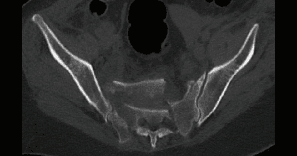
Figure 1: (Preoperative) Axial computed tomography (CT) imageof the pelvis demonstrating bilateral vertically oriented fractures through the sacral alae with an associated transverse fracture component involving the sacral body. The fracture configuration is consistent with a U-shaped (H-type) sacral fracture pattern, indicative of spinopelvic instability, with clear cortical disruption extending across both sacral wings into the central sacrum.
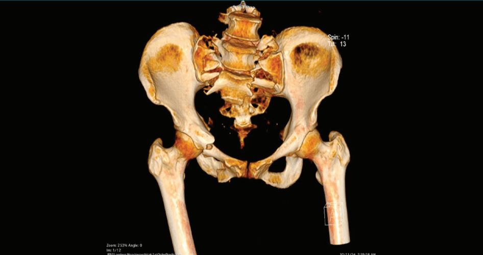
Figure 2: (Preoperative) Three-dimensional (3D) volumerendered computed tomography (CT) image of the pelvis demonstrating findings as described in Figure 1. The 3D reconstruction clearly delineates the extent of cortical disruption and fracture morphology involving the sacrum in relation to the pelvic ring.
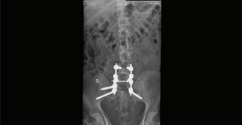
Figure 3: Postoperative anteroposterior radiograph of the lumbosacral spine and pelvis demonstrating posterior spinopelvic stabilisation. Bilateral pedicle screws are seen in the lower lumbar spine with extension into the sacrum and iliac bones, connected by longitudinal rods and a transverse connector, consistent with lumbopelvic fixation. The construct provides stabilisation of the previously identified U-shaped sacral fracture, with satisfactory alignment and appropriate positioning of the fixation construct.
Chest: No abnormalities.
Abdomen: Retroperitoneal haemorrhage with fat stranding; no visceral injury.
Right ankle X-ray: Displaced fracture of the calcaneum.
Definitive management
Following stabilisation, the patient was transferred to the Intensive Care Unit (ICU). A multidisciplinary team involving emergency medicine, orthopaedics, spine surgery, neurosurgery, anaesthesiology, and critical care was engaged. After ruling out cervical spine injury, central venous pressure (CVP) monitoring and arterial line placement were performed.
Surgical interventions
- 1. Lumbopelvic fixation (L4–S2) with L5–S1 laminectomy in view of neurological deficit, epidural haematoma, and S1 nerve root injury (Figure 3).
- 2. Right calcaneal fracture management: Initial debridement and stabilisation followed by definitive fixation using a sinus tarsi approach with K-wires and a lateral plate.
Postoperative course
Postoperative care included haemodynamic and neurological monitoring, pain management, psychological evaluation and counselling, and early physiotherapy and rehabilitation. The patient demonstrated gradual neurological recovery and was shifted to the ward. She was discharged with advice on wound care, medications, and rehabilitation. On follow-up, the patient showed complete neurological recovery with no residual disability.
Discussion
Sacral fractures are a critical subset of pelvic injuries, occurring in up to 45% of pelvic ring disruptions.2 These fractures are often associated with high-energy trauma and carry a significant risk of morbidity due to associated injuries and neurological involvement.6 Vertical sacral fractures are commonly linked to vertical shear mechanisms and frequently result in instability of the pelvic ring.1 However, bilateral vertical sacral fractures without transverse components are rare and represent a highly unstable injury pattern.3
Clinical challenges
These injuries are frequently underdiagnosed during initial trauma evaluation due to altered sensorium, the presence of multiple distracting injuries, and limitations of plain radiography. Delayed diagnosis can result in uncontrolled haemorrhage and irreversible neurological damage.4
Role of imaging
CT polytrauma protocols with three-dimensional reconstruction are now considered the gold standard for evaluating complex pelvic injuries. They allow accurate fracture classification, detection of associated injuries, and preoperative planning. This approach is significantly more efficient compared to sequential imaging modalities, especially in unstable patients.5
Management principles
Management of unstable pelvic fractures involves early resuscitation, following Advanced Trauma Life Support (ATLS) guidelines (ACS, 2018)7 pelvic binder application to reduce haemorrhage, definitive stabilisation with lumbopelvic fixation to provide mechanical stability8,9, and early surgical fixation to reduce complications. Neurological decompression may improve neurological outcomes in selected cases.10
Multidisciplinary care
Coordination between specialities is essential for optimal outcomes. Outcome depends on the timelines of intervention, severity of associated injuries, and the presence of neurological deficits.
This case demonstrates that early recognition, prompt stabilisation, and multidisciplinary intervention can result in excellent functional recovery, even in severe injury patterns.
Conclusion
This case highlights a rare and highly unstable pattern of bilateral vertical sacral fractures in the setting of high-energy trauma. Key learning points include the importance of early recognition in the ED, the critical role of CT polytrauma imaging, the need for rapid resuscitation and pelvic stabilisation, and the value of multidisciplinary management. The favourable outcome in this case underscores the importance of integrated trauma systems, a well-equipped ED, and coordinated care pathways in improving patient survival and functional recovery.
Krittika Bhatia, Niranjani V, Paresh Bang, Manoj Pahukar. Bilateral Vertical Sacral Fracture, A Highly Unstable Pelvic Injury in a Case of Accidental Fall from Height. MMJ. 2026, June. Vol 3 (2).
DOI: XXXX_XXXX_XXXX_XXXX
References
- Tile M. Pelvic ring fractures: should they be fixed? J Bone Joint Surg Br. 1988;70(1):1–
- Denis F, Davis S, Comfort T. Sacral fractures: an important problem. Retrospective analysis of 236 cases. Clin Orthop Relat Res. 1988;227:67–
- Bishop JA, Dangelmajer S, Corcoran-Schwartz I, et al. Sacral fractures: epidemiology and classification. J Orthop Trauma. 2010;24(8):e78–e
- Routt ML Jr, Simonian PT, Ballmer F. A rational approach to pelvic trauma: resuscitation and early definitive stabilization. Clin Orthop Relat Res. 1995;(318):61–
- Manson TT, Nascone JW, Sciadini MF, et al. Pelvic ring fractures and associated injuries in polytrauma patients. Injury. 2010;41(4):379–
- Starr AJ, Griffin DR, Reinert CM, et al. Pelvic ring disruptions: prediction of associated injuries and mortality. J Orthop Trauma. 2002;16(8):553–
- American College of Surgeons Committee on Trauma. Advanced Trauma Life Support (ATLS) Student Course Manual. 10th Edition. Chicago: American College of Surgeons; 2018.
- Keating JF, Werier J, Blachut P, et al. Early fixation of the vertically unstable pelvis: the role of lumbopelvic fixation. J Orthop Trauma. 1999;13(2):107–
- Käch K, Trentz O. Distraction spondylodesis of the lumbar spine in the treatment of sacral fractures. Spine (Phila Pa 1976). 1994;19(21):2425–
- Zelle BA, Gruen GS, Brown S, et al. Sacral fractures with neurological injury: is early decompression beneficial? Spine (Phila Pa 1976). 2004;29(17):E397–E404.