 WhatsApp Us (24/7)
 +91 926 888 0303 (24/7)


Abstract:
Hepatic artery pseudoaneurysm (HAP) is an uncommon but potentially fatal vascular complication of blunt abdominal trauma. It results from disruption of the arterial wall following traumatic injury and may present in a delayed manner, making diagnosis challenging in emergency settings. Delayed presentation is associated with a significant risk of rupture, haemorrhage, and haemodynamic deterioration. Early recognition with appropriate imaging and timely intervention are therefore essential to reduce morbidity and mortality. We report a case of a middle-aged male with a known history of hypertension and chronic kidney disease and was receiving Ayurvedic management, who presented to the emergency department (ED) weeks after a skid-related fall from a two-wheeler. The patient complained of right-sided chest pain, left upper abdominal pain, shortness of breath, and generalised weakness. Initial evaluation revealed severe anaemia, hyperkalaemia, acute kidney injury, and features characteristic of rhabdomyolysis. Contrast-enhanced computed tomography (CECT) polytrauma imaging demonstrated a Grade III liver laceration with active intraparenchymal bleeding and HAP, along with moderate haemoperitoneum, rightsided haemothorax, and multiple rib fractures. The patient underwent successful selective catheterisation and glue embolisation of the right HAP. Supportive management included transfusion of three units of leucodepleted packed red blood cells and one session of haemodialysis, resulting in clinical and biochemical improvement. This case was reported due to the rarity of delayed post-traumatic HAP and the diagnostic challenge associated with its presentation. It highlights the importance of maintaining a high index of suspicion and emphasises the role of multidisciplinary management in a tertiary care setting in achieving favourable outcomes.
Key words: Hepatic Artery Pseudoaneurysm, Blunt Abdominal Trauma, Delayed Presentation, Endovascular Embolisation, Polytrauma.
Introduction
Hepatic artery pseudoaneurysm (HAP) is a rare but potentially life-threatening vascular complication resulting from disruption of the hepatic arterial wall. Unlike true aneurysms, pseudoaneurysms do not involve all three layers of the vessel wall and are instead contained by the surrounding soft tissue or adventitia. HAPs account for a small proportion of visceral artery pseudoaneurysms but carry a significantly high risk of rupture, with mortality rates reported to be as high as 20%–40% in untreated cases.
The most common aetiologies of HAP include blunt or penetrating abdominal trauma, hepatobiliary interventions, surgery, pancreatitis, and iatrogenic vascular injury. 3 Among these, traumatic HAP following blunt abdominal trauma remains relatively uncommon and may present days to weeks after the initial injury. Delayed presentation occurs due to the gradual expansion of the injured arterial segment before clinical manifestation, making diagnosis particularly challenging in emergency settings. Patients may present with non-specific symptoms such as abdominal pain, anaemia, jaundice, gastrointestinal bleeding, or haemodynamic instability secondary to rupture. In some cases, the condition may remain clinically silent until a catastrophic haemorrhage occurs.
Contrast-enhanced computed tomography (CECT) angiography has emerged as the imaging modality of choice for diagnosis, allowing rapid identification of associated solid organ injury and vascular abnormalities. Endovascular intervention, including coil or glue embolisation, is currently considered the preferred treatment modality due to its minimally invasive nature, high technical success rate, and lower morbidity compared with open surgical repair.
We report a rare case of delayed post-traumatic HAP in a patient presenting weeks after blunt abdominal trauma with associated liver laceration, haemoperitoneum, severe anaemia, and metabolic derangements. This case is reported because of its delayed presentation, associated diagnostic challenge, and the successful multidisciplinary management in a tertiary care emergency setting.
Case Report
On arrival to the emergency department (ED), the patient was conscious and oriented to time, place, and person with a Glasgow Coma Scale (GCS) score of E4V5M6 (E = Eye response, V = Verbal response, M = Motor response), although he appeared acutely ill. He had a blood pressure (BP) of 170/100 mmHg, heart rate (HR) of 105 beats per minute, respiratory rate (RR) of 24 breaths/min, and oxygen saturation of 90%, while receiving oxygen at 3 L/min via nasal prongs. Respiratory system examination revealed bilaterally reduced air entry. Cardiovascular examination demonstrated normal heart sounds (S1 and S2). Abdominal examination revealed a soft but distended abdomen with generalised tenderness, while central nervous system examination was unremarkable.
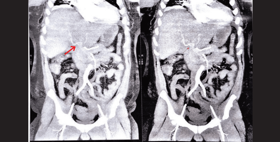
Figure 1: Contrast-enhanced computed tomography (CECT) abdomen demonstrating Grade III liver laceration with hepatic artery pseudoaneurysm (red arrow) and active intraparenchymal contrast extravasation.
A CECT polytrauma scan demonstrated a Grade III liver laceration with active intraparenchymal bleeding and features suggestive of a HAP, associated with moderate haemoperitoneum (Figure 1). Additional findings included right-sided haemothorax with multiple right-sided rib fractures (Figure 2).
Initial laboratory investigations revealed severe anaemia with a haemoglobin level of 5.5 g/dL. Renal function tests were markedly deranged, with serum creatinine elevated to 16.9 mg/dL. Significant electrolyte abnormalities were noted, including hyponatraemia with serum sodium of 126.9 mEq/L and severe hyperkalaemia with serum potassium of 7.4 mEq/L. The overall clinical and biochemical profile was suggestive of advanced chronic kidney disease with superimposed acute kidney injury, associated hyperkalaemia, and rhabdomyolysis in the setting of trauma.
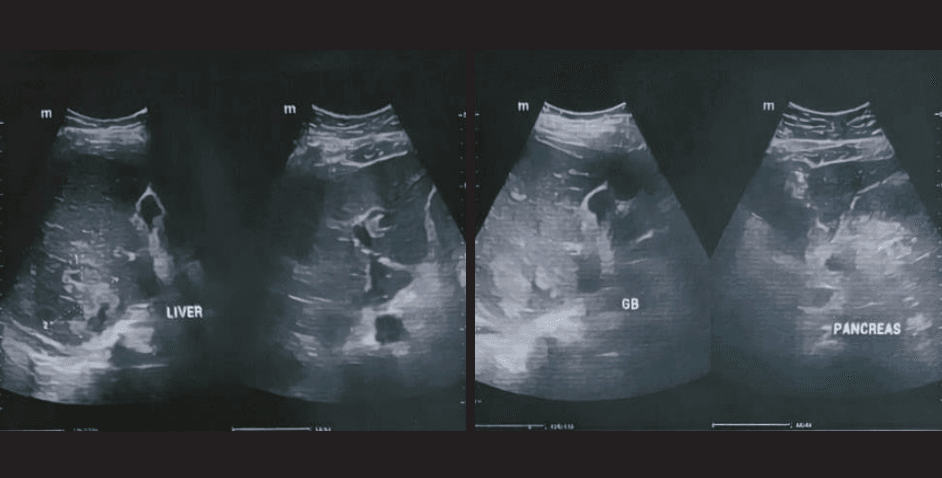
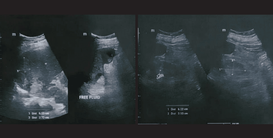
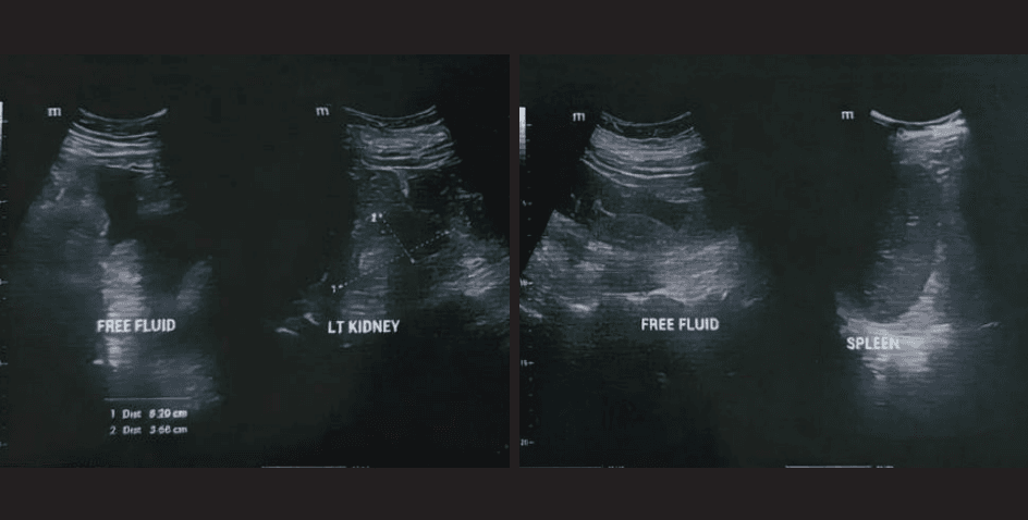
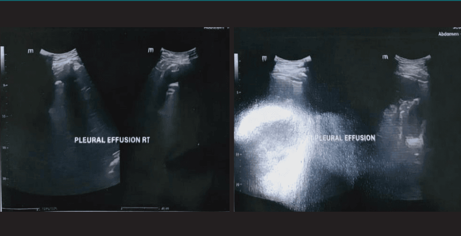
Figure 2: Ultrasonography (USG) of the whole abdomen — Focused assessment with sonography in trauma (FAST) revealed mild free fluid in the pelvis, bilateral paracolic gutters and perihepatic space with internal echoes suggestive of haemoperitoneum. Right-sided pleural effusion with minimal left-sided pleural effusion and features demonstrating posttraumatic liver contusion were also noted.
Management
The patient underwent selective catheterisation of the right hepatic artery with 30% glue embolisation, successfully excluding the pseudoaneurysm. Post-embolisation, the patient received three units of leucodepleted packed red blood cells, resulting in an improvement in haemoglobin to 10 g/dL. Due to a persistent electrolyte imbalance, one session of haemodialysis was performed, after which serum potassium decreased to 5.8 mEq/L and serum creatinine decreased from 16.9 mg/dL to 13 mg/dL. The patient was managed in the Intensive Care Unit (ICU) with close monitoring and supportive care. He remained haemodynamically stable and was breathing comfortably on room air throughout the hospital course.
Discussion
Post-traumatic HAP is a rare but serious complication of blunt abdominal trauma. Delayed presentation may lead to life-threatening haemorrhage if not recognised early. CECT imaging plays a pivotal role in diagnosis, while endovascular embolisation has emerged as the treatment of choice due to its high technical success rate and minimally invasive nature. Management is further complicated in patients with underlying chronic kidney disease, often necessitating renal replacement therapy.
Patient Consent
Written informed consent was obtained from the patient for publication of this case report and accompanying images.
Conclusion
Delayed presentation of HAP following blunt abdominal trauma requires a high index of suspicion. Successful management depends on early imaging, prompt endovascular intervention, and a multidisciplinary approach in a tertiary care setting. Timely referral to an appropriate tertiary care centre plays a crucial role in achieving favourable outcomes.
Nishit Mashru, Aaditya Yadav, Chintala Sai Venkata Chiranjeevi, Dheeraj Bhaskaran Nair. Delayed Presentation of Post-Traumatic Hepatic Artery Pseudoaneurysm Following Blunt Abdominal Injury. MMJ. 2026, June. Vol 3 (2).
DOI: XXXX-XXXX
References
- Abbas MA, Fowl RJ, Stone WM, et al. Hepatic artery aneurysm: factors that predict complications. J Vasc Surg. 2003;38:41–5.
- Stanley JC, Wakefield TW, Graham LM, et al. Clinical importance and management of splanchnic artery aneurysms. J Vasc Surg. 1986;3:836–40.
- Belli AM, Markose G, Morgan R. Interventional radiology in the management of hepatic artery pseudo aneurysms. Cardiovasc Intervent Radiol. 2012;35:789–98.
- Gaba RC, Couture PM, Lakhoo J, et al. Endovascular management of visceral artery pseudoaneurysms. J Vasc Interv Radiol. 2013;24:1386–92.
- Sarin S, Kumar A, Choudhury A. Management of visceral artery pseudo aneurysms: a single-center experience. Indian J Radiol Imaging. 2015;25:303–8.