 WhatsApp Us (24/7)
 +91 926 888 0303 (24/7)


Abstract:
Bifid renal collecting system is one of the most common congenital abnormalities. Management of renal calculi in patients with such an abnormality is complex. We hereby describe a patient with a partially duplex collecting system presenting with a renal calculus in each moiety. The patient underwent multipuncture tubeless percutaneous nephrolithotomy (PCNL) by taking punctures to access the stone in each moiety, followed by antegrade double-J stent insertion.
Key words: Bifid, Multipuncture, Percutaneous Nephrolithotomy (PCNL).
Introduction
Since the turn of the century, the management of patients with urolithiasis has evolved dramatically, with multiple treatment options. These include endourological treatments as well as percutaneous procedures. The choice of treatment depends upon technical considerations as well as patient choice. Congenital renal tract abnormalities may further complicate this decision. Duplex collecting systems are among the most common congenital renal abnormalities, with an incidence of 0.8%.1 This abnormality arises during the fourth week of gestation, when the ureteric bud, the precursor to the ureter, duplicates or splits.2 The duplex system may be complete, wherein two collecting systems drain via two independent ureters into the bladder or elsewhere, or partial, in which the two ureters fuse prior to their entry into the bladder. Though patients with duplex kidneys are more likely to be affected by vesicoureteric reflux and pelvic-ureteric junction (PUJ) obstruction, they often remain asymptomatic, with the abnormality being diagnosed incidentally due to other presentations such as urolithiasis. Retrograde intrarenal surgery, percutaneous nephrolithotomy (PCNL), and other modalities can be options for tackling renal calculi in duplicated systems. 3,4 We report the management of symptomatic urolithiasis in a partial duplex renal system with multipuncture access percutaneously, i.e. multipuncture PCNL.
Case Report
A 41-year-old male with a history of right urolithiasis treated with ureterorenoscopic lithotripsy (URSL) five years previously, presented with new onset intermittent right flank pain without signs of sepsis. On clinical examination, he was exquisitely tender at the right renal angle. Blood tests revealed no abnormality in renal function. The patient underwent a computed tomography (CT) urogram as the first-line imaging modality, which showed a 12 × 9 × 15 mm right renal calculus with 1560 HU in the PUJ and an inferior pole calyceal non-obstructive calculus of size 8 × 5 × 6 mm with attenuation value of 1390 HU, along with a partially duplicated renal system with both calyceal systems uniting at the PUJ to form a single ureter (Figures 1 and 2).
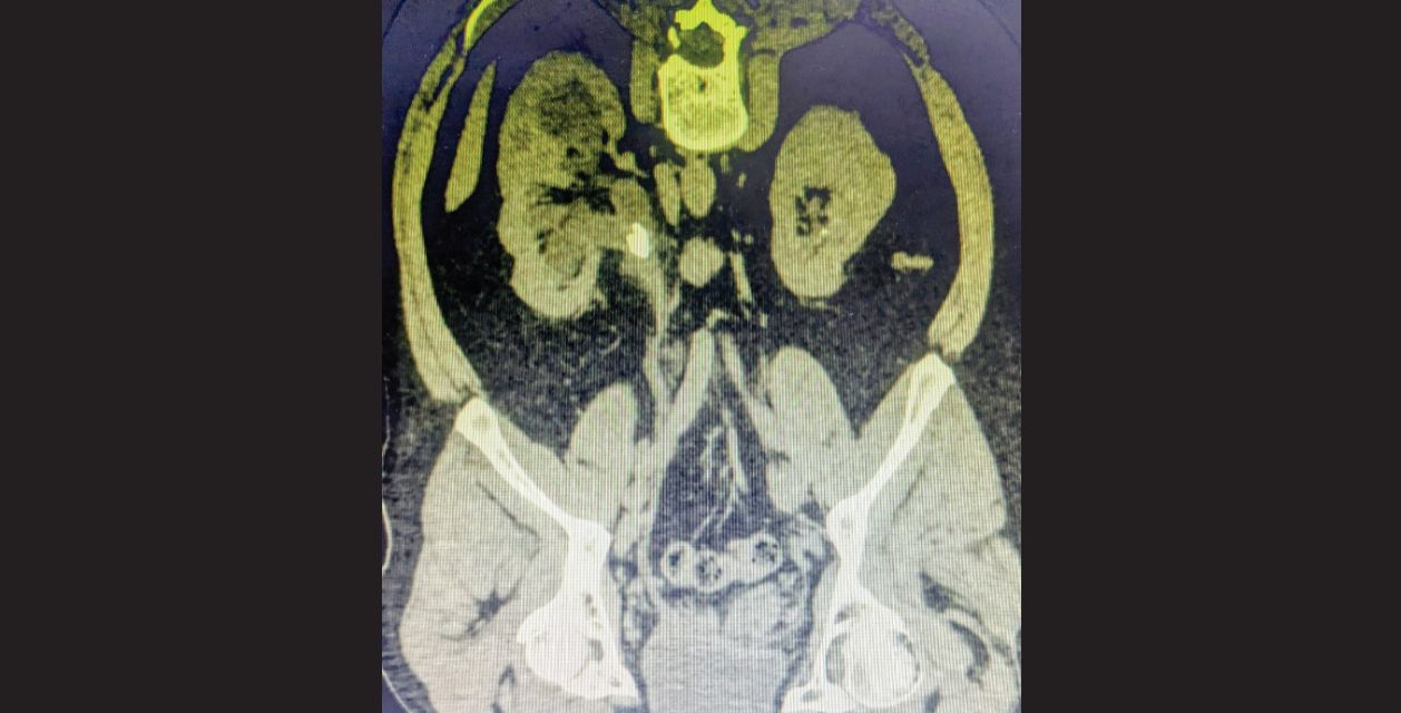
Figure 1: Reconstructed computed tomography (CT) cuts showing right-sided pelvic-ureteric junction (PUJ) calculus.
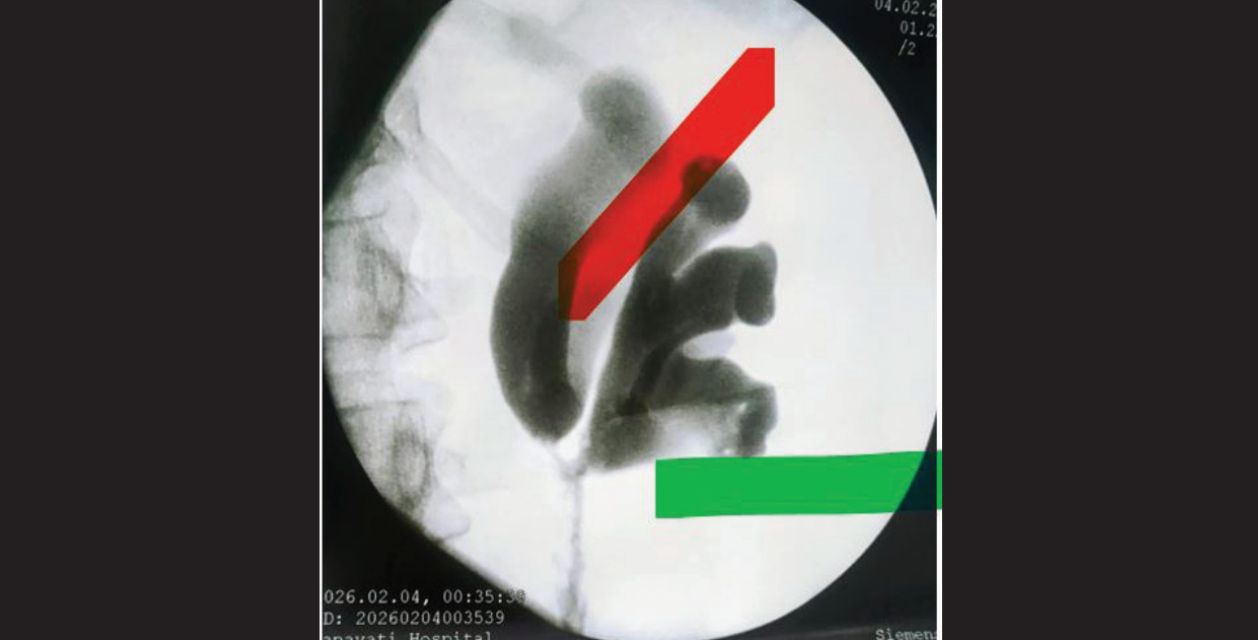
Figure 2: Intraoperative right-sided retrograde pyelogram suggestive of a partially duplex collecting system joining at the pelvic-ureteric junction (PUJ).
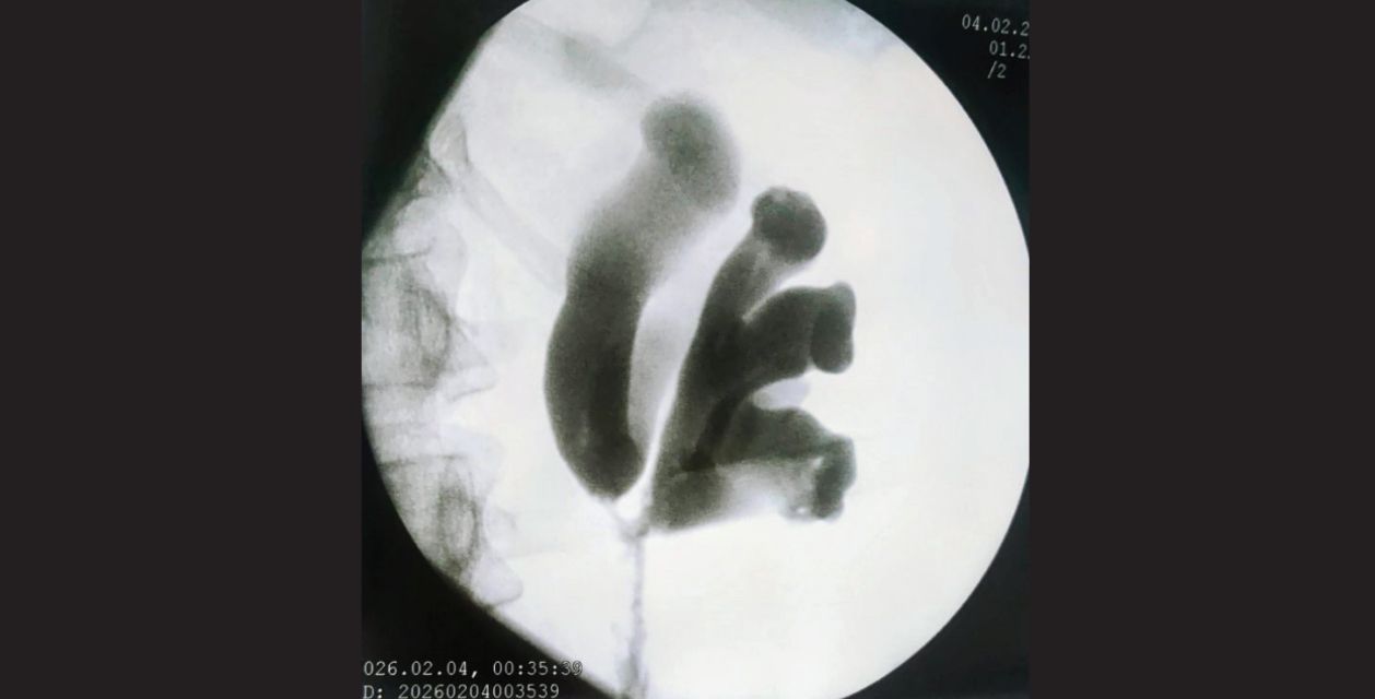
Figure 3: Schematic markings over retrograde pyelogram to show the multipuncture accesses taken. Green marker: Inferior calyceal/moiety access; Red marker: Superior calyceal/moiety access.
A second superior access to the superior moiety was required as a fragment of the PUJ calculus entered into it during lithotripsy. Hence, two separate accesses were gained by taking puncture into each moiety and subjecting each stone to lithotripsy separately (Figure 3). An antegrade double-J (DJ) stent was left in the lower moiety, as it had greater dilatation and was expected to have difficulty in drainage compared to the superior, straighter moiety. No nephrostomy tube was left, thus concluding multipuncture tubeless percutaneous nephrolithotomy (PCNL). The patient recovered well following this procedure and was discharged on the second day without complications after the Foley catheter removal. Three weeks later, the DJ stent was removed after confirming no residual radio-opaque calculi in a follow-up CT kidney, ureter, and bladder (KUB) plain.
Discussion
Non-contrast CT KUB is considered to be the gold standard imaging modality for calculi. Where a duplex collecting system is affected by renal stones, a CT intravenous urogram (IVU) should be performed, specifically to delineate the correct altered anatomy. 5,6 This more readily identifies duplex ureters and may visualise the level of ureteric fusion, further aiding endourological management. This also ensures that the operator is not misled by an apparently normal ureteroscopy (URS), which may have occurred in this case had we not known to seek an additional ureteric orifice. Endourological management of ureteric and renal calculi, including shockwave lithotripsy, URSL and PCNL, is now well established. However, new methods for applying these technologies in the management of complex renal calculi are emerging. We describe a case where selective puncture of a particular renal moiety was performed with percutaneous nephrostomy and antegrade stent insertion. This technique may prove useful where attempts to identify a calculus in a duplex system with URS alone have failed. This case also highlights the importance of the relationship between urology and radiology in the planning and management of such complex cases.
Declarations
Informed consent: Verbal informed consent was obtained from the patient.
Conflict of interests: The author(s) declare(s) that there is no conflict of interest.
Conclusion
Management of renal calculi in partially duplex collecting systems presents unique anatomical and technical challenges. This case demonstrates that multipuncture tubeless PCNL is a safe and effective approach for achieving complete stone clearance in such complex scenarios. Preoperative imaging to delineate anatomy and careful intraoperative planning are essential for successful outcomes. This technique may serve as a valuable alternative when conventional endourological approaches are insufficient.
Sanjay S. Nabar, Sachin V. Pahade, Vrushabh B. Petkar. Bifid Renal Pelvis in a Single
Kidney: A Different Approach to Managing Renal Calculi in a Partially Duplex Collecting
System — A Case Report. MMJ. 2026, June. Vol 3 (2).
References
- 1. Malek RS, Kelalis PP, Stickler GB, et al. Observations on ureteral ectopy in children. J Urol. 1972;107(2):308–13.
- 2. Rehman S, Ahmed D. Embryology, Kidney, Bladder, and Ureter. [Updated 2023 Aug 8]. In: StatPearls [Internet]. Treasure Island (FL): StatPearls Publishing; 2026 Jan-. Available from: https://www.ncbi.nlm.nih.gov/books/NBK547747/. Accessed on: 10th March 2026.
- 3. Bist A, Upadhyaya M, Giri D, et al. Recurrent renal calculi in a bilateral partial duplex collecting system: a rare case managed with suction RIRS. Ann Med Surg (Lond). 2025;87(9):6206–10.
- 4. Elhadi M, Bonomaully M, Sheikh MI, et al. Two pelvises, one stone: A different approach for management of calculi in a duplex renal collecting system. Afr J Urol. 2018;24(1):34–6.
- 5. Gong H, Gao L, Dai XJ, et al. Prolonged CT Urography in duplex kidney. BMC Urol. 2016:16(1).
- 6. Gunawardena S, Ranasinghe WK, McCahy P. Beware the stone in the duplex: use of CT IVP in detecting calculi in duplex ureteric system. J Clin Urol. 2016;9(2):128–30.