 WhatsApp Us (24/7)
 +91 926 888 0303 (24/7)


Abstract:
Acute gastric volvulus and strangulated paraoesophageal hernias (PEHs) have similar clinical histories, with individuals presenting with severe epigastric abdominal pain and lower chest pain. Development of gastric ischaemia and necrosis will present with abdominal pain and signs of peritonitis. PEHs represent 5%–15% of hiatal hernias, where part or all of the stomach protrudes through the oesophageal hiatus alongside the oesophagus, classified into types II–IV based on extent. Predominantly affecting older adults over 60 years of age, risk factors include obesity, advanced age, and increased intra-abdominal pressure from coughing or straining. Many remain asymptomatic and are discovered incidentally on imaging. But symptomatic cases manifest with postprandial chest or epigastric pain, dysphagia, early satiety, regurgitation, heartburn, nausea, or shortness of breath due to compression. Complications pose significant risks such as gastric volvulus, incarceration, strangulation, bleeding ulcers, or perforation, potentially leading to acute emergencies.
Key words: Abdominal Pain, Paraoesophageal Hernia, Gastric Volvulus, Nissen’s Fundoplication, Cruroplasty.
Introduction
Gastric volvulus is an uncommon but potentially life- threatening condition characterised by abnormal rotation of the stomach, leading to luminal obstruction and possible vascular compromise. 1 Although it can occur as a primary event due to congenital or acquired laxity of the gastric ligaments, secondary gastric volvulus is more frequently associated with underlying structural abnormalities, particularly diaphragmatic defects such as paraoesophageal hernia. Among the spectrum of hiatal hernias, paraoesophageal hernias (classified as type II–IV) are distinguished by herniation of the gastric fundus alongside a normally positioned gastro-oesophageal junction or by progressive migration of additional abdominal viscera into the thoracic cavity. This anatomical distortion predisposes the stomach to abnormal mobility and torsion, creating a favourable environment for the development of gastric volvulus.
The pathophysiology of gastric volvulus in the setting of paraoesophageal hernia involves displacement of the stomach into the mediastinum, elongation and attenuation of supporting ligaments, and disruption of normal anatomic fixation. Two principal types of volvuli have been described — Organoaxial, in which rotation occurs along the stomach’s longitudinal axis connecting the gastroesophageal junction and pylorus; and Mesenteroaxial, in which rotation occurs along the transverse axis. 2 Organoaxial volvulus is more commonly associated with paraoesophageal hernias and carries a higher risk of strangulation, ischaemia, and perforation. Clinically, presentation may range from intermittent, nonspecific upper gastrointestinal (GI) symptoms to acute surgical emergencies. Chronic or intermittent volvulus may present with dysphagia, postprandial fullness, anaemia from mucosal ulceration, or respiratory symptoms due to mediastinal compression. Given the potential for rapid progression to gastric ischaemia and necrosis, timely diagnosis is critical.
Advances in imaging, particularly contrast-enhanced computed tomography and upper GI contrast studies, have significantly improved diagnostic accuracy by delineating the abnormal orientation of the stomach and identifying associated complications. Management strategies depend on the acuity of presentation and patient stability but typically involve urgent surgical intervention in acute cases, with reduction of the volvulus, assessment of gastric viability, and definitive repair of the paraoesophageal hernia to prevent recurrence.
This study presents a case of gastric volvulus occurring in the context of paraoesophageal hernia, highlighting the clinical presentation, radiologic findings, surgical management, and relevant literature. Through this case, we aimed to underscore the importance of early recognition and definitive treatment of this rare yet serious complication of paraoesophageal hernia.
Case Report
A 72-year-old gentleman presented to the emergency department with complaints of abdominal pain for seven days associated with vomiting and obstipation. On primary survey, the patient had a pulse rate of 88 beats per minute (bpm), a respiratory rate of 18 breaths per minute, and SpO 2 of 92% on room air. On abdominal examination, the abdomen was distended with epigastric tenderness on palpation.
After resuscitation, the patient underwent basic radiological investigation, which showed “obscured dome of diaphragm and costophrenic angle” on the right side. An oral positive contrast-enhanced whole abdomen with pelvis scan was done (Figure 1), which revealed a “Large hiatus hernia with gastric volvulus and distention of the stomach with collapse of the rest of the bowel. Minimal fluid and fat stranding are seen surrounding the herniated stomach with almost entire distended stomach in the chest.”
With the above evidence of hiatus hernia with gastric volvulus, the patient was planned for an emergency surgery.
During the procedure, we found the entire stomach and the contents of the hernia sac along with the greater omentum. After a careful and meticulous adhesiolysis, the content was reduced into the abdomen, along with the excision of the sac (Figure 2). Intraoperatively, an upper GI endoscopy was performed. Once the content was reduced, crural repair was done with non-absorbable Ethibond suture (Figure 3), and a composite mesh was placed over the crura in an onlay fashion, secured with Prolene stitches. The lower part of the oesophagus was mobilised, and a 360-degree Nissen’s fundoplication was done in a tension-free manner (Figures 4 and 5). After fixing the stomach in anatomical position, an anterior wall gastropexy was done.
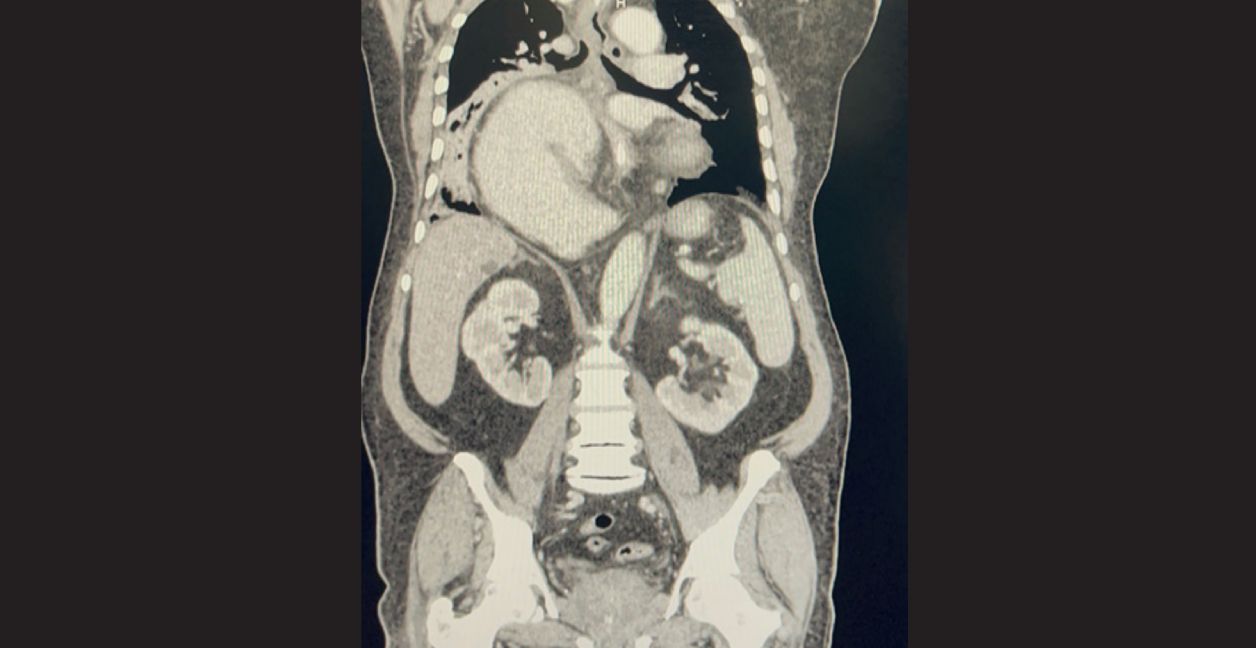
Figure 1: Gastric volvulus mimicking coffee bean appearance with the entire stomach in the right thorax, compressing the right lung.
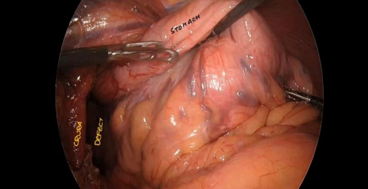
Figure 2: Reducing the content (stomach) from the sac.
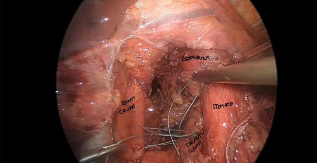
Figure 3: Repairing the crura (Cruroplasty).
Follow-up
Post-operatively, the patient was managed symptomatically, along with total parenteral nutrition, incentive spirometry and chest physiotherapy. Scheduled antiemetics were administered to prevent post-operative nausea and vomiting. Follow-up radiology scans show no abnormality
Discussion
Paraoesophageal hernias are relatively rare and typically occur in elderly patients. The various presenting symptoms are non-specific and often occur in combination. These include symptoms of gastro-oesophageal reflux disease (GERD) in 26%–70% of cases, microcytic anaemia in 17%–47%, and respiratory symptoms in 9%–59%.4 Acute complications such as gastric volvulus with incarceration or strangulation are rare (estimated incidence of 1.2% per patient per year). Before the advent of minimally invasive techniques, open surgeries via transabdominal and transthoracic entry were the indicated management of primary hiatal hernia (PHH). Since its introduction in 1992, laparoscopic PHH repair (LPHHR) has emerged as the forefront of PHH treatment.5 As most of the paraoesophageal hernias are associated with GERD symptoms, fundoplication is warranted.6 The concept of radial tension, caused by a very wide hiatus, and axial tension, caused by an apparent short oesophagus, are widely accepted among authors as the main incriminating factors in the process of recurrence after surgical repair.7 Radial tension can be managed with cruroplasty, and axial tension can be reduced by mobilising the distal part of the oesophagus. Advancements in mesh technology, including the advent of biologic and biosynthetic meshes, have contributed to a safer profile with fewer complications, better biocompatibility, and improved tissue integration.8 Studies showed that anterior gastropexy will decrease the chances of recurrences.9
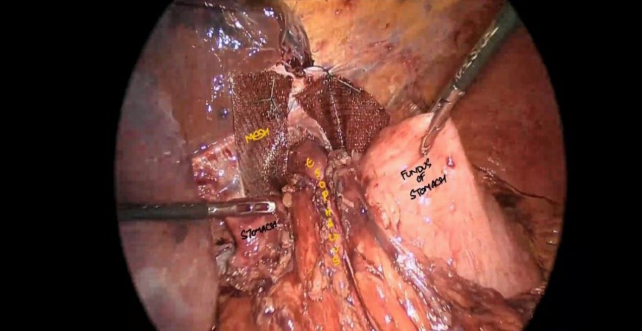
Figure 4: Performing Shoe sign manoeuvre for fundoplication, mesh is placed in situ.
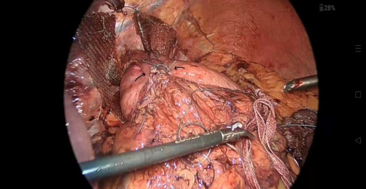
Figure 5: Nissen’s fundoplication (360-degree wrap).
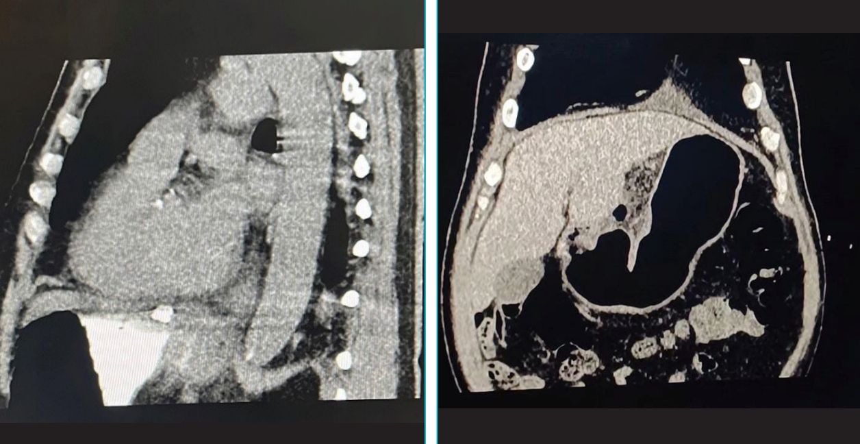
Figure 6: Post-operative oral contrast scan showing stomach below the diaphragm and contrast in wrap.
Conclusion
Gastric volvulus associated with paraoesophageal hernia is a rare but serious condition that demands prompt recognition and intervention to prevent life-threatening complications. This case underscores the importance of considering this diagnosis in elderly patients presenting with non-specific upper GI symptoms.
Early imaging and timely surgical management, including reduction of the volvulus and definitive hernia repair with fundoplication and gastropexy, are key to achieving favourable outcomes and minimising recurrence.
Vikas Panwar, Vedant Rai, Rittik Chakravorty. A Rare Case of Gastric Volvulus with
Paraoesophageal Hernia. MMJ. 2026, June. Vol 3 (2).
References
- Rashid F, Thangarajah T, Mulvey D, et al. A review article on gastric volvulus: a challenge to diagnosis and management. Int J Surg. 2010;8(1):18–24.
- Chau B, Dufel S. Gastric volvulus. Emerg Med J. 2007;24:446–7.
- Shivanand G, Seema S, Srivastava DN, et al. Gastric volvulus: acute and chronic presentation. Clin Imaging. 2003;27(4):265–8.
- Collet D, Luc G, Chiche L. Management of large para-esophageal hiatal hernias. J Visc Surg. 2013;150(6):395–402.
- Cuschieri A, Shimi S, Nathanson LK. Laparoscopic reduction, crural repair, and fundoplication of large hiatal hernia. Am J Surg. 1992;163(4):425–30.
- Solomon D, Bekhor E, Kashtan H. Paraesophageal hernia: to fundoplicate or not? Ann Transl Med. 2021;9(10):902.
- Bradley DD, Louie BE, Farivar AS, et al. Assessment and reduction of diaphragmatic tension during hiatal hernia repair. Surg Endosc. 2015;29(4):796–804.
- Lopez SZK, Gómez SG, Arenas SG, et al. Impact of mesh use in paraesophageal hernia repair: a review of short- and long-term outcomes. Int Surg J. 2025;12(7):1230–3.
- Ponsky J, Rosen M, Fanning A, et al. Anterior gastropexy may reduce the recurrence rate after laparoscopic paraesophageal hernia repair. Surg Endosc. 2003;17(7):1036–41.