 WhatsApp Us (24/7)
 +91 926 888 0303 (24/7)


Abstract: This study describes a rare case of omental implantation of gestational tissue with a mesenteric defect in an intestinal segment and large bilateral multiseptate clear ovarian cysts in a 31-year-old patient who presented 32 days after laparoscopic salpingectomy for a ruptured tubal ectopic pregnancy, manifesting with acute abdomen. The patient underwent partial omentectomy, bilateral ovarian cystectomy, and segmental intestinal resection with side-to-side anastomosis. Serial β-HCG monitoring post-partial omentectomy revealed a rapid decline, confirming successful removal of trophoblastic tissue. Histopathological examination confirmed omental pregnancy. This case underscores the importance of caution during laparoscopic procedures to prevent omental implantation, advocating for meticulous extraction of trophoblastic tissue, preferably using a tissue retrieval bag, and diligent postoperative monitoring with serial β-HCG assays to ensure complete resolution.
Key words: Ectopic Pregnancy, Omental Pregnancy
Introduction
Ectopic pregnancies are becoming more prevalent, with rates rising from 0.5% three decades ago to 1-2% in recent years. Although rare, persistent trophoblastic tissue can occur as a complication of laparoscopic salpingectomy, with only two prior cases reported in the literature. [1,2] This condition arises from the incomplete removal of gestational tissue. Omental implants following surgery to remove an ectopic pregnancy are uncommon and may pose challenges in detection, often manifesting as sudden lower abdominal pain and bleeding from implant sites, difficult to identify via ultrasound. Here, we present a unique case of persistent ectopic pregnancy in a patient with omental implants at the lateral port site, following a prior laparoscopic salpingectomy. Additionally, the patient exhibited bilateral large ovarian masses and a mesenteric defect in a segment of the small intestine. This case highlights potential complications associated with minimal access surgery for ectopic pregnancies.
Case report
A 31-year-old woman, Mrs. X, G5P0A4, with previous 4 spontaneous early abortions reported in Gynae casualty with 5 weeks of amenorrhea and severe pain in the abdomen for 1 day. She gave a history of fainting, sweating, and inability to sit for 1 day. Upon examination, she displayed tachycardia but otherwise stable vital signs. Her β-HCG level was 4303 mIU/ml, and her recent hemoglobin level, measured three days prior, was 13.2 gm%. However, upon admission, her hemoglobin had dropped significantly to 10.5 gm%. The ultrasound imaging revealed a right-sided adnexal mass with a massive hemoperitoneum. The uterus showed a thickened endometrium and no gestational sac. Based on these findings, a diagnosis of a ruptured ectopic pregnancy was made, necessitating immediate surgical intervention.
During laparoscopic surgery, approximately 1.5 liters of blood was drained from the abdomen. Left-sided (not right as diagnosed on ultrasound) ruptured tubal ectopic was seen. A left salpingectomy was done. The right tube and both ovaries were normal size and healthy. An intraabdominal drain was kept. 3 units of packed red cells were transfused. The post-operative period was smooth and the patient was discharged on Day 3 of surgery. At the time of discharge, her β-HCG level had decreased to 837.1 mIU/ml, and her hemoglobin level was 10.3 gm%.
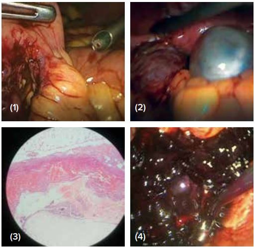
Figure: (1) Section shows omental tissue with areas of heamorrhage, chorionic villi with hydropic changes and trophoblastic proliferation (2) Omental implantaion of gestational tissue (3) Both sided ovarian cysts seen (4) Picture showing the mesentric defect
One week later, Mrs. X returned to the outpatient clinic without any notable complaints. The histopathology report confirmed the diagnosis of tubal ectopic pregnancy. During her visit, she received counseling regarding additional treatment options, particularly considering her history of recurrent pregnancy losses. She was advised to schedule a follow-up appointment in four weeks' time.
One-month post-surgery, Mrs. X presented to the emergency department with recurring pain, primarily on the right side, near the site of the previous drain port. Examination of the port sites revealed no abnormalities. She was subsequently admitted, and a complete blood count (CBC), β-HCG, and ultrasound were performed.
The ultrasound revealed bilateral well-defined multiloculated cystic lesions, measuring 72x61mm in the right ovary and a 41x40mm anechoic cyst with internal septations in the left ovary. Significant free fluid and internal debris were observed in Morrison's pouch and the pouch of Douglas. Subsequent MRI confirmed these findings, showing bilateral multiseptated ovarian cysts along with mild to moderate ascites. The CA125 level was measured at 19.3U/mL, but the origin of the elevated β-HCG could not be determined. The patient was managed conservatively with analgesics and broad-spectrum antibiotics. On day 2, the ultrasound showed slight regression in free fluid though the size of ovarian masses on both sides remained the same. The patient was symptomatically better. On day 3, though the pain was better, suspecting that the postoperative collection was getting infected the patient was taken up for surgery the same day.
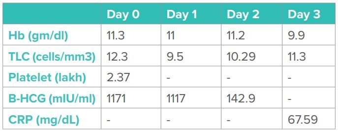
Table 1: Laboratory Parameter of the patient during the course of disease.
On Diagnostic laparoscopy
1. The omentum was pulled up & adherent towards the 10mm port on the left side lateral port (previous drain port)
2. No significant intra-abdominal fluid collection was seen
3. Bilateral thin-walled ovarian cysts were present. Right ovary 7x8cm ( 3-4 multicystic), left ovary 5x9cm with clear fluid
4. The right tube was healthy
The omentum was gently pulled down from the port side. Omental caking with hematoma was seen. Partial omentectomy was done and brought out through a tissue retrieval bag. Bilateral ovarian cystectomy was done. Both were clear cysts. While exploring, a mesenteric defect in the small intestine was detected. A surgical opinion was taken. The segment was looking dusky and devascularized so segmental resection and side to side anastomosis done.
An intraabdominal drain was kept. The postoperative period was smooth. Β-HCG on Day 2 postop dropped down to 142.9 mIU/ml. The drain was removed on Day 4 postop day and the patient was discharged on Day 5. The β-HCG on the day of discharge was 36.23 mIU/ml. The patient had an uneventful recovery and has been followed till β-HCG dropped below 10 and menses resumed. In the histopathology report, omentum showed trophoblastic proliferation with hydropic changes and products of conception. The ovarian tissue and bowel loop were unremarkable.
Discussion
The occurrence of pelvic and omental trophoblastic implantation following assisted reproduction is unprecedented. This case underscores the importance of post-operative monitoring through serial serum β-HCG measurements for ectopic pregnancies managed via laparoscopy until full resolution, as abdominal pregnancies, albeit rare, pose a significantly higher mortality risk compared to non-abdominal pregnancie.[3] Omental pregnancy represents the rarest variant of abdominal pregnancy and can be categorized as primary or secondary. Secondary omental implants are identified when there is no histological evidence of trophoblast invasion or neovascularization into the surrounding tissue.[4] It represents an uncommon complication of laparoscopic interventions for ectopic pregnancies, potentially stemming from a ruptured ectopic pregnancy, as seen in this instance. These trophoblastic implants persist in secreting β-HCG, hence resulting in elevated or stabilizing levels of β-HCG following the removal of the ectopic pregnancy.
Seventeen instances of omental ectopic pregnancies, including the primary omental pregnancy detailed by Onan et al., have been documented in the medical literature.[5] Of these cases, only two occurred subsequent to laparoscopic salpingectomy, as reported by Thatcher et al. in 1989 and Doss et al. in 1998.[1,2] In Thatcher's case, the patient experienced pain and escalating β-HCG levels following the laparoscopic removal of an early, unruptured ectopic pregnancy. A subsequent laparotomy revealed multiple pelvic trophoblastic tissue implants. In Doss's case, an unruptured ectopic pregnancy was identified during laparoscopy. The tube was excised laparoscopically, and although the patient was asymptomatic, her quantitative β-HCG levels plateaued four weeks post-surgery. An ultrasound scan suggested the presence of a right adnexal mass (later identified as a hemorrhagic corpus luteal cyst), prompting an exploratory laparotomy due to suspected residual ectopic pregnancy. Laparotomy revealed multiple peritoneal implants and extensive omental deposits. Consequently, a partial omentectomy and excision of peritoneal implants were performed without complications.
The combination of factors such as pneumoperitoneum, positive intra-abdominal pressure, and the Trendelenburg position used during laparoscopy, along with the omentum's scavenging function, may increase the risk of omental implantation following laparoscopic surgery, as noted by Pal et al. in 2003.[6] It is crucial to systematically examine the pelvis, paracolic gutters, and sub-diaphragmatic regions and to employ copious irrigation and a reverse Trendelenburg position to aid in the removal of any migrated tissue in the upper abdomen, as recommended by Pal et al. These secondary trophoblastic implants establish a new blood supply and can result in secondary hemorrhage. Patients may or may not exhibit symptoms; however, most commonly present with severe abdominal pain and intra-abdominal bleeding. Hemorrhagic shock stands as the leading cause of mortality in cases of omental pregnancy, as highlighted by Onan et al. in 2005.[5]
Omental implantation of ectopic gestational tissue following laparoscopic salpingectomy, although rare, is recognized, emphasizing the importance of thorough patient follow-up with serial β-HCG testing for a minimum of 51 days. Despite being more commonly associated with conservative surgeries like salpingostomy, studies have shown that over half of omental implantation cases occur post-salpingectomy. These implants often go undetected on ultrasound examination.
The omentum's extensive blood supply makes it an ideal site for the implantation of ectopic gestational tissue. Various precautions are suggested, including the aspiration of blood products and tissue fragments, reducing the use of the Trendelenburg position, careful removal of trophoblastic tissue, and utilizing a tissue retrieval bag.
During laparoscopic surgery, positive intra-abdominal pressure and the Trendelenburg position may contribute to the upward migration of trophoblast remnants. The omentum's scavenging action and its tendency to adhere to trocar insertion sites theoretically facilitate neovascularization and the sustenance of parasitic trophoblasts.
Careful use of suction under direct visualization is recommended, particularly in cases of hemoperitoneum, to prevent potential damage to the intestines. Bowel manipulation should be performed with caution, and a comprehensive examination for any injuries should be conducted at the conclusion of the surgery
In conclusion, this case presents a rare complication arising from the laparoscopic removal of an ectopic pregnancy. It underscores the significance of careful intraoperative measures during the laparoscopic treatment of ectopic pregnancies and prompts consideration regarding postoperative monitoring with serial HCG tests. When patients exhibit symptoms of intra-abdominal hemorrhage after ectopic pregnancy removal or tubal abortion, the possibility of secondary implantation of trophoblastic tissue should be taken into account.
CONCLUSION:
- It was concluded that the patient had a persistent ectopic pregnancy in the omentum or omental implantation It is likely that implantation of omentum occurred during salpingectomy or inadequate removal of gestational tissue especially in the presence of massive hemoperitoneum following tubal rupture.
- The mesenteric defect in the intestinal segment could not be explained. It was an old lesion and the mesenteric segment could have got weakened while pulled up alongside the omental ectopic tissue. Trauma during the first surgery was another theoretical possibility that couldn’t be ruled out.
- The bilateral ovarian cysts could represent continuous stimulation to the ovaries in view of persistent trophoblastic activity.
References
- Doss BJ, Jacques SM, Qureshi F et al. Extratubal secondary trophoblastic implants: clinicopathologic correlation and review of the literature. Human Pathology 1998;29:184–187.
- Thatcher SS, Grainer DA, True LD et al. Pelvic trophoblastic implants after laparoscopic removal of a tubal pregnancy Obstetrics and Gynaecology 1989;74:514–515.
- Atrash HK, Friede A, Hogue CJR 1987 Abdominal pregnancy in the United States: frequency and maternal mortality. Obstetrics and Gynaecology 1987;69:333–337.
- Berghella V, Wolf SC. Does primary omental pregnancy exist? Gynaecologic and Obstetric Investigation 1996;42:133–136.
- Onan MA, Turp A.B, Saltik A et al. Primary omental pregnancy: Case report Human Reproduction 2005;20:807–809.
- Pal L, Parkash V, Rutherford TJ. Omental trophoblastic implants and hemoperitoneum after laparoscopic salpingostomy for ectopic pregnancy. Journal of Reproductive Medicine 2003;48:57–59.