 WhatsApp Us (24/7)
 +91 926 888 0303 (24/7)


Abstract: Contrast-enhanced mammography (CEM) is emerging as a viable alternative to contrast-enhanced breast MRI, effectively reducing examination cost. Intravenous iodinated contrast material is used in CEM to enhance the visualization of tumor neovascularity. After injection, imaging is performed with dual-energy digital mammography, which helps provide a low-energy image and a recombined or iodine image that depict enhancing lesions in the breast. The utility of Contrast enhanced Mammography is under study and is substituting as Poor Man MRI, easier to interpret with wider accessibility available.
Key words: Contrast Enhanced mammography, Dual Energy mammography, MRI, Neovascularity, Breast cancer
Abbreviations: Contrast-enhanced Mammography - CEM, Magnetic Resonance Imaging - MRI, Full-field Digital Mammography - FFDM, Digital Breast Tomosynthesis - DBT, Low-energy (LE), Mediolateral Oblique - MLO View, Cranial Caudal (CC) View
Introduction
Contrast-enhanced mammography (CEM) is an emerging imaging technique that helps to improve diagnostic accuracy over conventional mammography and a good alternative to contrast-enhanced breast Magnetic Resonance Imaging (MRI) and importantly reducing examination cost. Intravenous iodinated contrast materials are used in CEM to look at the visualization of malignant neovascularity. After injecting contrast material, imaging is done with dual-energy digital mammography, which provides a low-energy image and recombined or iodine image that depicts enhancing lesions in the breast. CEM has proved to increase accuracy compared with digital mammography and ultrasound (US) in women with abnormal screening mammographic findings or symptoms of breast cancer. It has also gained significance comparable with breast MRI in preoperative staging of patients with breast cancer and in monitoring response after neoadjuvant chemotherapy. There are also early encouraging results from trials evaluating CEM in the screening of women who are at an increased risk of breast cancer. Although CEM is a promising and developing tool, it slightly increases radiation dose and carries a small risk of adverse reactions to contrast materials. This review details our hospital experience in CEM technique, diagnosis and screening.
Indications for contrast enhanced mammography-
- Contrast enhanced mammography can be used for preoperative assessment of extent of local disease using CEM over MRI (Figures 1 and 2).
- CEM can be used as a screening tool in women with high and intermediate risk of breast cancer (Figure 3).
- CEM can accurately monitor response to neoadjuvant chemotherapy in patients with breast cancer surgery (Figure 4)
Cases
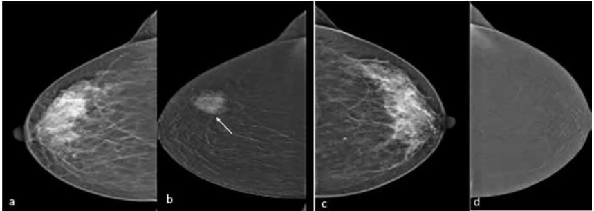
Case 1: Patient presented with suspicious right breast lump. Low dose mammogram right breast-mediolateral oblique (MLO) view (a) revealed dense breast. CE mammogram -recombined image (b) revealed complete suppression of background parenchyma and lesion with spiculated margins seen (arrow in b). Low dose mammogram left breast-MLO view (c) revealed dense breast with no enhancing lesion on CE recombined image (d).
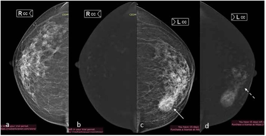
Case 2: Lump in left breast. Low dose Mammogram image of right breast craniocaudal view (CC) a. revealed dense breast with no enhancing lesion on Contrast enhanced recombined image (b). Low dose Mammogram image of left breast CC view c. showed a spiculated lesion on the inner aspect (arrow). Contrast enhanced recombined image (d) showed non-mass enhancement in segmental distribution associated with the mass (dashed arrow) revealing true extent of the disease.
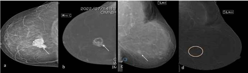
Case 3: Patient presented with bilateral breast lumps. Low dose mammogram image of right breast CC view a. revealed irregular lesion with spiculated margins in right breast (arrow). CE recombined image (b) showed heterogeneous peripheral enhancement of the lesion (arrow). Low dose mammogram image of left breast -MLO view (c) revealed a density (arrow) which did not enhance on CE recombined image (d), likely fibro glandular Parenchyma ruling out multicentricity and bilaterality.
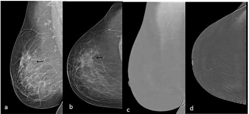
Case 4: Patient was a known case of carcinoma right breast, post neoadjuvant chemotherapy.
Low dose mammogram-MLO and CC views (a, b) revealed asymmetric density in upper outer quadrant (arrows).
CE-recombined images showed no contrast enhancement ruling out residual disease. Patient underwent mastectomy and no residual disease was seen.
Discussion
Contrast-enhanced mammography (CEM) is a developing technique that uses iodinated contrast materials for the visualization of breast tumour neovascularity in a fashion similar to MRI.1 This helps the malignant tumor to be visualised even in dense breast tissue. Full-field digital mammography (FFDM) and digital breast tomosynthesis (DBT) depend on anatomic changes in the breast caused by breast cancer. Currently, they are important in breast cancer screening and in assessment of symptomatic patients, although when done alone, they have limited accuracy and sensitivity in women with dense breasts.1,2 A more recent study3 using digital mammography showed a reduction of sensitivity with increased breast density, sensitivity of 79.9% overall, 100% for fatty breasts, 83.9% for breasts with scattered fibroglandular tissue, 72.9% for heterogeneously dense breasts, and 50% for extremely dense breasts. Although MRI can be used to better evaluate women with dense breasts and those at an intermediate risk of breast cancer, CEM is less expensive and can be accessible to a larger number of women.
In CEM, a dual-energy mammogram is acquired, around 2 minutes after the intravenous injection of iodinated contrast material. The radiologist is given - two images per breast and per view—one like a regular mammogram and the other highlighting areas of contrast uptake. CEM has the advantage of depicting both anatomic and local changes in breast perfusion, presumably caused by tumor neoangiogenesis. In a study involving 89 women with dense breasts and 100 lesions4,5, CEM in addition to mammography improved sensitivity from 71.5% to 92.7% and specificity from 51.8% to 67.9%.
The purpose of this review is to highlight its use in diagnostic and screening settings, using it as a tool over MRI in diagnosis, follow up of breast cancer cases post-chemotherapy.
A low-osmolarity iodine-based contrast material is given intravenously before image acquisition. The iodine concentration of the contrast material can vary from 300 mg/mL to 370 mg/ mL, total volume of 1.5 mL/kg of body weight (with a maximum of 150 mL) is administered. Image acquisition begins 2 minutes after contrast material injection. Standard craniocaudal and mediolateral oblique views of each breast are taken. During CEM, low-energy (LE) and high-energy images are obtained in quick succession as the breast remains compressed. Considering these settings, the low energy image may replace the FFDM image with a similar efficacy.
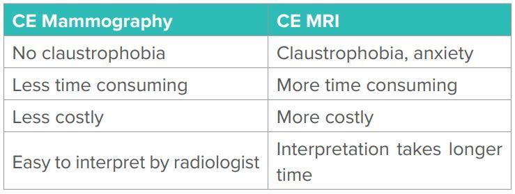
Table 1: Comparative features between CE Mammography and CE MRI.
Limitations
The most feared limitation of CEM is the possibility of developing contrast material reactions, which can be mild to severe reactions. CEM requires an increased radiation dose varying from 20% to 80% depending on system settings, breast thickness and type of mammographic device used.
CONCLUSION:
- CEM is becoming more commonly used, with an increasing number of units installed, examinations performed, and studies published. Since the advent of CEM, DBT has been increasingly used for routine mammography, where it may eventually replace two-dimensional mammography to some extent.
- CEM is easy to perform in everyday clinical practice and is useful in indications including abnormal screenings, symptomatic patients, preoperative staging of breast cancer, evaluation of response to neoadjuvant chemotherapy, screening women with dense breasts, and screening women at an increased risk of developing breast cancer. The use of CEM will further expand, especially where breast MRI availability is limited.
Acknowledgments:
None
Funding source:
None
Conflict of interest:
None
References
- Patel BK, Lobbes MBI, Lewin J. Contrast Enhanced Spectral Mammography: A Review. Semin Ultrasound CT MR 2018;39(1):70–79.
- Kuhl C. The current status of breast MR imaging. Part I. Choice of technique, image interpretation, diagnostic accuracy, and transfer to clinical practice. Radiology 2007;244(2):356–378.
- Weigel S, Heindel W, Heidrich J, Hense HW, Heidinger O. Digital mammography screening: sensitivity of the programme dependent on breast density. Eur Radiol 2017;27(7):2744–2751.
- Cheung YC, Lin YC, Wan YL, et al. Diagnostic performance of dual-energy contrast-enhanced subtracted mammography in dense breasts compared to mammography alone: interobserver blind-reading analysis. Eur Radiol 2014;24(10):2394–2403.
- Francescone MA, Jochelson MS, Dershaw DD, et al. Low energy mammogram obtained in contrast-enhanced digital mammography (CEDM) is comparable to routine full-field digital mammography (FFDM). Eur J Radiol 2014;83(8):1350– 1355.