 WhatsApp Us (24/7)
 +91 926 888 0303 (24/7)


Abstract:
Background: Background: For many years, absent or hypoplastic nasal bone during prenatal scans has been reported to be associated with increased risk of chromosomal abnormalities. We aimed to study the association of isolated absent or hypoplastic nasal bone with chromosomal abnormalities and genetic syndromes.
Methods: a retrospective observational study conducted at BLK Max foetal medicine unit, in which women with absent or hypoplastic nasal bone during second trimester scan between 16-24weeks of gestation were recruited over a period of 2 years, from June 2021 to June 2023. Nasal bone was defined as absent when no calcification was seen below the skin of the nasal bridge in mid sagittal view of facial profile, while defined as hypoplastic nasal bone when nasal bone length was <5th centile for gestation according to gestation corrected Indian charts. Absent or hypoplastic nasal bone was defined as isolated - when there was absence of other second trimester soft markers, no other structural abnormalities were seen on the ultrasound and biochemistry screen was low risk.
Results :A total of 3008 women were scanned during this period, among these 21 (0.69%) foetuses were found to have absent or hypoplastic nasal bone during second trimester scan. Thirteen (61.9%) fetuses were defined to have isolated absent (n-9) or hypoplastic (n-4) nasal bone which were included in the final analysis. Out of 13, 11 women opted for invasive testing where fluorescence in situ hybridisation (FISH) for 5 common aneuploidies and Microarray (750K) were sent on the amniotic fluid as a unit protocol, and 2 parents opted against any further genetic testing. In all tested cases, FISH and chromosomal microarray reports were reported as normal. All pregnancies were continued in our study group and had favourable foetal outcomes.
Key words: Absent Nasal Bone, Hypoplastic Nasal Bone, Second Trimester Scan, Genetic Abnormalities
Introduction
Nasal bone evaluation during prenatal ultrasounds plays a significant role in screening for aneuploidies. The absence or non -visualisation of nasal bones during antenatal ultrasounds, more during second or third trimesters is considered as a significant risk factor for increasing the background risk of foetus having chromosomal abnormalities.1 The most common chromosomal abnormality reported or studied with nasal bone absence or hypoplasia is trisomy 21, also many studies have shown association with other syndromes as well like Edward’s syndrome, sex chromosomal abnormalities and rare syndromes due to microdeletions or microduplications have also been reported.2,3 However, there is growing evidence which shows that if nasal bones are absent or hypoplastic in isolation, even during second trimester scan, chances of foetus having chromosomal aneuploidies or genetic syndromes are less.4,5 There is clear evidence which shows that if nasal bones are absent or hypoplastic and there are additional ultrasound abnormalities, prenatal diagnostic testing should be considered. The area of debate comes when nasal bones absence or hypoplasia are only ultrasound abnormalities, and should such cases be offered only advanced screening tests like cell free foetal DNA on maternal blood/non-invasive prenatal screening (NIPS) or prenatal diagnostic testing.
The aim was to study the association of isolated absent or hypoplastic nasal bone with chromosomal abnormalities and genetic syndromes (i.e. aneuploidies and copy number variations).
Methods
This was a retrospective observational study carried out at BLK MAX Foetal medicine unit, a tertiary care referral centre. Women with foetuses diagnosed with absent or hypoplastic nasal bone during second trimester ultrasound between 16-24weeks of gestation were recruited from June 2021 to June 2023. Astraia software gmbh (Version 1.25.2, Germany), computerized ultrasound data base, was used to evaluate all the records. All the scans which include detailed anatomical survey were performed by Fetal Medicine Foundation UK accredited sonographer on GE Voluson E10 using transabdominal transducer C2-9 or RAB6-D.
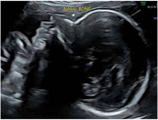
Figure 1: Mid sagittal facial profile showing normal nasal bone
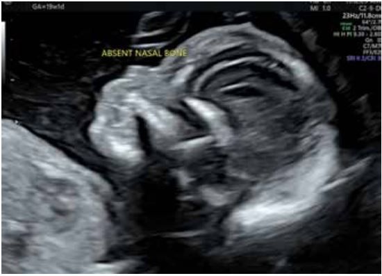
Figure 2: Mid sagittal facial profile showing absent nasal bone
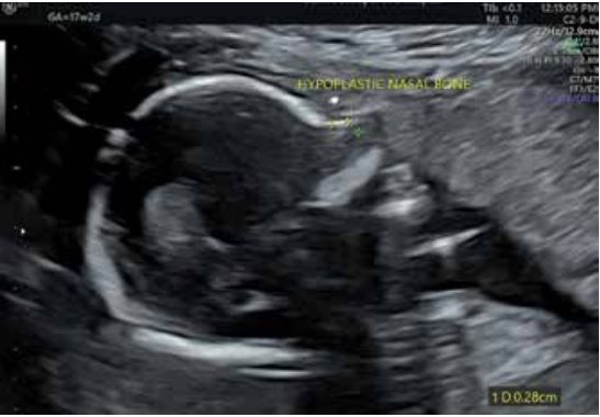
Figure 3: Mid sagittal facial profile showing hypoplastic nasal bone (nasal bone length less than 5th centile as per Indian charts)
Nasal bone was imaged as per standard protocol in the midsagittal facial profile view (Figure 1, normal nasal bone).6 Absent nasal bone was defined as no calcification or white line was seen below the skin of the nasal bridge in mid sagittal view of facial profile (Figure 2). Nasal bone was defined as hypoplastic in the second trimester scan (Figure 3) when nasal bone measurement in the midsagittal facial profile view was <5th centile according to gestation corrected Indian charts.7
Absent or hypoplastic nasal bone was defined as isolated
1. When there was absence of other second trimester soft markers like ventriculomegaly, nuchal oedema, renal pelvis dilatation, short femur and/or short humerus, intracardiac echogenic focus, echogenic bowel and aberrant right subclavian artery.
2. There were no other structural abnormalities seen on the ultrasound.
3. Serum biochemistry screen, where available, low risk for common aneuploidies i.e. trisomy 21, 18 and 13.
As a unit protocol, all women with foetuses diagnosed with absent or hypoplastic nasal bone during second trimester ultrasound were offered prenatal diagnostic testing i.e. amniocentesis. Though, the option of cell free foetal DNA was also discussed, amniocentesis was recommended in all cases. The amniotic fluid in all cases was tested for Fluorescence in situ hybridisation (FISH) and Microarray 750K, to rule out aneuploidies of chromosome 21, 18, 13 and sex chromosomes and atypical aneuploidies, copy number variations, sub-microscopic deletions and duplications, respectively. Results of these tests along with pregnancy outcomes were obtained from the database and analysed.
The primary objective of the study was to evaluate the association of isolated absent or hypoplastic nasal bone during second trimester scan with chromosomal abnormalities and genetic syndromes (i.e. aneuploidies and copy number variations).
Ethical clearance was obtained from the Institutional ethics committee for the reanalysis of de identified data.
Data analysis was carried out using SPSS statistics software IBM version 23.0 (Armork, NY: IBM Corp.). All continuous variables were tested for normality assumptions using Kolmogorov Smirnov test. Descriptive statistics such as mean, standard deviations and range values were calculated for normally distributed data. Categorical variables were expressed as frequency and percentages.
Results
A total of 3008 women had second trimester ultrasound (between 16-24weeks) during the study period, among these 21 (0.69%) foetuses were diagnosed to have absent or hypoplastic nasal bone during the second trimester scan (Figure 4). Among these 21 foetuses, 8 (38.1%) were excluded as they were associated with other structural abnormalities (n-5) or had presence of other second trimester soft tissue markers (n-3). Thirteen (61.9%) foetuses were defined to have isolated absent (n-9) or hypoplastic (n-4) nasal bone which were included in the final analysis. The outcomes were available for all the recruited women.
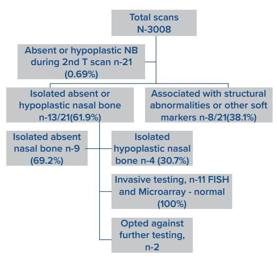
Figure 4: Flow chart showing recruited study group
Basic characteristics of the recruited women are mentioned in Table 1. All were spontaneously conceived pregnancies, only 1 was a twin pregnancy (one foetus among twin pair had absent nasal bone, which was included in the study), rest all were singleton pregnancies. Biochemical screening was categorized into: - not available (neither double or quadruple test done), low risk (< 1in 1000 risk for trisomy 21), intermediate risk (1in 250 to 1in 1000 risk for trisomy 21) and high risk (> 1in 250 for trisomy 21).
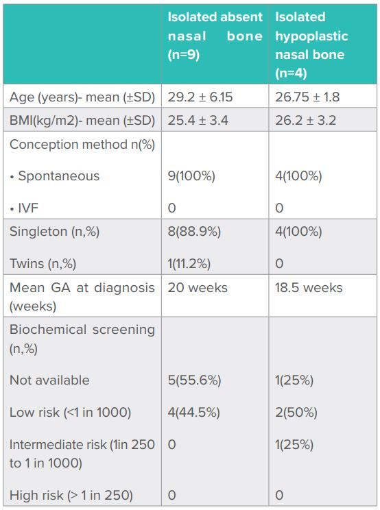
Table 1:Showing baseline characteristics of recruited women in the study
Out of 13 women who were included into the study, 11(84.6%) women opted for invasive testing i.e. amniocentesis to rule out chromosomal aneuploidies and genetic syndromes reported with absent or hypoplastic nasal bone. As a unit protocol, amniotic fluid in all cases was tested for FISH and Microarray (750K). There were no procedure related complications. Two (15.3%) women, both with hypoplastic nasal bone in foetus, opted against any further genetic testing. None of the recruited patients in our group opted for non-invasive prenatal testing. In all cases where invasive testing was done, FISH and chromosomal microarray reports were reported as normal for aneuploidies including common as well as atypical aneuploidies. None of the tested sample was positive for microdeletions or microduplications or copy number variations. All women continued the pregnancy in our study group and none of the baby, including two where parents opted against further antenatal genetic evaluation had dysmorphic features at birth, and all were apparently well postnatally.
Discussion
The results of our study adds to the evolving evidence which suggests that when absent or hypoplastic nasal bones are identified as an isolated finding during the second trimester scan the overall chances of fetus having chromosomal abnormalities like trisomy 21 or Down syndrome remain low only.
The landmark meta-analysis of second trimester soft markers for trisomy 21 by Agathokleous et al.2, which was published more than a decade back, had shown that absent or hypoplastic nasal bones increases pre-test odds of trisomy 21 by 6-7-fold. The odds or likelihood ratios of second trimester ultrasound markers published in this article are still used in counselling women and for risk recalculation for trisomy 21. Though, most of the studies included in this meta-analysis where aneuploidy was confirmed either there was presence of additional ultrasound abnormalities or there were multiple soft markers in foetuses.
The more recent published literature has studied the association of isolated absent or hypoplastic nasal bones during 2nd trimester scan with aneuploidies. Data published by a tertiary referral hospital in India where 142 women with absent hypoplastic nasal bone (AHNB) were recruited. Ninety-three women out of the 142 (65.5%) had an isolated AHNB, and out of which aneuploidy was reported only in 1.07% (1/93), which had high risk on biochemical screening as well. Authors concluded that isolated AHNB with low risk in biochemical screening is rarely associated with aneuploidy.8 A study done by Chanchal et al.5 had also shown that fetuses where absent nasal bones had additional ultrasound findings and/or had high risk on either dual or triple/quadruple markers had confirmed aneuploidy (8/26 fetuses- 7 with trisomy 21 and in trisomy 18), and rest had normal outcomes.
Dilemma in counselling comes as few studies have shown association of nasal bone abnormalities with microdeletions or microduplications or copy number variations which are diagnosed with advanced molecular genetic testing i.e. microarray. Li et al.9 had shown 10 /132 foetuses which had nasal bone abnormalities were diagnosed with copy number variations on microarray testing. However, most of these fetuses had additional ultrasound abnormalities as well. In our study group, none of the foetuses with isolated nasal bone abnormality had abnormally reported microarray, and these findings are consistent with Lostchuck’s study.10 Though, small sample size and retrospective study bias remain the main limitations of our study.
The parents with ultrasound findings where nasal bones remain unossified or measures less than the 5th centile according to the gestation plotted charts even during second trimester scan, with low-risk serum screening can be counselled that risk of common aneuploidies like trisomy 21 remains low. However, there is still limited data about the association of these findings in isolation with other genetic syndromes related to pathogenic or likely pathogenic copy number variations. Non-invasive prenatal testing by cell free fetal DNA in maternal blood has emerged as a promising test for Down syndrome with good detection rate, however its validation for atypical aneuploidies and submicroscopic genetic abnormalities is still not there and it is still considered as a screening test. The option of prenatal diagnostic testing i.e. amniocentesis where fetal DNA can be tested for additional genetic abnormalities as well should be discussed with the family.
CONCLUSION:
This case stresses the need of careful morphological examination and to investigate for mastocytosis in patients especially in the presence of longstanding skin allergy and prominent eosinophilia, even with the co-existence of parasitic infection and allergic sensitization. In our case, the bone marrow aspiration showed only eosinophilic prominence with only a few mast cells however trephine biopsy examination clinched the diagnosis.
Acknowledgments:
None
Funding source:
None
Conflict of interest:
None
Ethical approval:
Written informed consent is obtained from all the patients, visiting our centre, about the use of their personal data for future studies. Also, for doing reanalysis of de identified data permission was taken from the Institutional ethics committee
References
- Prabhu, Kuller M, J.A, Biggio JR. Society for maternal-fetal medicine consult series 57: evaluation and management of isolated soft ultrasound markers for aneuploidy in the second trimester. Am. J. Obstet, Gynecol 2021; 225 (4): B2-B15.
- Agathokleous M, Chaveeva P, Poon LC, Kosinski P, Nicolaides KH. Meta-analysis of second-trimester markers for trisomy 21. Ultrasound Obstet Gynecol 2013; 41: 247–261.
- Moczulska H, Serafin M, Wojda K, Borowiec M, Sieroszewski P. Fetal nasal bone hypoplasia in the second trimester as a marker of multiple genetic syndromes. J.Clin. Med 2022; 11(6): 1513.
- Ting YH, Lao TT, Lau TK, et al. Isolated absent or hypoplastic nasal bone in the second trimester fetus: is amniocentesis necessary? J Matern Fetal Neonatal Med. 2011; 24: 555.
- Singh C, Thakur S, Arora N, Khurana D. Revisiting absent nasal bone in the second trimester. J Clin Ultrasound. 2020;1–5.
- Fetal Medicine Foundation Nasal Bone. Fetal Medicine Foundation Website;2012. http://www.fetalmedicineusa. com/nasalbone.php.
- Narayani BH, Prathima R. Mid-second Trimester Measurement of Nasal Bone Length in the Indian Population. The Journal of Obstetrics and Gynecology of India. July– August 2013; 63(4):256–259.
- Prasad CS, Kunjukutty R, Krishnan V. Fetal absent/ hypoplastic nasal bone: a single center follow up study from a tertiary referral hospital in India. Int J Reprod Contracept Obstet Gynecol. 2020 Nov;9(11):4606-4611.
- Li H, Yao Y, Zhang C, Qin Y, Zeng L, Song J et al. Prenatal diagnosis and outcomes in 320 fetuses with nasal bone anomalies. Genetics of Common and Rare Diseases. Volume 14-2023.
- Lostchuck E, Hui L. Should second-trimester hypoplastic nasal bone be sole indication for diagnostic testing with chromosomal microarray analysis? Ultrasound Obstet Gynecol. 2019; 53:848.