 WhatsApp Us (24/7)
 +91 926 888 0303 (24/7)


Abstract: Thrombotic microangiopathies (TMAs) are a spectrum of disorders characterized by hemolytic anemia, thrombocytopenia, and formation of microthrombi in blood vessels, resulting in tissue injury due to compromised blood flow. This classical presentation of TMA is characterized by varying degrees of microangiopathic hemolytic anemia (MAHA), thrombocytopenia, and ischemic damage to the brain, kidneys, heart, lungs and GI tract.
TMA can be classified into different types based upon the etiology and pathogenesis like thrombotic thrombocytopenic purpura (TTP), Complement mediated TMA (atypical Hemolytic uremic syndrome [aHUS]), infection associated TMA (shiga toxin producing E.coli, streptococcus pneumoniae) secondary to systemic causes like malignancy, autoimmune diseases and medications. In this article, we discuss a case of a female diagnosed with aHUS with acute kidney injury complicated by hypertension and severe anemia. As aHUS is known to be associated with high mortality and morbidity, prompt recognition of aHUS is crucial for efficient treatment. Here we have discussed the clinical presentation, histopathological hallmarks, and the diagnostic approach for TMA.
Key words: Thrombotic Microangiopathies (TMA’s); Atypical Hemolytic Uremic Syndrome (aHUS)
Introduction
Thrombotic microangiopathies (TMAs) encompass a range of disorders marked by microangiopathic hemolytic anemia, thrombocytopenia, and the formation of microthrombi in tiny blood vessels, leading to tissue injury due to compromised blood flow.1 This situation can result in the classic presentation of TMA, characterized by varying degrees of microangiopathic hemolytic anemia (MAHA), thrombocytopenia, and ischemic damage to organs like the brain and kidneys, furthermore it may also affect numerous other organ systems including the heart, lungs, and gastrointestinal tract.2,3 TMA can be classified into different types based upon the etiology and pathogenesis like TTP, complement mediated TMA (atypical HUS), infection associated TMA(shiga toxin producing E.coli, streptococcus pneumoniae) secondary to systemic causes(malignancy autoimmune diseases and medications). We report a case of an Indian female diagnosed with atypical Hemolytic uremic syndrome (aHUS) with acute kidney injury complicated by hypertension and anemia.
Atypical hemolytic uremic syndrome (aHUS) is an extremely rare thrombotic microangiopathy (TMA) caused by complement dysregulation, characterized by non-immune hemolytic anemia, thrombocytopenia, and acute kidney failure.4
During childhood, the condition occurs with similar frequency in both boys and girls, with a ratio of 1.3 to 1, but in adulthood, there is a higher prevalence among females, with a ratio of 2 to 1. Familial forms account for approximately sixteen percent of cases. The typical age of onset is 21 years, with males typically affected at around 10 years and females at around 25 years. Onset before six months of age often suggests aHUS rather than STEC-HUS, with febrile infections in the respiratory or gastrointestinal tract frequently triggering the condition.5,6,7
The occurrence of aHUS was equally common in adulthood (58.4%) and childhood (41.6%). By specific ages, the proportions of patients developing the condition were 23%, 40%, 70%, and 98% by ages 2, 18, 40, and 60 years, respectively. Children had a higher mortality rate compared to adults (6.7% versus 0.8% at 1 year) (P=0.02), but adults were more likely to progress to end-stage renal disease (ESRD) following the initial aHUS episode. While children's prognosis is heavily influenced by genetic factors, adults face a poorer renal prognosis overall.
The primary cause of aHUS is the dysregulation of the alternative complement pathway, leading to inflammation, activation, and injury to endothelial cells through the assembly of the membrane attack complex. Mutations in genes related to the complement pathway or the presence of autoantibodies against complement factor H (CFH) have been detected in 60–70% of patients.8,9,10
Historically, aHUS has been divided into primary when there are inherent irregularities in the alternative complement pathway or when other factors commonly associated with secondary aHUS are excluded. Secondary aHUS is identified when it is prompted by known diverse conditions or triggers, such as autoimmune illnesses, malignancies, transplantation, pregnancy, specific drug administration, or infections.11,12
Initially, aHUS often begins with nonspecific symptoms like tiredness, paleness, or lethargy. These symptoms can develop into indications of acute kidney injury (AKI), such as decreased urine output, uremia, and fluid overload. The likelihood of advancing to stage 3 or 4 chronic kidney disease (CKD) and end-stage renal disease (ESRD) is significant in individuals with aHUS. In contrast to typical HUS, patients with aHUS frequently do not recover kidney function without medical intervention, if left untreated, approximately half of aHUS cases progress to needing dialysis, with a mortality rate of 25%.13,14,15 Treatment is usually by therapeutic plasma exchange or by using eculizumab which are expensive treatment modalities in developing countries thereby making it a challenge to manage Atypical HUS.
Case presentation
A 35 years old female was hospitalized with complaints of headache; vomiting and giddiness since 20 days.She had a history of recently diagnosed hypertension.
At presentation, the patient was conscious, oriented and dehydrated.
Vital signs were as follows: pulse rate—98bpm, blood pressure—140/80 mm Hg, respiratory rate—20/minute, and SpO2 of 97% on room air. The patient was afebrile. Respiratory and cardiovascular examinations were unremarkable except for mild epigastric tenderness.
She had done routine investigations from outside which showed serum creatinine of 3.66mg/dl (baseline unknown).
Patient was admitted to the ER and investigations were done as shown in table1: revealed severe renal impairment with creatinine levels elevated.
USG whole abdomen showed Cholelithiasis with no features of acute cholecystitis. Gall bladder sludge. Bilateral grade III medical renal disease. Bilateral ovarian haemorrhagic cysts.
| Blood counts | haemoglobin—10.9-6 gm/dL, platelet— 135,000, white blood cell (WBC) counts 6,600 |
| Erythrocyte sedimentation rate—20mm/hour, C-reactive protein—76.39 mg/L LDH levels—>461 U/L, Haptoglobin <30mg/dl Reticulocyte count 6.6%, Corrected reticulocyte count 4.47 % | |
| Direct and indirect Coombs test | negative C3—127 mg/dL, C4—38 mg/dL |
| Serology | nonreactive for hepatitis B, human immunodeficiency virus, and hepatitis C |
| Renal function | Urea—124 mg/dL, creatinine—8.1 mg/dL, sodium 131 mmol potassium—4.7 mmol/L |
| Liver function tests | Total bilirubin—0.7 mg/dL, direct bilirubin—0.1 mg/dL, AST—30 IU/L ALT—12 IU/L, ALP—90 U/L |
| Coagulation profile | Prothrombin time—11.3 seconds, activated partial thromboplastin time—31.7 seconds, international normalized ratio—0.98 |
| Urinalysis | Specific gravity—1.020, urine protein 2+, RBC—high number |
| Peripheral smear | RBC–Anisocytosis (+), Microcytosis (+), Hypochromia (+) WBC—normal limits platelets—reduced in number |
Table 1: Laboratory investigations
Given the patient's deteriorating renal function and severe uraemia, urgent haemodialysis was initiated. During dialysis, the patient's symptoms improved transiently, but there was no sustained improvement in renal function, so further investigations were done as shown in Table 2.
| Anti Nuclear Antibodies | Negative |
| ANCA -C, ANCA P | Negative |
| Intact PTH | 202 pg/ml |
| Phosphorus | 6.4mg/dl |
Table 2: Additional laboratory investigations
During the course of the treatment she remained hypertensive despite multiple antihypertensives, forced diuresis, and negative fluid balance.
She also received several blood transfusions due to the persistence of anaemia.
Despite being managed symptomatically her renal function was deteriorating and urine output was extremely reduced therefore renal biopsy was done to identify the aetiology and as the results were awaited she was started on Intravenous solumedrol 500 mg once daily for 3 days suspecting lupus nephritis, rapidly progressive glomerulonephritis.
Kidney biopsy showed 38 glomeruli of which 3 glomeruli were globally sclerosed and 15 glomeruli showed ischemic wrinkling capillary loops. In addition, few glomeruli show fibrillary appearance and mesangiolysis indicating acute thrombotic microangiopathy. Few vessels showed endothelial swelling, fibrinous necrosis and mucointimal oedema indicating acute to subacute thrombotic microangiopathy. No vasculitis was seen.
Immunofluorescence: studies depicted Albumin – Negative IgG- Negative
IgA- Negative IgM – 1+ granular in mesangium. C3 – NegativeC1q - Negative
Kappa and Lambda- Show no light chain restriction in tubular casts.
Overall features are suggestive of acute to subacute chronic thrombotic microangiopathy affecting glomerular and vascular compartment and acute tubulointerstitial nephritis ( ATIN).
She was started on hemodialysis and arteriovenous fistula was created for further requirement of hemodialysis. The patient's clinical condition improved, and the laboratory values stabilized and she was discharged.
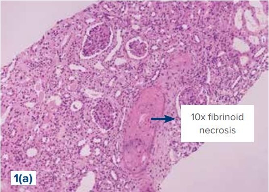
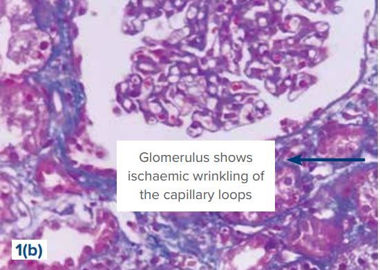
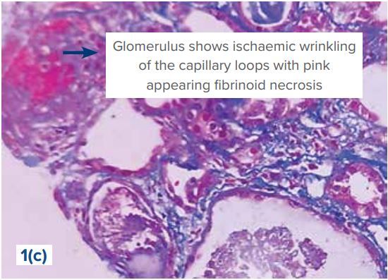
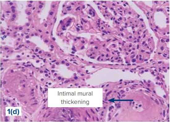
Figure 1(a,b,c,d): Histopathological changes in Thrombotic microangiopathies (TMAs)
Discussion
The diagnostic criteria for atypical hemolytic uremic syndrome (aHUS) involve the presence of microangiopathic hemolytic anemia characterized by increased lactate dehydrogenase and decreased haptoglobin levels, along with thrombocytopenia and organ dysfunction. This occurs alongside normal ADAMTS13 activity and the absence of Shigatoxin-associated E. coli infection. Additionally, about 20% of patients may exhibit extrarenal manifestations, such as involvement in the cerebral, cardiac, ocular, neurologic, gastrointestinal, and pulmonary systems.16
Suggested tests for investigating a suspected or confirmed thrombotic microangiopathy during the diagnostic process given in table 3.17
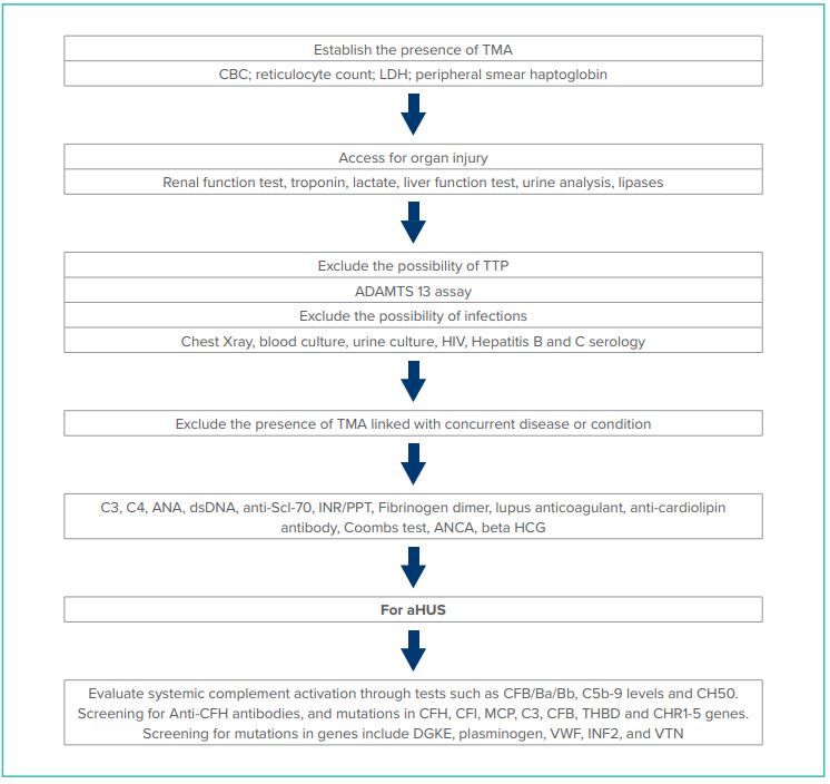
Table 3: Flowchart for diagnosis of Thrombotic micro angiopathy (TMA)
While deficiencies in the complement system are pivotal in the development of aHUS, it's important to note that normal levels of complement components C3 and/or C4 do not necessarily exclude the presence of aHUS.16
Therefore, it is not recommended to solely use complement activation biomarkers for definitively diagnosing aHUS. Our patient does not exhibit elevated serum levels of C3 and C4.In order to simplify the early diagnosis and effective management of this condition, the Joint Committee of the Japanese Society of Nephrology and the Japan Pediatrics Society (JSN/JPS) has established diagnostic
criteria for aHUS, focusing on three key clinical features. The first feature involves thrombocytopenia, identified by a platelet count below 150,000/μL. The second feature is microangiopathic hemolyticanemia, characterized by low hemoglobin levels (below 10 g/dL), increased lactate dehydrogenase levels, reduced haptoglobin levels, and the presence of fragmented red blood cells observed in the peripheral blood smear. The third feature is acute renal failure, as per the Kidney Disease: Improving Global Outcomes international guidelines. In our case, the patient presented with thrombocytopenia, severe hemolyticanemia with negative Coombs test, elevated lactate dehydrogenase, and acute renal injury, aligning with the diagnosis of atypical hemolytic uremic syndrome according to the aforementioned guidelines.18
The primary focus of treatment for aHUS is supportive care aimed at managing acute renal damage and systemic complications. To mitigate the effects of aHUS, acute kidney injury, and potential multisystem organ failure, it is vital to regulate fluid and electrolyte balance. Patients with severe anemia may require packed red blood cell transfusions. Specific therapeutic options include plasma exchange and the complement inhibitor eculizumab. There has been ongoing debate regarding the suitability of kidney transplantation for treating end-stage renal disease in aHUS patients. Understanding the diagnostic and treatment approach for aHUS is crucial, as early detection and intervention can significantly reduce disease morbidity and mortality.
CONCLUSION:
Recent medical literature suggests that thrombotic microangiopathy (TMA) may occur across diverse age groups, emphasizing the importance of early diagnosis for timely therapeutic measures. In our patient's case, there were no preceding infections or medication histories that could account for TMA. Therefore, it is advisable to investigate young individuals presenting with unexplained hypertension or TMA. Genetic testing should be considered, as genetic causes may also lead to recurrence post-renal transplantation. However, genetic testing was not pursued due to financial constraints. Plasma exchange (PLEX) was contemplated but not initiated due to the progression of the disease to end-stage renal disease (ESRD), necessitating dialysis owing to chronic histopathological findings. Although eculizumab, an alternative therapeutic option, was available, the patient's family declined further intervention due to financial limitations.
The existence of TMA can pose significant challenges due to its rarity, elevated morbidity, and mortality rates, as well as the necessity for specialized diagnostic testing and urgent therapeutic action. However, adopting a systematic approach to this clinical scenario can aid healthcare providers in promptly arriving at an accurate diagnosis and administering optimal treatment.
References
- Tsai HM. Untying the knot of thrombotic thrombocytopenic purpura and atypical hemolytic uremic syndrome. Am J Med 2013;126:200–9. [PubMed] [Google Scholar].
- Arnold DM, Patriquin CJ, Nazy I. Thrombotic microangiopathies: a general approach to diagnosis and management. CMAJ. 2017;189:E153-E159.
- Chapman K, Seldon M, Richards R. Thrombotic microangiopathies, thrombotic thrombocytopenic purpura, and ADAMTS-13. Semin ThrombHemost. 2012;38(1):47-54.
Am J Kidney Dis. 81(5):591-605. Published online December 10, 2022.doi: 10.1053/ j.ajkd.2022.10.014. - Fakhouri, F, Zuber, J, Frémeaux-Bacchi, V, and Loirat, C. Haemolytic uraemic syndrome. Lancet. (2017) 390:681–96. doi: 10.1016/S0140-6736(17)30062-4.
- Loirat, C, and Frémeaux-Bacchi, V. Atypical hemolytic uremic syndrome. Orphanet J Rare Dis. (2011) 6:60–30. doi: 10.1186/1750-1172-6-60.
- Licht C, Ardissino G, Ariceta G, Cohen D, Cole JA, Gasteyger C, et al The global aHUS registry: methodology and initial patient characteristics. BMC Nephrol. 2015;16:207. https:// doi.org/10.1186/s12882-015-0195-1.
- Karpman D, Loos S, Tati R, Arvidsson I. Haemolytic uraemic syndrome. J Intern Med. 2017 Feb;281(2):123–48. https://doi. org/10.1111/joim.12546.
- Sakari, JT. HUS and atypical HUS. Blood. (2017) 129:2847–56. doi: 10.1182/blood-2016-11-709865.
- Noris, M, Caprioli, J, Bresin, E, Mossali, C, Pianetti, G, Gamba, S, et al. Relative role of genetic complement abnormalities in sporadic and familial aHUS and their impact on clinical phenotype. Clin J Am Soc Nephrol. (2010) 5:1844–59. doi: 10.2215/CJN.02210310.
- Fremeaux-Bacchi, V, Fakhouri, F, Garnier, A, Bienaimé, F, Dragon-Durey, MA, Ngo, S, et al. Genetics and outcome of atypical hemolytic uremic syndrome: a nationwide French series comparing children and adults. Clin J Am Soc Nephrol. (2013) 8:554–62. doi: 10.2215/CJN.04760512.
- Campistol, JM, Arias, M, Ariceta, G, Blasco, M, Espinosa, L, Espinosa, M, et al. Actualizaciónensíndromehemolíticourémicoatípico: diagnóstico y tratamiento. Documento de consenso. Nefrologia. (2015) 35:421–47. doi: 10.1016/j.nefro.2015.07.005.
- Goodship, THJ, Cook, HT, Fakhouri, F, Fervenza, FC, Frémeaux-Bacchi, V, Kavanagh, D, et al. Atypical hemolytic uremic syndrome and C3 glomerulopathy: conclusions from a “kidney disease: improving global outcomes” (KDIGO) controversies conference. Kidney Int. (2017) 91:539–51. doi: 10.1016/j.kint.2016.10.005.
- Raina R, Grewal MK, Radhakrishnan Y, Tatineni V, DeCoy M, Burke LL, Bagga A. Optimal management of atypical hemolytic uremic disease: challenges and solutions. Int 12. Nephrol Renovasc Dis. 2019;12:183-204.
- Yerigeri K, Kadatane S, Mongan K, Boyer O, Burke LLG, Sethi SK, Licht C, Raina R. Atypical Hemolytic-Uremic Syndrome: Genetic Basis, Clinical Manifestations, and a Multidisciplinary Approach to Management. J MultidiscipHealthc. 2023;16:2233- 2249.
- Raina R, Vijayvargiya N, Khooblall A, Melachuri M, Deshpande S, Sharma D, Mathur K, Arora M, Sethi SK, Sandhu S. Pediatric Atypical Hemolytic Uremic Syndrome Advances. Cells. 2021 Dec 18;10(12).
- Hofer J, Rosales A, Fischer C, Giner T. Extra-renal manifestations of complement-mediated thrombotic microangiopathies. Front Pediatr. (2014) 2:97. doi: 10.3389/fped.2014.00097 PubMed Abstract | CrossRef Full Text | Google Scholar.
- McFarlane PA, Bitzan M, Broome C, et al. Making the Correct Diagnosis in Thrombotic Microangiopathy: A Narrative Review. Canadian Journal of Kidney Health and Disease. 2021;8. doi:10.1177/20543581211008707.
- Sawai T, Nangaku M, Ashida A, et al. Diagnostic criteria for atypical hemolytic uremic syndrome proposed by the joint Committee of the Japanese Society of nephrology and the Japan pediatric society. Pediatr Int. 2014; 56(1): 1-5. doi:10.1111/ ped.12274.