 WhatsApp Us (24/7)
 +91 926 888 0303 (24/7)


Abstract: Incidentally detected hypercalcemia is now a more common finding with expanded indications for blood investigations. Identification of patients requiring follow up investigation to clarify the cause of hypercalcemia is stressed, to facilitate early detection and treatment and prevent end organ damage and bone resorption. Here, we discuss a spectrum of cases of Primary, Secondary and Tertiary hyperparathyroidism.
Key words: Hyperparathyroidism, Primary hyperparathyroidism, Secondary hyperparathyroidism, Tertiary Hyperparathyroidism, hypercalcemia
Introduction
Hyperparathyroidism is defined as increased parathyroid gland activity which maybe due to an ‘intrinsic’ abnormality altering the excretion of parathyroid hormone (primary or tertiary hyperparathyroidism) or from an ‘extrinsic’ abnormality affecting calcium homoeostasis stimulating production of parathyroid hormone (secondary hyperparathyroidism).1
Isolated Hypercalcemia can be a vexing problem. Early hyperparathyroidism may be asymptomatic or may cause few subtle symptoms which are often missed.
Here we attempt to elucidate the common presentations of primary, secondary and tertiary hyperparathyroidism by detailing a case series, treated by our team.
Case 1: A 32-year-old housewife presented to the emergency with complaints of diffuse, dull mild to moderate non-radiating abdominal pain of insidious onset since 15 days. This was accompanied by generalized body ache, anorexia, nausea and recurrent vomiting containing food particles since one week. She developed continuous high-grade fever with chills and dysuria since 2 days.
Patient had an antecedent history dating back to 8 years with complaints of frequent urination due to which she was home bound, to maintain a proximity to a washroom. She also had polydipsia. She also complained of generalized bony pains.
Family members had noticed behavioural changes with irritability and low mood of 2-3 months duration. Patient had been seeking medical care from multiple medical practitioners but had failed to reach a diagnosis.
Her past medical history included a right renal calculus of 10mm, recurrent urinary tract infection (UTI) with bilateral pyelonephritis and hypothyroidism. She had been treated for pulmonary tuberculosis as a teenager.
On presentation, the patient was conscious, oriented to time place and person with relevant and clear speech. She had a pulse rate: 98/min, blood pressure 126/70mmhg, SpO2: 96% on room air and Temperature:101.2oF. She had no pallor, icterus, clubbing, cyanosis, goitre, oedema or lymphadenopathy. Abdominal exam revealed diffuse tenderness in peri-umbilical and infraumbilical region. Rest of the systemic exam was normal.
Patient was extensively investigated. She was found to have raised serum calcium (15.8 mg/dl) with ionized calcium of 1.78 mg/ dL. TSH was 9.47mIU/L and intact PTH was 1004pg/ml. Alkaline phosphatase was 260IU/L. Her serum amylase and lipase were elevated at 241 U/L and 301U/L respectively suggestive of pancreatitis. Serum magnesium was low at 1.4 mg/dL, vitamin D levels of 13.89 ng/ml. TSH was high (9.47Miu/l) and thyroid peroxidase antibody was 264.7IU/ml. Ferritin was high (1655 ng/ml) indicating acute inflammatory state. Urinalysis showed pyuria, hematuria, 1+ proteinuria and budding yeast cells. Urine culture was sterile since patient was on antibiotics. Urine spot calcium-9.11mmol/24 hours, Ca/creat ratio-0.55. DEXA Scan revealed a T-score at wrist (-7.5), LS spine (-2.5), femur neck (-3.2) respectively (s/o severely low BMD).
Ultrasound neck showed a lobulated hypoechoic lesion of 2.8 x 1.3cm postero-inferior to the right lobe of thyroid suggestive of parathyroid adenoma with left lobe of thyroid showing small mixed echogenic nodules, largest being 6 x 5mm.
Tc99 Sestamibi scan delayed images (20 min, 1hr, 2hr and 3 hrs.) showed uniform washout of tracer from both lobes of thyroid with a focal area of persistent uptake in area just inferior to the lower pole of right lobe of thyroid. A diagnosis of large right inferior parathyroid adenoma was made. (Figure 1)
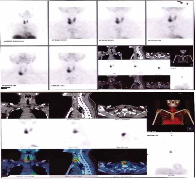
Figure 1: SPECT-CT of neck showing a soft tissue density nodule (~3.1 x 1.3 cm) just inferior to lower pole of right lobe with increased radiotracer uptake, suggestive of a right inferior parathyroid adenoma
Patient was managed conservatively for pancreatitis and UTI. Subsequently, she was taken up for parathyroidectomy. Right inferior enlarged parathyroid gland was identified and removed and sent for HPE. Rest 3 parathyroid gland were identified and preserved. Post-operative period was uneventful. She was closely monitored for serum calcium and serial intact parathyroid hormone (iPTH) measurements which were 403.5pg/ml(28th September 2022 before Surgery)- 269pg/ml(28th September 2022 after surgery)- 126.4pg/ml (29thSeptember 2022). On 04.03.23 iPTH was 172.9pg/ml, serum calcium 10.3 mg/dL.
Calcium supplementation was done and patient was closely monitored for hypocalcemia. After discharge, the patient was treated by the urology team for pre-existing nephrocalcinosis.
Case 2: A 23-year-old vegan female, with a desk job presented with complaints of bodyache and generalized weakness of one-month duration. Patient had persistent lethargy with painful lower limbs and back, especially pain concentrated in the thighs. She had difficulty climbing stairs. Patient had very limited sunlight exposure.
On examination, there was notable tenderness over bilateral shoulders, humeri and both femurs.
On Investigation (Table 1)
| Vitamin D | <2 ng/ml |
| Ionized Calcium | 1.2 mg/dl |
| Alkaline Phosphatase | 573 IU/L |
| iPTH | 571 pg/ml |
Table 1: Investigations at the time of presentation
X rays of the wrist, humerus/arm and skull were within normal limits. Ultrasound neck showed no parathyroid adenoma.
DEXA scan Spine and Femur - Spine(T-score -2) and Femur(T-score -2.5)showed osteopenia and osteoporosis respectively.
Tc99 Bone Scan - metabolic bone disease with focal areas of uptake in the 5th rib and right inferior pubic ramus-likely pseudo-fractures. (Figure 2)
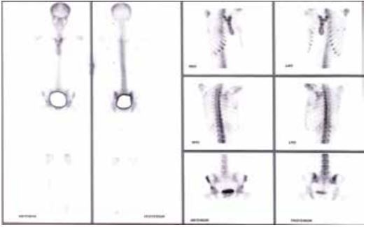
Figure 2: Tc99 Bone Scan showing pseudo-fractures 5th rib and right inferior pubic ramus
Patient was diagnosed as having secondary hyperparathyroidism due to severe dietary vitamin D and calcium deficiency. Patient was supplemented with Calciferol and Cholecalciferol along with calcium.
At a 1 month follow up on out-patient department (OPD) basis, patient reported symptomatic improvement with reduced body ache and generalized lethargy and weakness (Table 2).
| iPTH( 1 month) | 85 pg/ml |
| iPTH( 6 months) | 42 pg/ml |
Table 1: iPTH levels on follow up
Case 3: A 48-year-old housewife, a known case of Chronic Kidney Disease on maintenance Hemodialysis underwent renal transplant in February 2020. She had Secondary Hyperparathyroidism which was conservatively managed. Post transplant her calcium and PTH levels became normal.
On OPD follow-up over the next few years, she was found to have hypercalcemia (11.3 mg/dl) and her PTH levels (252.5 ng/L) were also raised. Raised serum calcium levels were managed conservatively. Ultrasound (USG) Neck was done but no abnormality was detected. Serial monitoring showed repeated raised levels of Serum Calcium and PTH. Later, magnetic resonance imaging (MRI) Neck/Thyroid was done which revealed well defined oval soft tissue lesion in postero-inferior aspect of the left thyro-esophageal groove suggestive of parathyroid adenoma.
The patient underwent Radio-Frequency Ablation (RFA) of the nodule on 10 June 2022 but even after the ablation, the PTH levels remained elevated (171.5 ng/L in September 2022 and 188.6 ng/L in January 2023).
Myocardial perfusion imaging (MIBI) scan was done in August 2022 which showed atypical pattern of washout seen from the left lobe of thyroid, predominantly in the upper polar region (Figure 3).
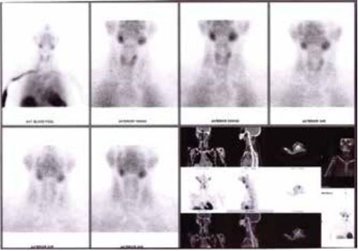
Figure 3: Shows atypical pattern of washout seen from the left lobe of thyroid, predominantly in the upper polar region
Discussion
We have described the various cases of primary, secondary and tertiary hyperthyroidism which we encountered in our department recently. Primary hyperthyroidism is diagnosed in a patient with hypercalcemia with elevated or normal concentrations of parathyroid hormone which are inappropriate to the serum calcium concentrations.1
The most frequent presentation of primary hyperparathyroidism is an asymptomatic rise in serum calcium. Classically, the presentation of hyperparathyroidism was described as moans, groans, stones and psychotic overtones, referring to the tendency for formation of calculi and bony pains due to accompanying osteopenia. Our patients had most of these clinical manifestations. The common radiological features found in hyperparathyroid patients are subperiosteal resorption of the distal phalanges, bone cysts, salt-and-pepper degranulation of the skull, tapering of the distal clavicle and brown tumors associated with fractures, deformities and bony pains. Collectively, the radiological findings are referred to osteitis fibrosa cystic.2
Renal involvement in primary hyperparathyroidism ranges from renal calculi (symptomatic/asymptomatic), nephro-calcinosis to deranged renal function as was seen in case 1.3,4 Neuromuscular involvement may present as proximal myopathy.5 Gastrointestinal symptoms include acid peptic disease and pancreatitis.6 Hypertension and accelerated atherosclerosis are commonly encountered cardiovascular complications.7 Neurological manifestations include anxiety, difficulty concentrating and cognitive decline.8,9
Asymptomatic primary hyperparathyroidism is defined as raised serum calcium on investigations but showing no overt signs of the disease or target organ manifestations other than hypercalcaemia.10 Primary hyperparathyroidism is most commonly found to be caused by single benign parathyroid adenoma (approximately 80% of patients). Multinodular or multiglandular disease is seen in about 15–20% of patients.11 Diagnosis of primary hyperparathyroidism rests on documenting raised corrected serum calcium or raised ionized calcium values. Serum phosphorus levels are usually in the low normal range. A raised or normal parathyroid hormone concentration in setting of raised ionized calcium, essentially confirms the diagnosis of primary hyperparathyroidism.12 Differential diagnosis of primary hyperparathyroidism include Familial hypocalciuric-hypercalcaemia, secondary hyperparathyroidism, multiple endocrine neoplasia type 1 or 2 and parathyroid cancer.1
Secondary hyperparathyroidism (SHPT) is found in response to dysregulation of serum calcium and phosphate levels with deficiency of bioactive vitamin D (as deficiency or as a result of a failure to bioactivation). This was witnessed in case 2. It is commonly seen with progressive renal failure as an adaptive response, but can also be seen with severe calcium and vitamin D deficiency. Raised serum phosphate levels lead to raised Phosphatonin fibroblast growth factor 23 (FGF-23), and reduced synthesis of calcitriol, the active form of vitamin D. This leads to upregulation and raised serum levels of parathyroid hormone (PTH) and parathyroid hyperplasia (with sustained stimulation).13,14 Although there is no current evidence of a phosphate receptor on the parathyroid gland,13 the pathogenesis of secondary hyperparathyroidism in renal dysfunction is through phosphorous homeostasis and FGF-23 and its receptor fibroblast growth factor receptor 1 (FGFR1).15 High phosphate enhances parathyroid hyperplasia. Studies in VDR knock-out mice suggest that vitamin D pathways play a secondary role in the development of parathyroid gland hyperplasia and that signaling through the CaSR is sufficient to prevent parathyroid hyperplasia in these animals.
Tertiary hyperparathyroidism is characterized by the ‘semiautonomous hypersecretion’ of PTH leading to hypercalcemia.16 The etiology has been postulated to be due to a monoclonal expansion of a clone of parathyroid cells, which have an altered calcium-sensing receptor (CASR) which induce secretion of PTH in spite of high serum calcium levels. Hyperphosphatemia has a direct stimulatory effect on the parathyroid gland cell resulting in nodular hyperplasia and increased PTH secretion.17 This is demonstrated in our post renal transplant case 3. With worsening renal function, the size of parathyroid glands increase. The glands are mostly enlarged due to polyclonal diffuse cellular hyperplasia, but there is monoclonal expansion of chief cells leading to nodule formation. Nodular hyperplastic tissue has lower VDRs and nodular hyperplastic glands have less VDRs and CASRs compared with diffusely hyperplastic glands, a fact that further exacerbates parathyroid gland resistance to calcitriol and calcium. Because the parathyroid glands are autonomously functioning, in some patients, PTH levels remain persistently high despite serum calcium levels that are within the reference range or even above normal after a renal transplant and so called tertiary hyperparathyroidism. Post-transplantation, that usually results in a return to normal biology of phosphorus homeostasis and an increase in 1,25-dihydroxyvitamin D production may not be sufficient to reduce PTH or serum calcium levels if the tissue mass is great enough to act semi-autonomously.
The gross biochemical differences between the various hyperparathyroidism types are tabulated below (Table 3).

Table 3: Differences between primary, secondary and tertiary hyperparathyroidism
CONCLUSION:
Hyperparathyroidism presenting with raised serum total and ionized calcium is also associated with elevated intact PTH. The underlying etiology and differentiation between types of hyperparathyroidism is essential for proper management of these patients.
Acknowledgements:
None
Conflict of interest::
None
Funding support::
None
References
- John P Bilezikian, Leonardo Bandeira, Aliya Khan, Natalie E Cusano,Hyperparathyroidism, The Lancet,Volume 391, Issue 10116,2018,Pages 168-178,ISSN 0140-6736, https://doi.org/10.1016/S0140-6736(17)31430- 7 (https://www.sciencedirect.com/science/article/pii/ S0140673617314307)
- Bandeira F, Cusano NE, Silva BC, Cassibba S, Almeida CB, Machado VC, Bilezikian JP. Bone disease in primary hyperparathyroidism. Arq Bras EndocrinolMetabol. 2014 Jul;58(5):553-61. doi: 10.1590/0004-2730000003381. PMID: 25166047; PMCID: PMC4315357
- Lila AR, Sarathi V, Jagtap V, Bandgar T, Menon PS, Shah NS. Renal manifestations of primary hyperparathyroidism. Indian journal of endocrinology and metabolism. 2012 Mar 1;16(2):258-62.
- Silverberg SJ, Clarke BL, Peacock M, Bandeira F, Boutroy S, Cusano NE et al. Current issues in the presentation of asymptomatic primary hyperparathyroidism: proceedings of the Fourth International Workshop. The Journal of Clinical Endocrinology & Metabolism. 2014 Oct 1;99(10):3580-94.
- Patten BM, Bilezikian JP, Mallette LE, Prince A, Engel WK, Aurbach GD. Neuromuscular disease in primary hyperparathyroidism. Annals of Internal Medicine. 1974 Feb 1;80(2):182-93.
- Abboud B, Daher R, Boujaoude J. Digestive manifestations of parathyroid disorders. World Journal of Gastroenterology: WJG. 2011 Sep 9;17(36):4063.
- Fitzpatrick LA, Bilezikian JP, Silverberg SJ. Parathyroid hormone and the cardiovascular system. Curr Osteoporos Rep. 2008;6(2):77-83.
- Walker MD, McMahon DJ, Inabnet WB, Lazar RM, Brown I, Vardy S et al. Neuropsychological features in primary hyperparathyroidism: a prospective study. The Journal of Clinical Endocrinology & Metabolism. 2009 Jun 1;94(6):1951-8.
- Walker MD, Rubin M, Silverberg SJ. Nontraditional manifestations of primary hyperparathyroidism. J Clin Densitom. 2013;16(1):40- 47.
- Silverberg SJ, Walker MD, Bilezikian JP. Asymptomatic primary hyperparathyroidism. Journal of Clinical Densitometry. 2013 Jan 1;16(1):14-21.
- Baloch ZW, Livolsi VA: Parathyroids: Morphology and Pathology. BilezikianJPMarcusRLevineMAMarocciCSilverbergSJPottsJT The parathyroids. 2015.ElsevierUSA:pp. 23-36.
- Bandeira F, Griz L, Chaves N, Carvalho NC, Borges LM, Lazaretti-Castro M et al. Diagnosis and management of primary hyperparathyroidism: a scientific statement from the Department of Bone Metabolism, the Brazilian Society for Endocrinology and Metabolism. Arquivos Brasileiros de Endocrinologia & Metabologia. 2013;57:406-24.
- Cunningham J, Locatelli F, Rodriguez M. Secondary hyperparathyroidism: pathogenesis, disease progression, and therapeutic options. Clinical Journal of the American Society of Nephrology. 2011 Apr 1;6(4):913-21.
- Slatopolsky E, Brown A, Dusso A. Pathogenesis of secondary hyperparathyroidism. Kidney Int Suppl. 1999;73:S14-S19.
- Martin KJ, Gonzalez EA, Slatopolsky E: The parathyroids in renal disease: Pathophysiology. In: The Parathyroids: Basic Clinical Concepts, 2nd edition, San Diego, Academic Press Inc., 2001, pp 625–634
- Jamal SA, Miller PD. Secondary and tertiary hyperparathyroidism. J Clin Densitom. 2013;16(1):64-68.
- Moallem E., Kilav R., Silver J., Naveh-Many T.: RNA-protein binding and post-transcriptional regulation of parathyroid hormone gene expression by calcium and phosphate. J BiolChem 1998; 273: pp. 5253-5259.