 WhatsApp Us (24/7)
 +91 926 888 0303 (24/7)


Abstract: We present the case of a 29-year-old female with a 2.5-month history of continuous fever, weight loss, vomiting, ataxia, tremors, altered consciousness, and motor deficits. Extensive investigations initially aimed at infectious aetiologies were unrevealing. However, the discovery of weakly positive anti- N-methyl-D-aspartate (NMDA) receptor antibodies in the cerebrospinal fluid (CSF) led to the diagnosis of anti-NMDA receptor encephalitis. The patient showed a positive response to immunomodulatory therapies but developed complex partial seizures and dyskinesias during the course of treatment. This case underscores the importance of considering autoimmune encephalitis in the differential diagnosis of refractory neurological symptoms, particularly when conventional investigations fail to yield a definitive diagnosis.
Key words: Anti NMDA Receptor Encephalitis, Neurological Symptoms, Diagnostic Challenge, Paroxysmal Kinesigenic Dyskinesia, Complex Partial Seizures, IVIG
Introduction
Anti- N-methyl-D-aspartate (NMDA) receptor encephalitis is a relatively newly recognized autoimmune disorder characterized by the production of antibodies against the NMDA receptor, leading to a range of neurological symptoms. Initially described in young women with ovarian teratomas, it has since been identified in individuals without tumours, leading to a broadening of its clinical spectrum. The diverse presentation of anti-NMDA receptor encephalitis often poses a diagnostic challenge, as it can mimic infectious, neoplastic, or psychiatric conditions. Here, we report a case of anti-NMDA receptor encephalitis presenting with atypical features, emphasizing the importance of a high index of suspicion for autoimmune encephalitis in patients with refractory neurological symptoms.
Case presentation
A 29-year-old-female, presented with complaints of fever with dry cough, vomiting and weight loss for 1 month. Laboratory tests were done which showed TLC 5700 C-reactive protein-25, with normal liver function tests (LFTs) and renal function tests (RFTs) (Figure 1). Malaria antigen, dengue NS1 antigen, WIDAL test, blood cultures, urine culture were all negative.
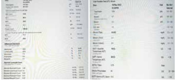
Figure 1: Complete Blood Count (CBC) and LFT showing nothing significant
After an extensive workup, the patient was suggested positron emission tomography-computed tomography scan (PET CT) and was started on empirical anti-tubercular therapy (ATT). Patient was discharged on ATT. Patient’s symptoms did not improve and she came back after 1 month with new onset ataxia, tremors, restlessness and altered sensorium. On examination, the Glasgow coma scale (GCS) (E4V5M6), 4/5 power in all 4 limbs, Babinski- flexor plantar response. PET CT was done which was normal. Magnetic resonance imaging (MRI) brain was done which was also normal (Figure 2).
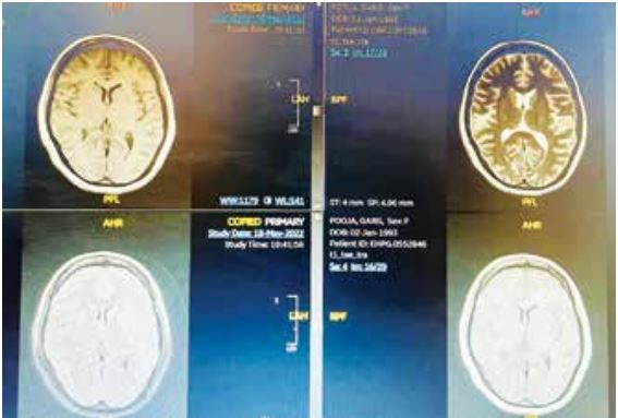
Figure 2: MRI Brain showing no significant changes
Cerebrospinal fluid (CSF) analysis was (Figure 3) done which showed normal glucose and total leukocyte count (TLC) but elevated protein. CSF paraneoplastic and meningitis panel were negative (Fig-3). CSF autoimmune encephalitis panel was sent which came to be weak positive (Fig-3).
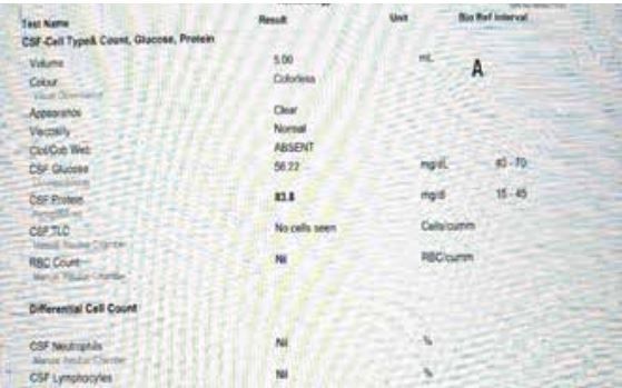
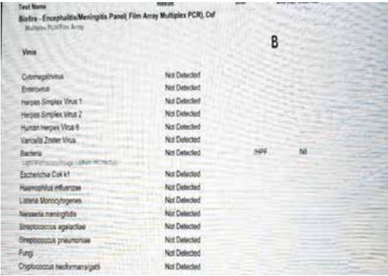
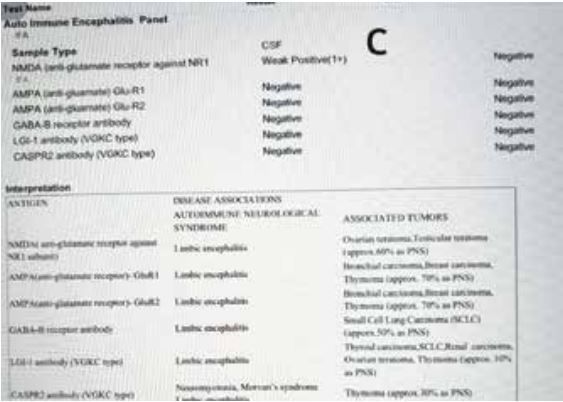
Figure 3: A-CSF Analysis showing CSF glucose, CSF protein and CSF TLC normal. B- Encephalitis/Meningitis panel negative. C-Auto immune encephalitis panel showing NMDA weak positive
In view of anti-NMDA, auto immune encephalitis Injection Methyl prednisolone was given and intravenous immunoglobulin (IVIG) was started in view of. Total dose of IVIG given was 110 gm. Electroencephalogram (EEG) revealed 7-8 hertz and 20-30 microvolt theta, alpha activity seen with bifrontal beta with intermittent sharp and slow activity seen from left occipital area. Patient had Oro alimentary and bimanual automatisms with behavioural arrest suggestive of complex partial seizure, patient was started on carbamazepine. Patient also developed paroxysmal kinesigenic dyskinesia which responded well with clonazepam and carbamazepine.
| Intravenous immunoglobulins (IVIG: 2gm/ kg + methylprednisolone 1 gm iv daily for 5 days followed by Prednisolone 1 mg/kg/day in a tapering dose) |
| For Agitation: Clonazepam: 0.5 mg once daily |
| For Abnormal movements: Carbamazepine 100mg twice daily |
| Additional consideration: Early physical therapy |
Table 1: Therapy and dosage for anti-NMDAR encephalitis
Discussion
The protracted course of symptoms and the lack of response to conventional antimicrobial therapies prompted further investigation into autoimmune aetiologies. Weakly positive anti-NMDA receptor antibodies detected in the CSF were consistent with a diagnosis of anti-NMDA receptor encephalitis. The presence of these antibodies suggests an autoimmune mechanism underlying the patient's neurological symptoms.1 Although the MRI brain did not show any lesions, the absence of radiological abnormalities does not exclude the diagnosis of anti-NMDA receptor encephalitis, as imaging findings can be normal or nonspecific in up to 50% of cases.2 This highlights the importance of considering autoimmune encephalitis in the differential diagnosis of patients with refractory neurological symptoms, even in the absence of characteristic imaging findings.
The patient's positive response to pulse steroids and IVIG supports the autoimmune nature of her condition, as immunomodulatory therapies are the cornerstone of treatment for anti-NMDA receptor encephalitis.3 However, the development of complex partial seizures and dyskinesias during the course of treatment underscores the heterogeneity of the disease and the need for vigilant monitoring for potential complications.
Differential diagnosis
The differential diagnosis often includes a primary psychiatric disorder, drug abuse, neuroleptic malignant syndrome, or infectious encephalitis.6 In some instances, the diagnosis of rabies has been considered due to the presence of extreme agitation, prominent sialorrhea, and abnormal movements. In contrast to anti-NMDAR encephalitis in which the brain MRI is frequently normal,7 the MRI of patients with rabies often shows symmetric involvement of the grey matter of dorsal brainstem, thalamus, basal ganglia, or central region of the spinal cord.8 Prognosis is as follows: the recovery process from anti-NMDAR encephalitis can take many months, the symptoms may reappear in reverse order, and the patient may experience psychosis again leading many people to falsely believe the patient is not recovering. As the recovery process continues, the psychosis fades. Lastly, the person’s social behaviour and executive functions begin to improve.
CONCLUSION:
This case highlights the diagnostic challenges associated with anti-NMDA receptor encephalitis, particularly when presenting with atypical features and negative initial investigations. A high index of suspicion for autoimmune encephalitis is crucial in patients with refractory neurological symptoms, as early recognition and initiation of immunomodulatory therapies can significantly impact patient outcomes. Further research is needed to elucidate the pathophysiological mechanisms underlying the diverse clinical manifestations of anti-NMDA receptor encephalitis and to optimize treatment strategies for this debilitating condition.
References
- Dalmau J, Lancaster E, Martinez-Hernandez E, Rosenfeld MR, Balice-Gordon R. Clinical experience and laboratory investigations in patients with anti-NMDAR encephalitis. Lancet Neurol. 2011;10(1):63-74. doi:10.1016/S1474-4422(10)70253-2.
- Titulaer MJ, McCracken L, Gabilondo I, et al. Treatment and prognostic factors for long-term outcome in patients with anti-NMDA receptor encephalitis: an observational cohort study. Lancet Neurol. 2013;12(2):157-165. doi:10.1016/S1474- 4422(12)70310-1.
- Dalmau J, Gleichman AJ, Hughes EG, et al. Anti-NMDA-receptor encephalitis: case series and analysis of the effects of antibodies. Lancet Neurol. 2008;7(12):1091-1098. doi:10.1016/ S1474-4422(08)70224-2.
- Gresa-Arribas N, Titulaer MJ, Torrents A, et al. Antibody titres at diagnosis and during follow-up of anti-NMDA receptor encephalitis: a retrospective study. Lancet Neurol. 2014;13(2):167-177. doi:10.1016/S1474-4422(13)70282-5.
- Peery HE, Day GS, Dunn S, et al. Anti-NMDA receptor encephalitis. The disorder, the diagnosis and the immunobiology. Autoimmun Rev. 2012;11(12):863-872. doi:10.1016/j.autrev.2012.03.001.
- Gable MS, Gavali S, Radner A et al. Anti NMDA receptor encephalitis: report of ten cases and comparison with viral encephalitis. Eur J Clin Microbiol Infect Dis. 2009;28:1421–1429
- Dalmau J, Gleichman AJ, Hughes EG et al.Anti-NMDA– receptor encephalitis: case series and analysis of the effects of antibodies. Lancet Neurol. 2008; 7:10091–10098
- Laothamatas J, Sungkarat W, Hemachudha T. Neuroimaging in rabies. Adv Virus Res. 2011; 79:309–327