 WhatsApp Us (24/7)
 +91 926 888 0303 (24/7)


Abstract: Sarcomatoid carcinoma of the inferior vena cava (IVC) is an exceedingly rare malignancy often misdiagnosed as thrombotic conditions due to its mimicking features. We present a case of a 63-year-old male with non-specific symptoms, initially treated for thrombus, later diagnosed with sarcomatoid carcinoma of the IVC. Despite its rarity, clinicians must consider this malignancy in the differential diagnosis of IVC tumours, particularly in patients with atypical symptoms or inadequate response to conventional therapies. Further research is needed to establish standardized diagnostic and therapeutic guidelines for improved prognosis and survival rates.
Key words: Sarcomatoid Carcinoma, Inferior Vena Cava, Mesenchymal Tumor, Thrombus, Diagnosis, Treatment Options, Prognosis
Introduction
Sarcomatoid carcinoma of the inferior vena cava (IVC) is an exceedingly rare malignancy, characterized by aggressive behaviour and limited treatment options.1 Due to its rarity and non-specific symptoms, diagnosis is challenging and frequently delayed, often mimicking thrombotic conditions within the IVC.2 We present a case of sarcomatoid carcinoma in the IVC, emphasizing the importance of considering this malignancy in the differential diagnosis of IVC tumours.
Case presentation
A 63-year-old male was admitted with chief complaints of swelling in feet since 6-months, dry cough for 3 months, decreased appetite for 3 months, severe weakness for 1 month and shortness of breath on exertion for 1 month.
He was treated initially for the swelling in the feet in various hospitals in Dubai which improved for a short duration with the treatments, but reoccurred. He also started having abdominal distension which gradually increased. He was evaluated again in a hospital in Dubai and Computed Tomography (CT) Pulmonary Angiography and CT Abdomen was done which showed Intra-hepatic thrombus extending to IVC and Right Atrium query of pulmonary embolism was made. He presented to Max Hospital for further evaluation and management. Patient came walking to the hospital boarding flight himself.
In view of worsening symptoms whole body positron emission tomography (PET) CT (with contrast) was done, it showed: Grossly dilated IVC with a large F-18 fluorodeoxyglucose (FDG) avid intraluminal heterogeneously enhancing soft tissue lesion in intrahepatic IVC, extending into infrahepatic IVC, upto renal hilar level and suprahepatic IVC upto right atrium. FDG avid filling defects in segmental branches of bilateral pulmonary arteries in upper lobe and left lung lower lobe - likely tumor thrombosis. Despite anti coagulation symptoms did not improve and further worsened.
Ultrasound (USG) guided fine needle aspiration cytology (FNAC) and biopsy was taken from the IVC lesion FNAC report was suggestive of Malignant Mesenchymal Tumour.
Biopsy from IVC tumour. Histopathology Impression: Round cell neoplasm.
Tumour cells were positive for Vimentin and showed patchy positivity for Ki67, CK, CK18, CD68, Pax8 and negative for SMA, S100, CD34, HMB45, MDM2, CD45, CD23, CD21, CA1X, Hep-Par1, CD31, Desmin, CD20, CD3,CD117, Glypican, Arginase, CD10 and CK8.
Immunohistochemistry (IHC) Impression: Malignant round cell tumour, possibility of Sarcomatoid Carcinoma was suggested.
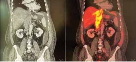
Figure 1: Grossly dilated IVC with a large FDG avid intraluminal heterogeneously enhancing soft tissue lesion in intrahepatic IVC, extending into infrahepatic IVC, upto renal hilar level and suprahepatic IVC upto right atrium.
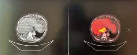
Figure 2: Grossly dilated IVC with a large FDG avid intraluminal heterogeneously enhancing soft tissue lesion in intrahepatic IVC.
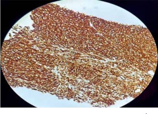
Figure 3: Immunohistochemistry showing tumor cells strongly positive for Vimentin
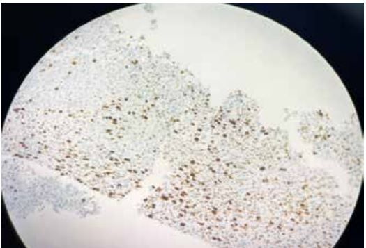
Figure 4: Immunohistochemistry showing Tumor cells patchy positive for Ki67
Discussion
Sarcomatoid carcinoma of the IVC poses diagnostic challenges due to its rarity and mimicking features of thrombotic conditions. Histopathological examination and immunohistochemistry play pivotal roles in differentiating it from benign thrombus. In our case, positive staining for Vimentin and patchy positivity for Ki67, CK, CK18, CD68, and Pax8, along with negative staining for SMA, S100, CD34, and other markers, supported the diagnosis.3
Treatment options for sarcomatoid carcinoma of the IVC remain limited, comprising surgical resection, chemotherapy, and radiotherapy.4 However, prognosis remains poor, with a high propensity for recurrence and metastasis.5 Novel therapeutic approaches, including targeted therapies and immunotherapy, warrant exploration to improve outcomes.
Despite advancements in diagnostic modalities and treatment strategies, standardized guidelines for the management of sarcomatoid carcinoma of the IVC are lacking. Further research and case reports are warranted to elucidate optimal diagnostic and therapeutic approaches, ultimately enhancing prognosis and survival rates for affected individuals.6
CONCLUSION:
Sarcomatoid carcinoma of the inferior vena cava presents a diagnostic challenge due to its rarity and mimicking features of thrombotic conditions. Clinicians must consider this malignancy in the differential diagnosis of IVC tumours, particularly in patients with atypical symptoms or inadequate response to conventional therapies. Further research is needed to establish standardized diagnostic and therapeutic guidelines, ultimately improving prognosis and survival rates.
References
- Srinivasan V, Mehta A, Levine E. Inferior Vena Cava Thrombosis. StatPearls [Internet]. Treasure Island (FL): StatPearls Publishing; 2022 Jan.
- Arshad HM, Fatehullah A, Razzaq S, Ali Z. Inferior Vena Cava Tumors: A Rare but Important Differential in the Setting of Deep Venous Thrombosis. Cureus. 2021 Mar;13(3):e14061.
- Rong J, Yu W, Rong Y, et al. Malignant mesenchymal tumor of the inferior vena cava: a case report and literature review. BMC Surg. 2017;17(1):67.
- Cahan WG. Radical resection of tumors of the inferior vena cava with reconstruction of the right atrium: report of a case. Ann Surg. 1951;133(4):637-643.
- Dedeilia A, Aravantinos G, Patra E, Pavlidis N. Inferior vena cava tumor thrombus from transitional cell carcinoma of the bladder. Indian J Urol. 2012;28(1):109-111.
- Krishnamurthy J, Rangan V, Reddy G, et al. Sarcomatoid renal cell carcinoma with tumor thrombus in inferior vena cava - a rare entity. Urol Case Rep. 2021;38:101732.