 WhatsApp Us (24/7)
 +91 926 888 0303 (24/7)


Abstract: Submucosal tumors (SMTs) of different etiologies exist from esophagus to rectum. Esophagogastric junction (EGJ) is one of the known difficult locations for tumor resection. Although minimally invasive surgery (MIS) is a well-established approach for gastrointestinal surgery, there is no consensus that MIS for resection of SMTs around EGJ is superior to laparotomy.
Key words: Leiomyoma, Laparoscopic Resection, Gastroesophageal Junction
Introduction
Leiomyoma is the most common smooth muscle tumor of the stomach and represents 80% of all gastric stromal tumors. About one-third of these neoplasms are located at the gastroesophageal junction.1,2 Leiomyomas are classically intramural and solitary on Computed Tomography (CT) Scan with smooth outlines and are multilobulated. Presence of calcifications is pathognomonic for leiomyomas.3 They are often asymptomatic or minimally symptomatic.4 Surgical removal can be required when the lesion is large or rapidly increasing in size. Resection of leiomyoma of the gastroesophageal junction has been traditionally performed via laparotomy.5
Case report
A 62-year-old lady presented to the hospital with chief complaint of difficulty in swallowing, feeling of heaviness in chest after consuming meals and reflux of gastric contents since 5-6 months. No physical abnormalities were observed. Upper GI endoscopy revealed large submucosal mass along the lesser curvature starting from the Gastro-esophageal junction extending to the proximal body of stomach suggestive of Large gastrointestinal stromal tumour (GIST), rest of the stomach normal. (Figure 1)
Positron emission tomography (PET) CT scan showed metabolically active well defined mass along the gastric wall protruding into the gastric cavity with scattered calcifications. (Figure 2) As mass was enhancing on PET Scan, possibility of the lesion being a gastrointestinal tumour was considered. After further evaluation she was taken up for surgery.
As the lesion was very close to EGJ and on lesser curvature, preoperative endoscopy was done and tumour margins were marked by dye to determine the exact site of tumour on serosal surface that would aid in resection and help in preservation of EGJ.
Laparosopy revealed extent of tumour on surface marked by dye (Figure 3). Stomach was opened just adjacent to the tumour keeping a margin of around 1 cm. Whole of tumor was removed keeping a margin of at least 1 cm all around with harmonic scalpel. (Figure 4) Posterior and left lateral wall of EGJ could be preserved using this method. (Figure 5) Few small nodules were also seen along main tumour and were also resected. Whole defect was closed in single layer with V-Loc 3-0 continuous suture in single layer. (Figure 6) Insufflation test was done intraoperatively to rule out any leak from suture line. Feeding jejunostomy was also done.
Feeding through jejunostomy was started from POD 1. Oral gastrograffin study performed on post operative day (POD) 5 showed free flow of contrast across EGJ with no leakage of dye across suture line. Oral diet was started after dye study. She tolerated orally well without any complaints of reflux and was discharged on POD 6.
Histopathology revealed spindle cell tumour that was negative for C-kit, CD34 receptors favouring diagnosis of leiomyoma.
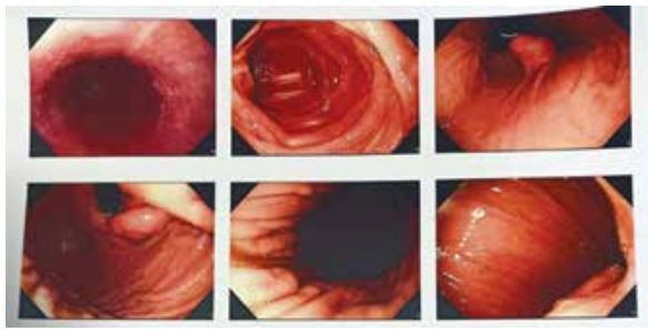
Figure 1: UGI endoscopy showing submucosal bulge near EGJ
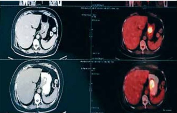
Figure 2: PET scan images showing hyperactive lesion along lesser curvature of stomach protruding into gastric cavity
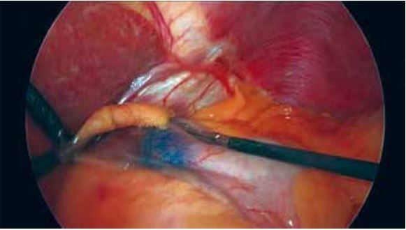
Figure 3: Methylene blue injected tumor
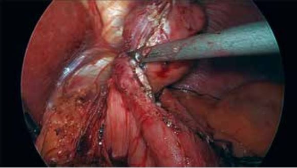
Figure 4: Tumor resection with margin
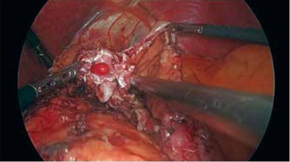
Figure 5: EGJ after resection of tumor showing Ryle’s tube in esophagus
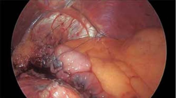
Figure 6: Reconstructed Gastro- esophageal junction
Discussion
The EGJ tumours pose a challenge to the surgeon due to a higher risk of deformity, stenosis and overall post-surgical complications that decrease life quality. Some surgical procedures suggested for EGJ tumours include open or laparoscopic approaches with or without fundoplication, laparoscopic trans gastric approach, combined endoscopic and laparoscopic approach, Proximal gastrectomy or even esophagectomy.6-11 There have been several series focusing on MIS for the resection of SMTs near the EGJ that concluded that it is a safe and rational approach.6-11
Huang et al.11 in their retrospective series of 909 patients of submucosal tumours (SMTs) divided the area of the EGJ based on endoscopic view into 4 zones for patients who underwent minor gastrectomy:
- zone 1 included tumours located at the ventral aspect (anterior wall) of the EGJ.
- zone 2 contained tumours located at the fundus (greater curvature) side of the EGJ.
- zone 3 included tumours located at the dorsal side (posterior wall) of the EGJ.
- zone 4 contained tumours located at the lesser curvature side of the EGJ area.
Majority of the tumours located in zone 2 were resected laparoscopically (82% versus 18%)
For SMTs in the zone 1, 3, and 4, they were more often resected by laparotomy
Excluding SMTs in the zone 2, the percentage of resection performed by laparotomy was 59%. They also observed that tumour with size more than 5 cms are more likely to undergo major gastrectomy or esophagogarectomy.
Herein we presented a case of large tumour, around 6 cms just below EGJ that was on lesser curvature thus making resection with stapler difficult. As tumour was endophytic, it was decided to mark the location of tumour preoperatively that helped us in siting exact location of gastrotomy near tumour and helped in resecting tumour with 1 cm margin thus saving EGJ at least more than 1/2circumference. Defect was closed and feeding jejunostomy was done as this site carries very high risk of leak. This technique also helped us in removing small lesions lying just adjacent to main lesion that became obvious when tumour was being resected thus providing complete resection of all tumour masses. Our patient did not have any symptoms of dysphagia or reflux postoperatively justifying our approach to resection.
References
- Appelman HD, Helwig EB. Gastric epithelioid leiomyoma and leiomyosarcoma (leiomyoblastoma). Cancer. 1976 Aug;38(2):708-28.
- Lee MJ, Lim JS, Kwon JE, Kim H, Hyung WJ, Park MS et al. Gastric true leiomyoma: computed tomographic findings and pathological correlation. Journal of computer assisted tomography. 2007 Mar 1;31(2):204-8.
- Jeong ID, Jung SW, Bang SJ, Shin JW, Park NH, Kim DH. Endoscopic enucleation for gastric subepithelial tumors originating in the muscularis propria layer. Surgical endoscopy. 2011 Feb;25:468-74.
- JL The,A Shabbir. Mastering Endo-Laparoscopic and Thoracic surgery; Chapter -Resection of Gastroesophageal junction tumors pp207- 211
- Shim CS, Jung IS. Endoscopic removal of submucosal tumors: preprocedure diagnosis, technical options, and results. Endoscopy. 2005 Jul;37(07):646-54.
- Shim JH, Lee HH, Yoo HM, Jeon HM, Park CH, Kim JG et al. Intragastric approach for submucosal tumors located near the Z-line: A hybrid laparoscopic and endoscopic technique. Journal of Surgical Oncology. 2011 Sep 1;104(3):312-5.
- Tagaya N, Mikami H, Kogure H, Kubota K, Hosoya Y, Nagai H. Laparoscopic intragastric stapled resection of gastric submucosal tumors located near the esophagogastric junction. Surgical endoscopy. 2002;16(1):177.
- Nguyen NT, Jim J, Nguyen A, Lee J, Chang K. Laparoscopic resection of gastric stromal tumor: a tailored approach. The American surgeon. 2003 Nov;69(11):946-50.
- Hwang SH, Park DJ, Kim YH, Lee KH, Lee HS, Kim HH et al. Laparoscopic surgery for submucosal tumors located at the esophagogastric junction and the prepylorus. Surgical endoscopy. 2009 Sep;23:1980-7.
- Song KY, Kim SN, Park CH. Tailored-approach of laparoscopic wedge resection for treatment of submucosal tumor near the esophagogastric junction. Surgical endoscopy. 2007 Dec;21:2272-6.
- Huang YC, Yeh CN, Chen MY, Wang SY, Liu KH, Tsai CY et al. Surgical options for submucosal tumors near the esophagogastric junction: does size or location matter?. BMC surgery. 2020 Dec;20:1-7.