 WhatsApp Us (24/7)
 +91 926 888 0303 (24/7)


Abstract:
Background: The coexistence of Rosai-Dorfman disease (RDD) with Langerhans cell histiocytosis (LCH) is very rare, as to date only a handful of cases have been reported. Though both the entities come under the spectrum of histiocytic disorders, the patient outcomes are very different for each one of them. RDD is a benign disorder with often a self-limiting course; whereas LCH has a more aggressive outcome. The pathobiology of this rare co-occurrence is still unknown and the topic of several theories.
Case presentation: We report a case of a 2-year-old female patient who presented with fever, pallor, lymphadenopathy and neck pain. X-ray radiograph and Computed Tomography (CT) scan chest showed multiple lytic lesions on vertebra. The biopsy finding of lymph node and bone marrow were consistent with a combined RDD-LCH disease.
Conclusion: Combined RDD-LCH presentation is very rare and it’s pathophysiology is still elusive and a topic of numerous conjectures.
Key words: Langerhans Cell Histiocytosis, Rosai-Dorfman Disease
Introduction
Langerhans cell histiocytosis (LCH) is a rare, aggressive disease characterized by clonal expansion of CD1a+/langerin+ dendritic cells causing tissue destruction and has a predilection for pediatric age group. On the other hand, Rosai-dorfmann disease (RDD) is characterized by benign histiocytic proliferation in lymph node sinuses. The coexistence of these two diseases is rare and only a handful of cases have been reported in the literature. We herein, describe a 2-year-old male patient who presented with RDD in the cervical lymph node and later found to have LCH in the bone marrow along with multiple lytic lesions in the vertebra.
Case
A 2-year-old female presented with fever and neck pain for 1.5 months. She also had complaint of breathlessness and pallor for last 1 month, for which she had one unit blood transfusion. On physical examination, she was found to have multiple palpable non tender left cervical lymphadenopathy but no organomegaly.
Primary investigations
Peripheral smear (post transfusion) showed moderate microcytic hypochromic anemia (Hb: 10.9gm/dl) with neutrophilic leucocytosis (Total Leukocyte Count (TLC): 38,000/cumm) and thrombocytosis (Total Platelet Count (TPC):6,26,000/cumm). Serum iron and transferrin saturation were low with normal.
Total iron binding capacity (TIBC) and ferritin levels. C-reactive protein (CRP) was markedly raised (55.49 mg/L). Ultrasound (USG) guided fine needle aspiration cytology (FNAC) done from the lymph node showed cellular smears with predominantly histiocytic cells in a polymorphous lymphoid background and few showing emperipolesis [FIGURE: 1A]. No well-defined granuloma or necrosis or evidence of malignancy was seen. Ziehl-Neelsen (ZN) stain for acid-fast bacilli (AFB) was negative. X-ray spine, positron emission tomography (PET) CT, Bone marrow examination and lymph node biopsy, were advised simultaneously.
On lateral x-ray of cervical spine, reduced C5 vertebral height was noted suggestive of collapse. PET-CT revealed multiple lytic lesions involving skull base, left scapula, sternum, multiple vertebrae including C5 collapse, sacrum, right ileum, right pubis and bilateral femur. Additionally, Para aortic and mesenteric lymph nodes along with right Para pharyngeal and bilateral cervical lymph nodes showed metabolically active disease.
Bone marrow aspirate showed cellular marrow with trilineage haematopoiesis and scattered histiocytic cells [Figures: 1B and C] and biopsy showed large sheets and aggregates of histiocytic cells [Figures: 1D and E]. Occasional histiocytic showed nuclear grooving. Few histiocytes show emperipolesis.
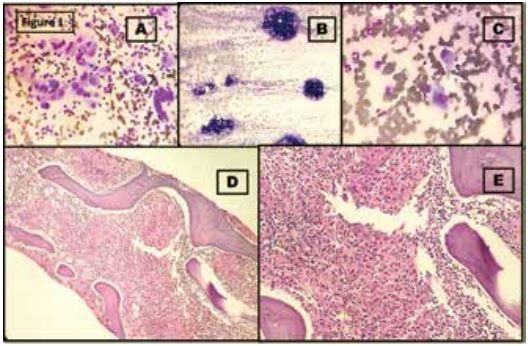
Figure 1: [A] High power (40X) view of lymph node FNAC showing histiocytic clusters in a background of lymphocytes and emperipolesis. [B] Wright- Giemsa stained scanner view of bone marrow aspirate. [C] Scattered histiocytes in bone marrow imprint. No nuclear groove is seen. [D] H&E stained bone marrow biopsy section scanner view. [E] High power (40X) magnification view of bone marrow biopsy showing infiltration of marrow by histiocytes.
Differential diagnosis and laboratory work up
A differential diagnosis of multisystem RDD and LCH were considered. CD1a immunohistochemistry was ordered on bone marrow biopsy and simultaneously lymph node biopsy were ordered to assess multisystem involvement.
Confirmation of the diagnosis
On immunohistochemistry, CD1a and S100 were diffusely positive in histiocytic cells in the bone marrow biopsy section [Figures 2 A and B], concluding a diagnosis of LCH. However, the cervical lymph node biopsy done afterwards showed a slightly different picture. Lymph nodal architecture was found to be effaced at places with large aggregates of histiocytic cells [Figure 2 C]. However, histiocytic cells lack typical LCH type morphology with nuclear grooving or coffee-bean type nucleus. The histiocytes are diffusely positive for CD68, but only a few clusters are showing positivity for CD1a [Figures 2 D, E and F]. A large number of histocytes are negative for CD1a and few of them showing emperipolesis.
Considering all investigational findings, a final diagnosis of LCH with concurrent RDD in cervical nodes was furnished.
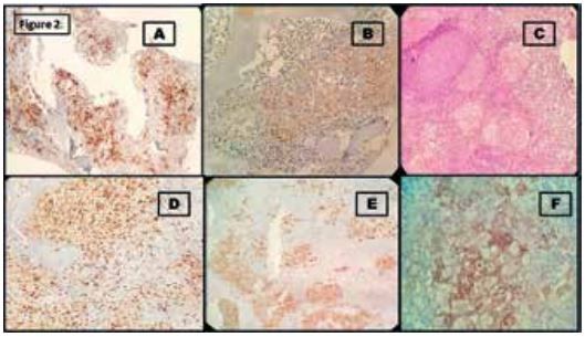
Figure 2: [A] 20X view of CD1a IHC on bone marrow biopsy. Histiocytic cells are positive for CD1a. B] 40X view of S-100 IHC on bone marrow biopsy. Histiocytic cells are positive for S100. [C] H&E stained section (40X) of lymph node biopsy shows lymph nodal architecture effacement and collection of histiocytes. [D, E, F] High power (40X) view of CD68, S-100 & CD1a IHC respectively on lymph node biopsy. Histiocytic cells are positive for CD68; however not all histiocytes are positive for CD1a. Only a few histiocytic aggregates are positive for CD1a, others are negative.
Discussion
Both RDD and LCH are rare diseases and their coexistence is even rarer.1 Less than 20 cases have been reported in the different literature, since the first case reported by Wang KH et al.2 The largest case series was reported by O’Malley DP et al.1 Every recurrence that was documented demonstrated that the pattern of connection was either synchronous, involving the same organ, or metachronous, involving the same organ or a different organ, with RDD appearing as a secondary event.3 The reported morphological pictures are often an admixture of signature features of both RDD and LCH.1,3
RDD is an abnormal proliferation of histiocytes and has been classified into variant subgroups i.e. classical, familial, extra nodal, neoplasia associated, and immune associated etc. Though emperipolesis is a prominent finding in RDD, the same
is nonspecific and have been documented in Erdheim-Chester disease, juvenile xanthogranuloma, and malignant histiocytosis.4 Unlike RDD, LCH usually show distinct morphological features like nuclear grooving in Langerhans cells and are often associated with abundant eosinophils and necrosis.
In our case, the first eye-catching feature was emperipolesis in lymph node FNAC and typical LCH type coffee bean nuclear groove was not well appreciated in bone marrow as well as in lymph nodes. But, at immunohistochemistry, histiocytic cells in bone marrow biopsy were strongly CD1a positive, only some cells with emperipolesis were negative for this marker. Examining CD1a + cells closely reveals that their shape is primarily round to oval, and strongly mimicking the “Banks cells” as proposed by O’Malley DP et al.1 These cells have morphological characteristics that resemble those of RDD, i.e. large round nuclei, open chromatin, abundant eosinophilic cytoplasm, but immunohistochemically it shows a strong CD1a expression. Between conventional LCH cells and RDD, these cells appeared to have transitional properties.1 These “Banks cells” are a major component in the bone marrow of our case. Therefore, we may describe our current instance as an RDD-LCH with "Banks cells," in sync with other authors. In our case, there was no accompanying eosinophilia or necrosis either, which are commonly found in LCH.
LCH typically presents in a continuum of systemic involvement that ranges from a solitary eosinophilic granuloma to widespread disseminated disease leading to multiorgan dysfunction.5 Our patient had multiple lytic bony lesions involving both axial and appendicular skeletal system in addition to cervical as well as retroperitoneal lymphadenopathy.
The pathobiology of LCH lies in BRAFV600E or alternative activating MAPK pathway gene mutations.6 In contrast, approximately half of the cases of RDD reveal a normal mutational profile and only 30% cases demonstrate somatic mutations involving NRAS, KRAS, MAP2K1, CSF1, and CBL genes. Among these genes, MA2P2K1 and KRAS have been found to be the most frequent genetic aberrancies.4,7
Prognosis in RDD is usually favorable in most of the cases; however, CNS involvement has been associated with variable outcomes.2 LCH on the other hand, has a relatively poor outcome with a significantly lower median overall survival in cases with multiorgan involvement. Late consequences are relatively higher with diabetes insipidus, orthopedic abnormalities, growth retardation, hearing loss, and neurodegeneration being the commonest morbidities.6
It is clear from numerous documented instances of RDD-LCH that the RDD evolved after the original LCH. Nevertheless in our instance, the RDD and LCH were identified concurrently in the lymph node and bone marrow, arguing against secondary histioctosis and in favour of the concurrent RDD-LCH spectrum diagnosis.
Learning points
A very small number of cases have been documented in the existing literature about the association between RDD and LCH. There are still many theories regarding the process underlying the coexistence of these entities, and further research is needed to solve this mysterious association.
Acknowledgments:
None
Funding source:
None
Conflict of interest::
None
References
- O'Malley DP, Duong A, Barry TS, Chen S, Hibbard MK, Ferry JA et al. Co-occurrence of Langerhans cell histiocytosis and Rosai-Dorfman disease: possible relationship of two histiocytic disorders in rare cases. Mod Pathol. 2010 Dec;23(12):1616-23.
- Wang KH, Cheng CJ, Hu CH, Lee WR. Coexistence of localized Langerhans cell histiocytosis and cutaneous Rosai-Dorfman disease. Br J Dermatol. 2002; 147(4):770–4.
- Cohen-Barak E, Rozenman D, Schafer J, Krausz J, Dodiuk-Gad R et al. An unusual co-occurrence of Langerhans cell histiocytosis and Rosai-Dorfman disease: report of a case and review of the literature. Int J Dermatol. 2014; 53(5):558–63.
- Elbaz Younes I, Sokol L, Zhang L. Rosai–Dorfman Disease between Proliferation and Neoplasia. Cancers. 2022; 14(21):5271.
- Haupt R, Minkov M, Astigarraga I et al; Euro Histio Network. Langerhans cell histiocytosis (LCH): guidelines for diagnosis, clinical work-up, and treatment for patients till the age of 18 years. Pediatr Blood Cancer. 2013;60(2):175-184.
- Héritier S, Barkaoui MA, Miron J et al. Incidence and risk factors for clinical neurodegenerative Langerhans cell histiocytosis: a longitudinal cohort study. Br J Haematol. 2018;183(4):608-617.