 WhatsApp Us (24/7)
 +91 926 888 0303 (24/7)


Abstract: Cardiovascular Disease (CVD) is a leading cause of morbidity and mortality in women worldwide. Biologic and sociocultural factors account for the variation in presentation and natural history of CVD in women. Research in the last two decades focusing on CVD in women has led to an improved understanding of conventional and novel risk factors, sex specific pathophysiology of Ischemic heart disease (IHD). The salient features of IHD in women are a higher prevalence of risk factors, angina, lower burden of obstructive coronary artery disease and a poorer prognosis in comparison with men. Evaluation and management of disease with higher prevalence in women including myocardial infarction with non obstructive coronary arteries (MINOCA), stress induced cardiomyopathy (Takotsubo syndrome) and spontaneous coronary artery dissection (SCAD) are discussed.
Key words: Cardiovascular Disease (CVD), Ischemic Heart Disease (IHD), Microvascular Coronary Dysfunction (MCD) Myocardial Infarction with Non Obstructive Coronary Arteries (MINOCA), Spontaneous Coronary Artery Dissection (SCAD)
Introduction
Cardiovascular disease (CVD) is the leading cause of mortality and a significant cause of morbidity in women.1 Women have been conventionally considered more protected from heart disease and have been ‘understudied, under-recognized, underdiagnosed and undertreated’. Biological differences between the sexes, as result of genetic, epigenetic and sex hormone-mediated factors, are complex and incompletely understood. In addition, social and cultural (gender) factors contribute to the variation in the prevalence, presentation and natural history of cardiovascular disease in women.2
Menopause marks an important cardiovascular biological transition, with an increase in CVD risk in women > 55 years age. Ischemic heart disease (IHD) better describes the expanded spectrum of disease in women - it includes atherosclerotic obstructive Coronary Artery Disease, Coronary Microvascular Dysfunction, endothelial dysfunction, vasomotor abnormalities, Spontaneous Coronary Artery Dissection (SCAD), Stress related Cardiomyopathy and Myocardial Infarction Normal Coronary Arteries (MINOCA). The salient features of IHD in women are a higher prevalence of angina, lower burden of obstructive coronary artery disease and a poorer prognosis in comparison with men.
Evolving knowledge of sex-specific presentations, improved recognition of conventional and novel risk factors, and expanded understanding of the sex-specific pathophysiology of ischemic heart disease have resulted in improved clinical outcomes in women.
Pathophysiology of IHD in women
It is known that women have smaller coronary arteries per 100 g of left ventricular mass and their arteries have increased stiffness. Plaque rupture with disrupted fibrous cap, large necrotic core with macrophages to be more prevalent in men and older women. Plaque erosion, is more common in younger women, there is an abundance of smooth muscle cells in a proteoglycan / collagen rich matrix, intimal thickening, absence of surface endothelium and lipid core. There is increased frequency of distal microembolisation from platelet rich debris. Plaque erosion is associated with less thrombus burden, less calcification, less percentage vessel stenosis, less overall plaque burden, minimal inflammation and negative arterial remodelling.3,4
Calcified nodule, that represent fibrocalcific plaques with little or no underlying necrotic core but there is disruption of the luminal surface ie absence of collagen and endothelium with luminal thrombosis overlying the eruptive dense calcified nodules.
The role of microvascular dysfunction, vascular inflammation, coronary reactivity, endothelial function, hormonal influences, oxidative stress and coronary size on development of IHD has been established.
In the presence of endothelial dysfunction, there is an increased vasoconstrictor response to autonomic stimuli and increased platelet aggregation, leukocyte migration and thrombus formation.
Microvascular Coronary Dysfunction (MCD)
The smaller coronary arteries that cannot be seen on angiography constitute the coronary microcirculation regulates coronary blood flow, growth, inflammation, coagulation and permeability. It is the adaptive properties of these vessel to metabolic stimuli that determine coronary blood flow and thereby the matching of myocardial perfusion to metabolic demand. Attenuated coronary dilation response to adenosine less than 2.5 fold is a marker of MCD.5
Cardiovascular risk factors in women
The classic risk factors for CVD are comparable in women and men, but gender differences in the prevalence of each risk factor and unique factors exist for women. Hypertension, diabetes mellitus and smoking are more potent risk factors for MI in women. Non traditional risk factors unique to women include early menopause or menarche, gestational diabetes mellitus, hypertension, pre eclampsia and eclampsia during pregnancy, and systemic inflammatory disorders. Women more often have multiple risk factors.
Established risk factors
Age: The prevalence of CVD increases with age, 1 in 8 women 45 – 64 years have CVD while 1 in 3 women older than 65 have CVD.6
Hypertension: Women older than 60 years have a higher prevalence and poorer control of hypertension than men. They are more likely to develop isolated systolic hypertension, reflecting aortic stiffness, and have a higher prevalence of stroke and heart failure with preserved ejection fraction. Hypertension rises two to three fold in women taking oral contraceptives.
Diabetes: The relative risk for CVD was 44% greater in women with DM than in similarly affected men. The risk of fatal CHD is 3.5 times that in a non diabetic woman. Gestational diabetes doubles the risk of developing diabetes and remains a life long risk factor. Diabetic women have a higher risk of developing HF, stroke, peripheral vascular disease compared with diabetic men and have higher mortality after myocardial infarction.7
Smoking: Although women smoke lesser than men, it may be more detrimental in them. The combination of smoking with oral contraceptive use has a synergistic effect on risk of acute myocardial infarction (MI), stroke, and venous thromboembolism.8,9
Dyslipidemia: Adverse changes in lipids accompany menopause, with increase in LDL cholesterol, Triglycerides and decrease in HDL. Dyslipidemia has the highest population-adjusted risk among women, at 47.1%, compared with all other known risk factors for CVD. Pharmacological therapy of hyperlipidemia for secondary prevention has clearly been shown to be equally effective in women and men for reduction of recurrent cardiac events and mortality.10,11
Family History: Premature CHD in a first degree female relative is a relatively more potent family history risk factor than in male relative.
Physical activity and fitness
Women are more likely to be sedentary, inactivity increases with age. Physical inactivity is associated with higher blood pressure, worse cholesterol levels, poorer glucose metabolism, poor mental health and obesity. Exercise capacity strongly and independently predicts all cause mortality in women.
Emerging risk factors
Metabolic Syndrome: Women are more likely than men to have metabolic syndrome, the relative risk of developing CVD is increased 2.6 fold. In the Framingham Heart Study, obesity increased the relative risk of CAD by 64% in women, as opposed to 46% in men.12
Autoimmune Disease: Women with autoimmune diseases to develop accelerated CVD.26 The female to male ratio for rheumatoid arthritis is 2.5:1 and for systemic lupus erythematosus is 9:1. Patients with rheumatoid arthritis have a 2- to 3-fold higher risk of MI and a 50% higher risk of stroke. Young women with SLE were 2.27 times more likely than their age matched peers without SLE, to be hospitalised because of AMI, 3.8 times more likely to be hospitalised because of congestive cardiac failure and 2.05 times more likely to be hospitalised because of cerebrovascular accident.13,14,15
Gestational Hypertension: Gestational Hypertension of any sort is associated with an increased risk of hypertension, chronic kidney disease, diabetes, stroke, CVD. Women with pre eclampsia, have approximately double the risk of subsequent ischemic heart disease, stroke and venous thromboembolic events. Pregnancy is a stress test for future cardiovascular events.16
Polycystic Ovarian Disease: Disruption of ovulatory cycling, indicated by estrogen deficiency and hypothalamic dysfunction or irregular menstrual cycling in premenopausal women is associated with an increased risk of coronary atherosclerosis and adverse CVD events. Polycystic ovary syndrome is prevalent in 10% to 13% of women and is linked with a clustering of risk factors, incident type 2 diabetes mellitus, and adverse IHD events.17
Breast Cancer Therapy: Survivors of breast cancer have an elevated risk of developing CVD. Exposure of the heart to ionising radiation during radiotherapy for breast cancer increases the rate of developing IHD. The risk is proportionate to the mean dose of radiation to the heart., with an increase in CVD events of 7.4% per 1 Gy of radiation. The mean radiation received is 4.9 Gy. The risk of IHD begins within a few years after exposure and appears to continue for 20 years after exposure. The absolute risk is highest in those women with pre existing CVD risk factors.18
Hysterectomy as a risk factor for coronary artery disease: Hysterectomy is the most common non obstetrical procedure performed on women. The Swedish hospital discharge registry data on 1,84,441 women who underwent hysterectomy between 1973 – 2003 has shown that hysterectomy in women aged 50 years or younger substantially increases the risk for cardiovascular disease later in life. Oopherectomy further adds to the risk of both coronary heart disease and stroke.19
Psychosocial factors
In the INTERHEART study, an aggregate exposure to psychosocial risk factors, including depression, perceived home/ work stress, low locus of control, and major life events, was significantly associated with AMI in women, with an adjusted odds ratio of 3.5.20
Among women with MI or other forms of ischemic heart disease, depression is associated with an ≈3-fold increased risk for death or subsequent cardiac events independently of severity of depression. those with higher social support experience better mental functioning, better quality of life, and fewer depressive symptoms at 12 months.21
Clinical spectrum of IHD
The Framingham study previously suggested that development of CAD in women lags approximately 10 years behind men, however current data shows women are susceptible to CAD at a much younger age. The mean age of women in the Kerala ACS registry was 64.4±11.7 years, they were older than men by 5 years (mean age in men 59.3 ± 11.9 years, p<0.001).22
Majority of patients present with chest pain, women are more likely to be overwhelmed with by a multiplicity of associated symptoms - back, neck and jaw pain, dyspnea, paroxysmal nocturnal dyspnea, nausea, indigestion,and weakness or fatigue. Women were more likely than men to present without chest pain (37% vs 27%) (85) Women more commonly present with NSTEMI and non obstructive coronary artery disease (CAD).
Women were more likely to delay seeking care, had more diabetes, hypertension and heart failure with preserved ventricular systolic function. Mechanical complications and HF are more likely to develop in women, whereas ventricular arrhythmias occur at similar rates in both sexes after an AMI. Women with ACS are treated less aggressively, with fewer catheter-based interventions, fibrinolytic and bypass surgical procedures, resulting in less favourable clinical outcomes with higher mortality and lower health-related quality of life. Compared with men, women with ACS and those after coronary revascularization have longer hospitalizations and higher in-hospital mortality, manifest more bleeding complications, and up to 30% more readmissions within 30 days after the index hospitalization.
Regardless of age, within a year of a first AMI, more women than men will die (26% of women and 19% of men); within 5 years of a first AMI, more women than men will die (47% of women and 36% of men), have heart failure (HF), or suffer from a stroke. Studies report a consistent high risk of mortality after AMI in younger women. The mechanisms, likely multifactorial, contributing to excess risk and inferior health among young women remain unclear.23,24,25,26
Ischemia With Non Obstructive Coronary Arteries (INOCA)
Cardiac ischemia without obstructive CAD is caused by CMD and coronary spasm that cause mismatch between blood supply and myocardial oxygen demand..INOCA is more common in women 45 – 55 years age, a prevalence 34.4% to 62% has been reported. Early diagnosis and treatment is essential because of the elevated risk of major adverse cardiac events.27
Myocardial Infarction with Non Obstructive Coronary Arteries (MINOCA)represents up to 10% of all acute MIs, and is diagnosed more frequently in women, particularly younger women.Conventional risk factors hyperlipidemia and diabetes are less frequent, a higher prevalence of hypertension is seen. It is caused by plaque erosion, distal embolisation and endothelial and microvascular vascular dysfunction that amplify the consequences of an upstream spastic or thrombotic event. Patients with MINOCA have heightened CVD risk - an all-cause in-hospital mortality of 0.9%, a 2% risk of death or reinfarction over 6 months and a 15% readmission rate within 6 months and 12-month mortality of 4.7%. Up to 25% of MINOCA patients report persistent angina following MI, and experience similar rates of heart failure,hospitalization, repeat testing due to angina than their counterparts with obstructive CAD.At 10 years, CVD death or MI occurred in 6.7% of those with no evident angiographic CAD and in 12.8% among those with non obstructive CAD.16
In the VIRGO study, 11.1% had MINOCA, women had 5 times higher odds of having MINOCA than men (14.9% versus 3.5%), were likely to be premenopausal. Non plaque mechanism was responsible in 25.1%, one and 12 month mortality was 1.1% and 0.6% respectively.28
Coronary artery spasm
CAS is a well-known phenomenon for recurrent chest pain episodes at rest with associated transient ST-segment elevation, but it is also a rare mechanism for AMI. The pathogenesis of CAS is multifactorial and includes vagal withdrawal, vascular smooth muscle hyperactivity, endothelial dysfunction, and an imbalance of the autonomic nervous system. Cigarette smoking is a major risk factor for CAS, and possible triggers include variation in autonomic activity, cocaine use, ephedrine alkaloids, and other drugs.
Tako Tsubo cardiomyopathy, is a clinical entity characterized by acute, rapidly reversible systolic and diastolic LV dysfunction, triggered by profound psychological or physical stress. Signs and symptoms of myocardial ischemia occur in the absence of obstructive coronary artery disease. A distinctive abnormality of LV contraction consists of akinesia or dyskinesia of the apical and / or mid ventricular segments of the left ventricle (“apical ballooning”), together with hypercontractility of the base. This distinctive contraction pattern usually recovers on follow up, is the sine qua non of Takotsubo Cardiomyopathy (TTC).
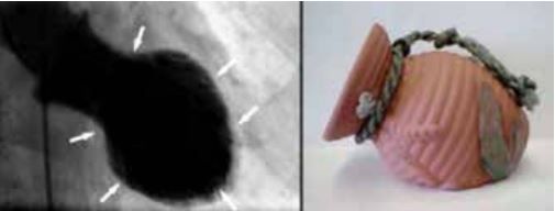
Figure 1: Left ventricular shape in Takotsubo cardiomyopathy resembles a Tako (octopus) tsubo (trap)
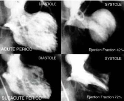
Figure 2: Left ventriculogram demonstrating apical ballooning in the acute stage and recovery after 2 weeks
Prevalence
TTC, accounts for1.7% to 2.2% of all patients admitted with a presumed diagnosis of acute coronary syndrome; this rate increases up to 6–7% if only females are considered. In the HORIZON – AMI trial, Takotsubo cardiomyopathy was diagnosed with a frequency of 0.5% of all patients and 2.1% of the female patients.
The cause of TTC remains unknown, the role of brain heart axis, exaggerated sympathetic stimulation and the negative inotropic effect of high-dose epinephrine on the LV myocardium have been postulated. The sympathetic innervation gradient has been reported in normal human hearts with a density of sympathetic nerve endings approximately 40% higher in the basal myocardium than in the apical myocardium and the left ventricular apex contains a higher density of β adrenoceptors, making it more susceptible to catecholamine-induced microvascular dysfunction and to direct myocyte toxicity. The heart stands out among organs of the body in terms of dependence on neuronal uptake for inactivating catecholamines in the extracellular fluid. Extremely high serum catecholamine levels observed in patients with TTC - there is a negative β2 adrenoreceptor mediated inotropic effect – which is more pronounced at the apex and completely reversible on removal of the catecholamine stimulus.
In the International Takotsubo Registry, of the 1750 patients 89.8% were women (mean age, 66.8±13.0 years), and 79.1% were women older than 50 years of age. The predominant symptom on admission was chest pain (75.9%), followed by dyspnea (46.9%) and syncope (7.7%). Physical triggers were more frequently present than emotional triggers (36.0% vs. 27.7%), whereas 7.8% of patients had both triggers. In 28.5% of patients, TTC occurred without any evident trigger. Apical TTC was identified in 81.7% of patients, whereas the mid-ventricular form was found in 14.6%, and basal and focal forms were diagnosed in 2.2% and 1.5%, respectively. 27.0% of patients with TTC had an acute, former, or chronic history of a neurologic disorder, 42.3% had received a diagnosis of a psychiatric illness, 21.8% of patients had a combined end point of serious in-hospital complications, cardiogenic shock in 9.9%, 17.3% required ventilatory support, ventricular tachycardia (3.0%), ventricular thrombus (1.3%), and ventricular rupture (0.2%) and mortality of 4.1%. Long-term follow-up of patients with TTC revealed a rate of death from any cause of 5.6% per patient-year and a rate of major adverse cardiac and cerebrovascular events of 9.9% per patient-year. Among these patients, the incidence of myocardial infarction was relatively low, given that the rate of stroke or transient ischemic attack was 1.7% per patient-year and the rate of recurrence of TTC was 1.8% per patient-year, with a span of 25 days up to 9.2 years after the first event.29
Spontaneous coronary artery dissection (SCAD)
Over the past decade, Spontaneous Coronary Artery Dissection (SCAD), has been recognised as a nonatherosclerotic, nontraumatic cause of acute coronary syndrome and sudden cardiac death. SCAD is characterised by the presence of blood entering and separating the layers of the coronary arterial wall to form a false lumen. This leads to external compression of the true coronary lumen restricting coronary blood flow and leading to coronary insufficiency and myocardial infarction.
Women comprise 87% to 95% of SCAD with a mean age of presentation between 44 and 53 years. Single-center studies have estimated a prevalence of SCAD as high as 4% of patients presenting with acute coronary syndrome (ACS) and the underlying cause of up to 35% of all ACS cases in women ≤ 50 years of age, with 20% to 25% of cases occurring in the peripartum period. The classic presentation is of a young healthy woman, without traditional ASCVD risk factors, and sudden onset of ACS.30
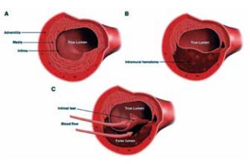
Figure 3: Cross-sectional views of the coronary artery. A, Normal coronary artery. B, Coronary artery with intramural hematoma. C, Coronary artery with intimal tear. Spontaneous coronary artery dissection is characterized by the spontaneous formation of an intramural hematoma, which can lead to compression of the true lumen and myocardial infarction. An intimal tear may be present
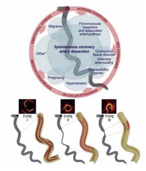
Figure 2: Associated Conditions, Inciting Factors, and Angiographic Diagnosis of Spontaneous Coronary Artery Dissection Hayes, S.N. et al. J Am Coll Cardiol. 2020;76(8):961– 84
If hormones do play a role, it is unknown whether it is the absolute levels and/or fluctuations in circulating estrogen and progesterone that affect the process. Temporally, SCAD has been reported to occur just before or during menstruation while taking hormonal contraceptives, postmenopausal hormone therapy and in women with a history of infertility and/or prior or current treatment for infertility.
Pregnancy-associated spontaneous coronary artery dissection (P-SCAD) may occur at any time during or after pregnancy; the majority (>70%) occur postpartum, most commonly within the first week. P-SCAD comprises <5% to 17% of SCAD cases overall and 14.5% to 43% of pregnancy-associated AMI.
P-SCAD patients tend to have more severe clinical presentations with impaired left ventricular function, shock, left main, and multivessel dissections. Both P-SCAD and non–P-SCAD patients are more frequently multiparous and report a higher prevalence of prior infertility treatment and pre-eclampsia.
SCAD can be associated with multifocal Fibro Muscular Dysplasia (FMD) that can affect nearly any arterial bed and can manifest as arterial stenosis, aneurysm, tortuosity, or dissection. Screening for extracoronary arteriopathies with brain to pelvis imaging is recommended.32,33
SCAD is diagnosed at coronary angiography. The Yip-Saw angiographic classification of SCAD divides it into three types: In type 1 SCAD, contrast penetrates into the false lumen and there may be dye ‘hang up.’ Less than one-third of SCAD presents as type 1 and can suggest late presentation. Type 1 is less likely to progress and best managed conservatively. Type 2 SCAD is the most common and appears as a long smooth narrowing, often tapering distally, either with distal reconstitution of a normal vessel or extending into the terminal branches. Type 3 SCAD mimics atherosclerosis and requires intracoronary imaging to make the distinction.
Three approaches to help distinguish SCAD from atherosclerosis in ambiguous cases include presence of luminal thrombus (atherosclerosis), intracoronary imaging, and coronary computed tomography angiography. The classic intravascular ultrasound feature of SCAD is the triple band (white-black-white) of the intimal-media membrane and is pathognomonic for SCAD. Optical coherence tomography is preferred due to its higher spatial resolution and better ability to visualize the intimal medial membrane, false lumen, and external elastic membrane.
The left anterior descending artery is the most commonly affected (32%–46% of cases). In the majority of cases, the mid to distal segments of coronary arteries are affected; in only <10% of cases are the proximal left anterior descending or circumflex, right coronary, or left main arteries affected. Multivessel SCAD occurs in 9% to 23% of cases.
The substantial rate of spontaneous vascular healing, suggests a role for conservative management in stable SCAD patients with preserved distal coronary flow. PCI for SCAD has been associated with lower technical success and higher complications and should be reserved for a select group of patients with ongoing acute ischemia.
The focus of management is on relieving symptoms with optimal medical therapy (e.g., blood pressure control, antianginals, analgesia). Recommended therapy includes a single antiplatelet, avoidance of statin, use of beta blockade, avoidance of systemic hormones including oral contraceptives. Statins were associated with recurrent SCAD; therefore, are recommended only when hyperlipidemia is documented. The length of hospital to be individualized according to perceived risk but erring toward longer admission than for uncomplicated atherosclerotic cases.
Patients should be encourages to pursue regular moderate exercise, they should avoid extreme endurance training, exercising to exhaustion, elite competitive sports, or vigorous exertion in extremes of ambient temperature. Additionally, patients should avoid lifting or carrying heavy objects that require straining or prolonged Valsalva
The post-discharge major adverse cardiac events (MACE) rate of 2.7% at 30 days following a median 4-day length of stay has been reported. There is a high recurrence rate of 17% (occurring solely in women), a 10-year mortality rate of 7.7%, and a high MACE (death, recurrent SCAD, MI, and HF) rate of 47.4%.31,34,35
CONCLUSION:
CVD is a leading cause of mortality in women. In an environment where cardiologists have traditionally been trained to equate IHD with angiographically defined obstructive CAD, failure to recognize unique aspects of IHD in women has contributed to less aggressive preventive and therapeutic interventions in women relative to men.
Thus, a paradigm shift beyond solely an anatomic description of obstructive CAD is needed to translate into earlier IHD risk detection and treatment for women. Women, including young women, must be educated that the female heart is vulnerable to coronary heart disease and that risk recognition and intervention when appropriate has the potential to delay or avert coronary events. Rarely do cardiac risk factors occur in isolation; rather, there is a synergistic action among several risks factors that exacerbates the disease burden. A holistic approach to prevention and treatment of disease will improve clinical outcomes for patients.
References
- Bots SH, Peters SAE, Woodward M. Sex differences in coronary heart disease and stroke mortality: a global assessment of the effect of ageing between 1980 and 2010. BMJ Glob Health. 2017;2:e000298. doi: 10.1136/ bmjgh-2017-000298.
- McSweeney JC, Rosenfeld AG, Abel WM, Braun LT, et al Preventing and experiencing Ischemic Heart Disease as a woman : State of the Science. A scientific statement from the American Heart Association Circulation2016;133:1302-1331.
- Virmani R, Burke AP, Farb A, Kolodgie FD. Pathology of the vulnerable plaque. J Am Coll Cardiol. 2006;47(suppl):C13–C18. doi: 10.1016/j. jacc.2005.10.065.
- Schwartz RS, Burke A, Farb A, Kaye D, Lesser JR, Henry TD, Virmani R. Microemboli and microvascular obstruction in acute coronary thrombosis and sudden coronary death: relation to epicardial plaque histopathology. J Am Coll Cardiol. 2009;54:2167–2173. doi: 10.1016/j. jacc.2009.07.042.
- Kothawade K, Merz CNB Microvascular dysfunction in women – pathophysiology, diagnosis and management Curr Probl Cardiol 2011;36:291-318.
- Lerner DJ, Kannel WB,.Patterns of coronary heart disease morbidity and mortality in the sexes : a 26 year follow up of the Framingham population. Am Heart J 1986 : 111: 383 – 90.
- Huxley R, Barzi F, Woodward M. Excess risk of fatal coronary heart disease associated with diabetes in men and women: meta-analysis of 37 prospective cohort studies. BMJ. 2006;332:73–78. doi: 10.1136/ bmj.38678.389583.7C.
- Huxley RR, Woodward M. Cigarette smoking as a risk factor for coronary heart disease in women compared with men: a systematic review and meta-analysis of prospective cohort studies. Lancet. 2011;378:1297–1305. doi: 10.1016/S0140- 6736(11)60781-2.
- Lidegaard O. Smoking and use of oral contraceptives: impact on thrombotic diseases. Am J Obstet Gynecol. 1999;180:S357– S363.
- Langendorfer A, Stein EA, Kruyer W, Gotto AM Jr. Primary prevention of acute coronary events with lovastatin in men and women with average cholesterol levels: results of AFCAPS/ TexCAPS. Air Force/Texas Coronary Atherosclerosis Prevention Study. JAMA. 1998;279:1615–1622.
- Pedersen TR, Kjekshus J, Berg K, Haghfelt T, et alScandinavian Simvastatin Survival Study Group. Randomised trial ofcholesterol lowering in 4444 patients with coronary heart disease: theScandinavian Simvastatin Survival Study (4S). 1994. Atheroscler Suppl. 2004;5:81–87. doi: 10.1016/j. atherosclerosissup.2004.08.027
- Wilson PW, D’Agostino RB, Sullivan L, Parise H, Kannel WB. Overweight and obesity as determinants of cardiovascular risk: the Framingham experience. Arch Intern Med. 2002;162:1867– 1872.
- del Rincón I, Polak JF, O’Leary DH, Battafarano DF, Erikson JM, Restrepo JF, Molina E, Escalante A. Systemic inflammation and cardiovascular risk factors predict rapid progression of atherosclerosis in rheumatoid arthritis. Ann Rheum Dis. 2015;74:1118–1123. doi: 10.1136/ annrheumdis-2013-205058.
- Gianturco L, Bodini BD, Atzeni F, Colombo C, Stella D, Sarzi-Puttini P, Drago L, Galaverna S, Turiel M. Cardiovascular and autoimmune diseases in females: The role of microvasculature and dysfunctional endothelium. Atherosclerosis. 2015;241:259– 263. doi: 10.1016/j. atherosclerosis.2015.03.044.
- Esdaile JM, Abrahamowicz M, Grodzicky T, Li Y, Panaritis C, du Berger R, Côte R, Grover SA, Fortin PR, Clarke AE, Senécal JL. Traditional Framingham risk factors fail to fully account for accelerated atherosclerosis in systemic lupus erythematosus. Arthritis Rheum. 2001;44:2331–2337.
- Bellamy L, Casas JP, Hingorani AD, Williams DJ. Pre-eclampsia and risk of cardiovascular disease and cancer in later life: systematic review and meta-analysis. BMJ. 2007;335:974. doi: 10.1136/bmj.39335.385301.BE.
- Bairey Merz CN, Johnson BD, Sharaf BL, et al. Hypoestrogenemia of hypothalamic origin and coronary artery disease in premenopausal women: areport from the NHLBI-sponsored WISE study J Am Coll Cardiol 2003;41:413-419.
- Darby SC, Ewertz M, McGale P, et al. Risk of ischemic heart disease in women after radiotherapy for breast cancer. N Engl J Med. 2013;368:987–998. doi: 10.1056/NEJMoa1209825.
- Ingelsson E, Lundholm C, Johansson ALV, Altman D, Hysterectomy and risk of cardiovascular disease a population based cohort study. European Heart Journal 2011; 32: 745-50.
- Yusuf S, Hawken S, Ounpuu S, Dans T, et al INTERHEART Study Investigators Effect of potentially modifiable risk factors associated with myocardial infarction in 52 countries (the INTERHEART study): case-control study. Lancet. 2004;364:937– 952.
- Xu X, Bao H, Strait K, Spertus JA, Lichtman JH, et al Sex differences in perceived stress and early recovery in young and middle-aged patients with acute myocardial infarction. Circulation. 2015;131:614–623. doi: 10.1161/ CIRCULATIONAHA.114.012826.
- Mohanan PP, Mathew R, Harikrishnan S, Krishnan M N, et al on behalf of the Kerala ACS Registry Investigators Presentation, management, and outcomes of 25 748 acute coronary syndrome admissions in Kerala, India: results from the Kerala ACS Registry European Heart Journal 2013 : 34(2) 121 – 9.
- Vaccarino V, Parsons L, Every NR, Barron HV, Krumholz HM. Sexbased differences in early mortality after myocardial infarction: National Registry of Myocardial Infarction 2 Participants. N Engl J Med. 1999;341:217–225. doi: 10.1056/ NEJM199907223410401.
- Nohria A, Vaccarino V, Krumholz HM. Gender differences in mortality after myocardial infarction: why women fare worse than men. Cardiol Clin. 1998;16:45–57.
- Vaccarino V, Krumholz HM, Berkman LF, Horwitz RI. Sex differences in mortality after myocardial infarction: is there evidence for an increased risk for women? Circulation. 1995;91:1861–1871.
- Vaccarino V, Rathore SS, Wenger NK, Frederick PD, Abramson JL, Barron HV, Manhapra A, Mallik S, Krumholz HM; National Registry of Myocardial Infarction Investigators. Sex and racial differences in the management of acute myocardial infarction, 1994 through 2002. N Engl J Med. 2005;353:671–682. doi: 10.1056/NEJMsa032214.
- Kunadian, V.; Chieffo, A.; Camici, P.G.; Berry, C.; Escaned, J.; Maas, A.; Prescott, E.; Karam, N.; Appelman, Y.; Fraccaro, C.; et al. An EAPCI Expert Consensus Document on Ischaemia with Non-Obstructive Coronary Arteries in Collaboration with European Society of Cardiology Working Group on Coronary Pathophysiology & Microcirculation Endorsed by Coronary Vasomotor Disorders International Study Group. EuroIntervention 2021, 16, 1049–1069. [CrossRef].
- D’Onofrio G, Safdar B, Lichtman JH, Strait KM, Dreyer RP, Geda M, Spertus JA, Krumholz HM. Sex differences in reperfusion in young patients with ST-segment-elevation myocardial infarction: results from the VIRGO study. Circulation. 2015;131:1324–1332. doi: 10.1161/CIRCULATIONAHA.114.012293.
- Templin C, Ghadri JR, Diekmann J, Napp LC, et al. Clinical Features and Outcomes of Takotsubo (Stress) Cardiomyopathy N Engl J Med 2015; 373:929-938.
- Vanzetto G, Berger-Coz E, Barone-Rochette G, Chavanon O et al. Prevalence, therapeutic management and medium-term prognosis of spontaneous coronary artery dissection: results from a database of 11,605 patients. Eur J Cardiothorac Surg. 2009;35:250–254. doi: 10.1016/j.ejcts.2008.10.023.
- Alfonso F, Paulo M, Lennie V, Dutary et alSpontaneous coronary artery dissection: long-term follow-upof a large series of patients prospectively managed with a “conservative”therapeutic strategy. JACC Cardiovasc Interv. 2012;5:1062–1070. doi:10.1016/j.jcin.2012.06.014.
- Michelis KC, Olin JW, Kadian-Dodov D, d’Escamard V, Kovacic JC. Coronary artery manifestations of fibromuscular dysplasia. J Am Coll Cardiol. 2014;64:1033–1046. doi: 10.1016/j. jacc.2014.07.014.
- Saw J, Ricci D, Starovoytov A, Fox R, Buller CE. Spontaneous coronary artery dissection: prevalence of predisposing conditions including fibromuscular dysplasia in a tertiary center cohort. JACC Cardiovasc Interv. 2013;6:44–52. doi: 10.1016/j.jcin.2012.08.017
- Shamloo BK, Chintala RS, Nasur A, Ghazvini M, Shariat P, Diggs JA, Singh SN. Spontaneous coronary artery dissection: aggressive vs. conservative therapy. J Invasive Cardiol. 2010;22:222–228.
- Hayes S.N., Kim E.S.H., Saw J., et. al.: Spontaneous coronary artery dissection: current state of the science: a scientific statement from the American Heart Association. Circulation 2018; 137: pp. e523-e557.