 WhatsApp Us (24/7)
 +91 926 888 0303 (24/7)


Abstract: One of the principles that drive reconstructive surgery is replacing ‘Like with Like’. Free vascularized bone flaps have become the inevitable reconstructive option when it comes to segmental mandibular resection. Free fibula Osteo-cutaneous Flap (FFOCF) is probably the best choice among the available bone flaps. Abundant bone stalk, multiple osteotomies, adequate soft tissue and pedicle length makes it a near universal answer to all defects. However, no plan is fool proof and there are scenarios which require a plan B. In this series, we talk about that plan B and situations which may require you to think beyond the conventional free fibula flap.
Vascular anomalies of the lower limb preclude usage of peroneal vessels. Complex oro-mandibular defects require the cutaneous paddle to be oriented in multiple planes. Septal attachment of the skin paddle with the fibula bone and variable perforator configuration prevents free mobility for such multi-component defects. The answer to these situations is the free scapula osteo-cutaneous flap (FSOCF) which provides us with an adequate bone stock although with limited osteotomies. The real advantage comes with the abundant soft tissue (Scapula, para-scapular cutaneous territory, Latissimus dorsi muscle and thoracodorsal artery perforator flap), yet allowing primary closure of the donor area. It’s an excellent flap to have in the armamentarium of any reconstructive surgeon which can bail you out from difficult situations. Possible drawbacks are patient position change, limited osteotomies and temporary restriction of shoulder mobility. The free scapula flap although not as widely used as compared to fibula flap, is a panacea for the head and neck reconstructive surgeon when use of free fibula flap is not possible. A thorough knowledge of alternatives of the free fibula flap is necessary for providing appropriate reconstructive solutions to patients with head and neck neoplasms.
Key words: Free Fibula Osteo-cutaneous Flap (FFOCF), Osteotomies, Free Scapula Osteo-cutaneous Flap (FSOCF)
Introduction
Successful reconstruction of head and neck defects requires knowledge of the structure and function of the maxillomandibular complex and surrounding soft tissue. Ablative surgeries for oral cancer which cause mandibular discontinuity lead to loss of support to the tongue and suprahyoid musculature, compromising the airway and causing severe functional and cosmetic morbidity for the patient.1 Bone reconstruction of mandibular defects is mandatory to ensure functionally sustainable and cosmetically pleasing results.1
The Free Fibula Osteo-cutaneous Flap (FFOCF) dominates the world of mandibular reconstruction because of ease of harvest, low morbidity of donor site, availability of excellent bone stalk, possible multiple osteotomies, sufficient pedicle length and intraoperative two-team surgical approach.2 This makes FFOCF the preferred choice in mandibular reconstruction among most of the reconstructive surgeons and rightly so. However, FFOCF has certain limitations preluding its blanket usage. Rare vascular anomalies, such as peronia magna, is an absolute contraindication for the use of FFOCF with a reported prevalence of 7%.3,4 Patients with severe atherosclerotic diseases of lower limbs and complex oro-mandibular defects also necessitates the need for an alternative bone flap. Deep Circumflex Iliac Artery (DCIA) flap, Free Scapula Osteo-Cutaneous Flap (FSOCF) or the Free Radial Artery Osteo-Cutaneous Flap are some of the options that have been proposed over the years.5 FSOCF is a viable alternative in reconstructing mandibular defect.5,6 It is a must know flap for all reconstructive surgeons as an alternative in baling you out from difficult situations.
Case 1: Vascular anomaly of the leg
A 52-year-old male patient was diagnosed with squamous cell carcinoma (SCC) of the right lower gingivo-buccal sulcus, clinically abutting the mandible without involving the outer skin or any palpable nodes. Computed Tomography (CT) scan pre-operatively showed bony erosions, hence stage 4, T4N0M0. The patient was planned for a segmental mandibulectomy from the left 1st Premolar to the right angle of mandible. Clinically pulsation of both the anterior tibial artery (ATA) and posterior tibial artery (PTA) were palpable around the ankle joint. A pre-operative ultrasonography was performed which showed presence of ATA and PTA with triphasic flow pattern. Reconstruction was planned with a left sided Osteo-cutaneous Free Fibula Flap (FFOCF). However, during the intra-operative flap harvest, absence of the PTA was noted. A much more detailed and comprehensive intra-operative ultrasonography showed distal division of the tibio-peroneal trunk just proximal to the ankle joint, hence giving a false clinical impression of normal vascular anatomy (Figure 1A).
Decision was taken to go ahead with the left sided Osteo-cutaneous Scapula Flap (Right handed individual). Reconstruction was planned with 2 bone segments (Centre and right lateral body) with a cutaneous paddle for intraoral defect. A lateral segment of 7cm and a center segment of 3.5 cm was needed with a skin paddle of 10 X 5 cm. We planned to harvest the lateral border of the scapula based on the circumflex scapular artery with the skin paddle supplied by the cutaneous perforator. Angle of scapula was also harvested based on the angular branch which was originating from the thoracodorsal artery (Figure 1B).
Single osteotomy was done and fixation was performed with a low profile 2.5 mm locking recon plate. Anastomosis was done with facial artery and a tributary of the internal jugular vein (IJV) using 8-0 ethilon, interrupted simple suturing technique. The cutaneous paddle was plugged in intraorally to reconstruct the mucosal defect. Healthy bright bleed was confirmed in the cutaneous paddle and both the bone segments.
Post-operative period was uneventful. Complete wound healing with suture removal was done at 2 weeks (Figure 1C). Patient continued to receive his planned radiotherapy. Donor site did not have any morbidity other than some early restriction in shoulder mobility. This was rectified in due course of time to full range of motion with supervised physiotherapy.
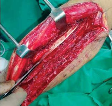
Figure 1A: Peronia Magna noted while dissecting free fibula flap
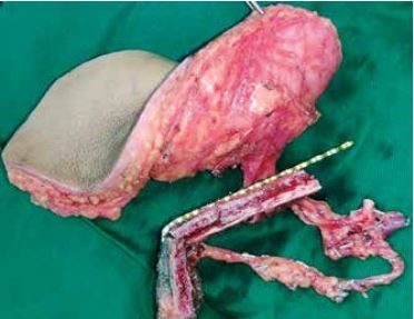
Figure 1B: Free Scapula Osteo-cutaneous flap showing two segments. 6cm segment from the lateral border of scapula along with the cutaneous skin perforator originating from the circumflex scapular artery. 3.5 cm bone segment from the angle of scapula supplied from the angular branch of the thoraco-dorsal artery
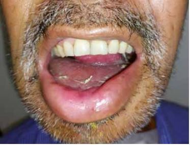
Figure 1C: Post-operative outcome
Case 2: Large soft tissue and less bone requirement
A 45-year-old male with left buccal mucosa SCC was planned for a bite composite resection (Segmental mandibulectomy + Upper alveolectomy + skin resection of 8 X 6 cms). The mandibular segment to be excised was from the left canine to the left angle (Figure 2A).
On analyzing the defect, bone requirement was a single straight segment of 6.5 cm. On the other hand, soft tissue requirement was significant including the entire buccal mucosa extending till hemipalate (anteriorly up to the maxillary canine) with a large skin excision. FFOCF for such defects can be challenging, in view of the variable perforator origin along the fibula and restricted skin mobility due to septal attachment. Hence, electively free scapula osteo-cutaneous flap was planned.
Skin perforator was marked with a handheld doppler. A cutaneous paddle of 20 X 6 cms parallel to the lateral border of the scapula and a 6.5 cm bone segment along the lateral border of the scapula was harvested. Bone was fixed with a 2.5 mm recon plate. Arterial anastomosis with facial artery and vein with IJV tributary was done. The long perforator length and a single point attachment on the bone segment allowed unrestricted mobility for this multiplanar complex oral defect (Figure 2B). Donor was closed primarily. Patient had an uneventful recovery and received timely adjuvant therapy (Figure 2C).
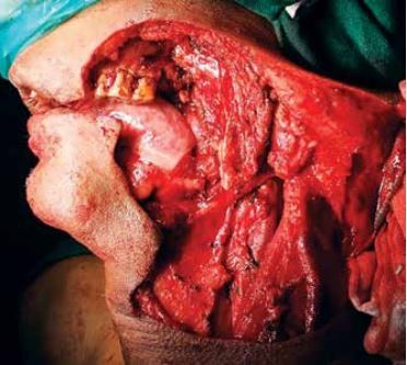
Figure 2A: Defect - Segmental mandibulectomy from left canine to left angle of mandible
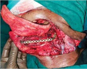
Figure 2B: C. Intra-operative fixation
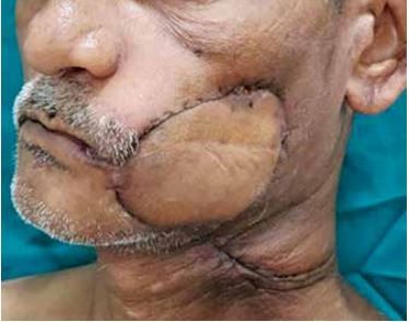
Figure 2C: Post-operative outcome
Discussion
Taylor et al. described the use of osseous free fibula flap for reconstruction of tibial defects7 following which Chen described the first fibula osteocutaneous free flap in 1983.8 Hidalgo reported the use of free fibula flap for mandibular reconstruction in 1989.9 In the past two decades the free fibula flap has become the gold standard in reconstruction of mandibular defects for several reasons. Large vascularized bone and multiple osteotomy makes it possible to reconstruct almost the entire mandible. The caliber of the peroneal artery and venae commitantes closely matches the dimensions of vessels in the head and neck region making microanastomosis convenient.10 Both the ablative and reconstructive surgeon can simultaneously operate thereby reducing the overall operative time. Bicortical nature of the fibula has shown its ability to successfully osteointigrate dental implants aiding in oromandibular rehabilitation.
In spite of these advantages the free fibula flap has some inherent limitations. Peronia Arteria Magna is a well-documented variation. Classifications for the branching pattern of the popliteal artery has been proposed by Lippert and later by Kim et al.12 In their review, Kim et al. found 75 variant cases from 1000 femoral angiograms and classified them as Class I (popliteal artery branches below the knee), Class II (Branching is proximal at or above knee) and Class III (Hypoplastic/ Aplastic tibial arteries with Altered Distal Supply). In class III peronia magna, the blood supply of the foot is completely dependent on the peroneal artery and harvesting the free fibula flap may lead to foot ischemia. There is no equivalent of Allens test in lower-limb, to ascertain the degree of dependence of the foot on the peroneal artery making pre-operative clinical evaluation difficult. Obtaining a pre-operative vascular imaging such as a CT angiography of lower limb is ideal, however not feasible economically in all cases. Although not foolproof, we perform CT angiography only when there is an absent ATA and PTA pulsation at the foot. Likewise, Clemenza et al. in their study have shown that clinical palpation remains the most widely used technique by surgeons. It should be noted that there are not enough published studies to verify the sensitivity and the specificity of CT angiography, MR angiography and Color Doppler techniques for preoperative vascular mapping.13,14 In our case, despite pre-operative color doppler we were unable to identify the aberrant low division of the tibio-peroneal trunk. The presence of peripheral vascular disease or symptoms such as claudication are also a contraindication to the use of this flap in view of severe atherosclerotic disease.
Complex oromandibular defects are multiplanar in nature involving buccal mucosa, floor of mouth, palate and skin. Although abundant skin paddle is available with FFOCF, it is adherent to the bone with a septum with short perforator length. This septal attachment prevents free mobility of the skin paddle in reconstructing 3D defects. This can be overcome by chimerism or use of multiple flaps, allowing independent mobility of each paddle. However, this is dependent on the anatomical location and origin of the perforators along the peroneal artery. Chimerism may sometimes also require performing multiple sets of anastomosis. Hence it may not be possible in all patients.
These situations make FFOCF either impossible (i.e. Vascular Anomalies like Peronea Magna) or less ideal (i.e. Complex oromandibular defects with large soft tissue requirement). Scapula Osteo-cutaneous flap provides respectfully sturdy bone with abundant soft tissue. The Skin paddle has much higher mobility and hence can be oriented for reconstructing multiplanar defects. The subscapular system allows for chimerism based on the combination of its components. Lateral scapular border (supplied by the circumflex scapular artery, CSA), scapular tip (angular artery), and rib (branch to serratus anterior) are the bone components. Lateral border of scapula can be harvested from 1 cm below the glenohumeral joint up to the scapular tip. The angular branch from the thoracodorsal pedicle allows for an osteotomy as well. The biggest advantage over FSOCF is the ample amount of soft tissue components that can be harvested from the same pedicle. The subscapular artery originates from the axillary artery which then divides into the CSA and TDA. Scapular fasciocutaneous and parascapular fasciocutaneous flaps (descending branch) are supplied by the CSA. Latissimus dorsi muscle and thoracodorsal artery perforator (TDAP) flap from the thoracodorsal artery. Approximately 7-10 cm of pedicle can be obtained from CSA to the fasciocutaneous components.15 Such a robust vascular anatomy allows the cutaneous or myofascial flap components to be oriented with a wide range of motion from the native bone. Also the large calibre size of the vessels spares them from intimal disease.16
Fujiki et al. compared use of scapula and fibula free flaps and showed that the overall complication rate and donor site morbidity was similar between the two.17 The main concern with use of scapula flaps remains shoulder morbidity; however, despite a deficit in the range of motion, activities of daily living are not significantly affected for patients.18 The ability to primarily close the donor site defect avoids use of skin grafts making donor site healing relatively uneventful. An added advantage of the abundance of soft tissue available in this flap is evident in lower rate of hardware exposure following radiotherapy compared with the fibula flap as reported by Tsang et al.19 Another advantage of the Free scapula flap is the greater bone height compared to FFOCF, matching with the native mandible resulting in better lip support.
While comparing post-operative morbidity risks associated with prolonged immobility in the postoperative period, such as deep vein thrombosis, atelectasis, and pneumonia, are minimized when the lower extremities are spared.17,18 A retrospective review of perioperative morbidity following scapular flaps found that the return to ambulation was 2.7 days, compared with the average of 4 to 6 days with fibula flaps.20 The shoulder does not require post-operative immobilization, but early physical therapy is recommended to prevent adhesive capsulitis and decreased range of motion. Patients most susceptible to perioperative complications, such as the elderly or patients with poor functional status, may be better served by scapular flaps to increase mobilization and functional status postoperatively.
CONCLUSION:
The scapular flap provides robust soft tissue and osseous donor option for oro-mandibular reconstruction. It is well suited for use in patients with vascular anomalies, complex oro-mandibular defects, elderly patients with peripheral vascular disease or patients with lower limb trauma. Defects that demand less osteotomies with significant soft tissue requirement are ideal with minimal donor morbidity. This flap should be in the repertoire of the modern head and neck reconstructive surgeon to deal with unforeseen complications.
References
- Urken ML. Oromandibular reconstruction in multidisciplinary head & neck reconstruction: a defect oriented approach. Philadelphia: Lippincott Williams & Wilkins; 2010. p. 135–233.
- Postablative Reconstruction Techniques for Oral Cancer D. David Kim, DMD, MD*, G.E. Ghali, DDS, MD, FACS.
- Ferri J, Piot B, Ruhin B, Mercier J. Advantages and limitations of the fibula free flap in mandibular reconstruction. J Oral Maxillofac Surg. 1997;55(5):440-449. doi:10.1016/s0278-2391(97)90685-6.
- Abou-Foul AK, Borumandi F. Anatomical variants of lower limb vasculature and implications for free fibula flap: Systematic review and critical analysis. Microsurgery 2016;36:165–72.
- Gibber MJ, Clain JB, Jacobson AS, Buchbinder D, Scherl S, Zevallos JP et al. Subscapular system of flaps: an 8-year experience with 105 patients. Head & Neck. 2015 Aug;37(8):1200-6.
- Brown J, Bekiroglu F, Shaw R. Indications for the scapular flap in reconstructions of the head and neck. British Journal of Oral and Maxillofacial Surgery. 2010 Jul 1;48(5):331-7.
- Taylor GI, Miller GD, Ham FJ. The free vascularized bone graft: a clinical extension of microvascular techniques. Plastic and reconstructive surgery. 1975 May 1;55(5):533-44.
- Chen ZW, Yan W. The study and clinical application of the osteocutaneous flap of fibula. Microsurgery. 1983;4:11–6.
- Hidalgo DA. Fibula free flap: a new method of mandible reconstruction. Plast Reconstr Surg. 1989;84(1):71-79.
- Wei FC, Mardini S. Flaps and reconstructive surgery. Saunders; 2009. p. 439–55.
- Kramer FJ, Dempf R, Bremer B. Efficacy of dental implants placed into fibula-free flaps for orofacial reconstruction. Clinical oral implants research. 2005 Feb;16(1):80-8.
- Kim D, Orron DE, Skillman JJ. Surgical significance of popliteal arterial variants. A unified angiographic classification. Annals of surgery. 1989 Dec;210(6):776.
- Clemenza JW, Rogers S, Magennis P. Pre-operative evaluation of the lower extremity prior to microvascular free fibula flap harvest. Annals of the Royal College of Surgeons of England. 2000 Mar;82(2):122.
- Kelly AM, Cronin P, Hussain HK, Londy FJ, Chepeha DB, Carlos RC. Preoperative MR angiography in free fibula flap transfer for head and neck cancer: clinical application and influence on surgical decision making. AJR Am J Roentgenol. 2007;188(1):268-274.
- Jacobson L, Dedhia R, Kokot N, Chalian A. Scapular osteocutaneous free flap for total lower lip and mandible reconstruction. Microsurgery. 2016 Sep;36(6):480-4.
- Bianchi B, Ferri A, Ferrari S, et al. Reconstruction of mandibular defects using the scapular tip free flap. Microsurgery 2015;35:101-6.
- Fujiki M, Miyamoto S, Sakuraba M, Nagamatsu S, Hayashi R. A comparison of perioperative complications following transfer of fibular and scapular flaps for immediate mandibular reconstruction. Journal of Plastic, Reconstructive & Aesthetic Surgery. 2013 Mar 1;66(3):372-5.
- Patel KB, Low TH, Partridge A, Nichols AC, MacNeil SD, Yoo J et al. Assessment of shoulder function following scapular free flap. Head & neck. 2020 Feb;42(2):224-9.
- Tsang GF, Zhang H, Yao C, Kolarski M, Gullane PJ, Irish JC et al. Hardware complications in oromandibular defects: comparing scapular and fibular based free flap reconstructions. Oral oncology. 2017 Aug 1;71:163-8.
- Kearns M, Ermogenous P, Myers S, Ghanem AM. Osteocutaneous flaps for head and neck reconstruction: A focused evaluation of donor site morbidity and patient reported outcome measures in different reconstruction options. Archives of plastic surgery. 2018 Nov;45(06):495-503.