 WhatsApp Us (24/7)
 +91 926 888 0303 (24/7)


Abstract:
Nutcracker syndrome (NCS) is a rare renal vein compression disorder. In its classical presentation, the left renal vein is compressed between the aorta and the superior mesenteric artery. It may present with symptoms like haematuria, proteinuria, pain in the left lumbar or lower abdomen, or symptoms due to pelvic congestion, or be asymptomatic. In suspected cases, the diagnosis can be established by radiological imaging.
We report a 23-year-old female who presented as a live kidney donor and was diagnosed with NCS during preoperative evaluation. Our case is unique because the donor presented not only with NCS but also with a left-sided early-branching renal artery and a single right renal artery, creating a surgical dilemma in selecting the appropriate side for nephrectomy.
In NCS, surgical treatment is recommended for patients with serious symptoms. In our case, the donor had both left-sided NCS and an early-branching left renal artery, with a single right renal artery, and she was asymptomatic for NCS. Instead of choosing the right side that could have made the recipient surgery easier, we made the surgically difficult decision and chose the left donor kidney with practically two renal arteries to avoid future prospective complications to the donor. In this article, we highlight the right approach to select the side of kidney in such a living donor.
Key words: Nutcracker Syndrome, Kidney Donor, Transplantation.
Introduction
Nutcracker syndrome (NCS) is a rare renal vein compression disorder. In its classical presentation, left renal vein is compressed between the aorta and the superior mesenteric artery (SMA).1,2 NCS may be seen in all age groups, but its prevalence peaks during the 2nd and 3rd decades.2,3 The compression over the renal vein may induce sufficient pressure in the affected kidney’s venous system, leading to proteinuria, microscopic haematuria and progressive loss of renal function.4 Patients with NCS usually seek medical attention with clinical symptoms such as pain in the left lumbar or lower abdomen, orthostatic proteinuria, gross or microscopic haematuria, gynaecological symptoms due to pelvic congestion in females and varicocele in males.5 Rarely, this can lead to renal vein thrombosis and pulmonary thromboembolism.6 If NCS is suspected, the diagnosis can be made by Doppler ultrasonography (US), computed tomography (CT), and magnetic resonance imaging (MRI).7,8
Case of a prospective kidney donor with NCS has rarely been reported in literature.9 Our case is further unique because not only did the donor have NCS, but also had left-sided early-branching renal artery with right single artery causing a surgical dilemma to choose between the two sides.
Case Report
A 23-year-old female underwent evaluation as a prospective kidney donor for her father. A detailed preoperative evaluation was performed. All the haematological, biochemical and immunological tests were within normal limits. Complement-dependent cytotoxicity (CDC) crossmatch tests were negative. The donor had no complaints or findings in her routine donor evaluation to suggest the NCS.
In the preoperative donor evaluation, there was no microscopic haematuria or proteinuria. The 24-hour urinary protein was 29 mg/day. The glomerular filtration rate (GFR) on diethylenetriaminepentaacetic acid (DTPA) renal scan was 116 mL/min, and both kidneys had equal differential renal function.
On CT renal angiography, both the right and left kidney size and parenchymal thickness were similar. However, there was an early branch arising from the upper border of the main renal artery and supplying the lower pole of the left kidney (Figure 1). In axial section images, it was determined that the left renal vein was compressed between the aorta and the SMA. The diameter of the left renal vein was 9.87 mm before the compression site, and 2.32 mm at the compressed region (Figure 2A). In sagittal plane images, the aortomesenteric angle (AMA) was measured as 14o (Figure 2B) and the distance between the aorta and the SMA was 3.1 mm.
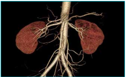
Figure 1: Computed tomography (CT) renal angiography showing left early division renal artery
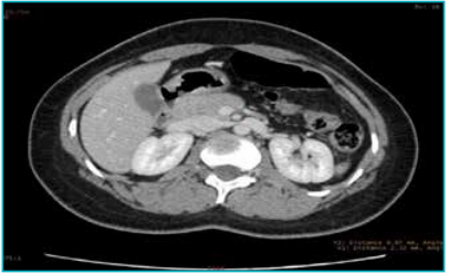
Figure 2A: Compression of left renal vein between the superior mesenteric artery (SMA) and the aorta seen on axial section.
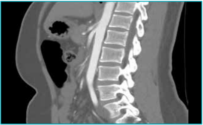
Figure 2B: Abdominal aortomesenteric angle (AMA) measurement on the sagittal plane.
Since there was early branching in renal artery, which would have become two arteries after harvesting, it required one additional anastomosis during transplantation. We still preferred to take the left side kidney, since there was significant renal vein compression by the SMA. The donor’s family was counselled about future risk to the donor because of NCS and the need to proceed with left side kidney donation, requiring additional arterial anastomosis. After consent, the donor was planned for the left laparoscopic nephrectomy
During the laparoscopic donor nephrectomy, mild dilatation was seen in the renal vein, left gonadal vein, left adrenal vein, and left lumbar vein. To avoid any harm to the kidney and the donor, the renal vein dissection towards the SMA was avoided. The kidney was harvested after securing the renal artery and renal vein and performing the transection of the renal vein proximal to the site of compression (Figure 3). The harvested left donor kidney was transplanted into the right iliac fossa of the recipient in the standard manner using the external iliac artery and vein. As the inferior epigastric artery was of very narrow calibre, the additional lower pole renal artery anastomosis was also performed with the external iliac artery (Figure 4). Both recipient and donor surgeries were uneventful intraoperatively, and postoperative recovery was normal.
Discussion
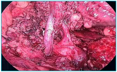
Figure 3: Laparoscopic view of early division of renal artery and renal vein.
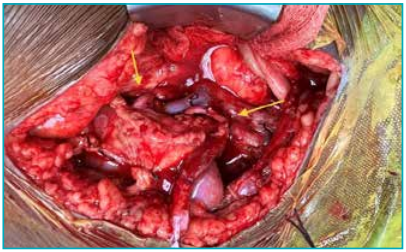
Figure 4: Two arterial anastomoses with the external iliac artery in the recipient.
Most patients with NCS are asymptomatic. In symptomatic individuals, the most frequent finding is microscopic haematuria because of rupture of thin-walled varicose veins that develop in the collecting duct system because of high pressure in the renal vein.1-4 Pain spreading from the left lumbar to the left inguinal region is another frequent finding. Patients may present with pain due to venous congestion, which is aggravated by walking, sitting, standing, and running.5 Depending upon the degree of pelvic congestion, varicocele in males and dyspareunia, dysuria, dysmenorrhea, and polycystic ovary in females can be detected.
The presence of proteinuria is another possible finding in NCS. The prevalence of proteinuria in NCS is 0.6%–10.7%.1 This protein leak in the calyceal system is due to increased pressure, and the degree of proteinuria is affected by postural changes. In our case, the donor was asymptomatic, and a 24-hour urine collection revealed proteinuria of less than 29 mg/day with no microscopic haematuria.
Low body mass index (BMI) is also correlated with NCS. Previously, it has been reported that intense weight loss reduces retroperitoneal fat tissue, causing the AMA to become narrower and, as a result, NCS may occur.1 In our case, the BMI of the donor was 21.6 kg/m2.
In NCS, the diagnosis is mainly established by imaging methods. Radiological findings should be supplemented by physiological flow information (ultrasound and/or catheter-based venography).2,7 In Doppler US, flow patterns of the main renal vein and collateral veins, varicose formations, retrograde blood flows may be evaluated, and vein diameters measured.7 In recent years, multi-detector CT angiography and MR angiography images have been reported to be highly efficient for diagnosis.8 In axial sections of CT, when comparing proximal normal renal vein diameter with the diameter of the narrow part, if the ratio is over 4:1 and if the AMA measurement on sagittal planes is narrower than 50o, then the diagnosis can be established with the accompanying symptoms.10 In the three-dimensional (3D) CT angiography, an AMA of 39.3 ± 4.3 degrees and a distance between the SMA and the aorta 3.1 ± 0.2 mm suggest NCS, while in normal individual, the AMA is 90 ± 10 degrees and the distance between the SMA and the aorta is 12 ± 1.8 mm.7 In our donor, the AMA is 14°, and distance between the SMA and the aorta is 3.1 mm.
For symptomatic NCS patients, treatment is recommended. The aim is to correct renal vein blood flow, treatment modalities include intravascular stenting, left renal vein bypass, left renal vein transposition, superior mesenteric artery transposition, renal-to-inferior vena cava shunt and rarely autotransplantation.1,3,5 After treatment, haematuria usually disappears within a few days.
A patient with NCS can also be a kidney donor as in this case,9 where the left kidney was chosen as a graft. In a routine donor, usually selection of kidney is based on simpler anatomy and differential function of both the kidneys. In this donor, both kidneys were equally functioning, but because of the right single renal artery and left early branching renal artery, the common selection would have been the right kidney. However, in view of the presence of NCS, we selected the left kidney, which needed one additional anastomosis with inferior epigastric artery. This avoided any future risk to the donor and risk of surgical correction later in life.
If the donor is symptomatic, most of the present symptoms will probably disappear after graft nephrectomy. Because the phenomenon does not cause a pathological problem at the glomerular and tubular level in the kidney, the graft function of the recipient will have an optimal outcome. In our case, postoperatively, GFR of the recipient remained at a fairly good level, and there was no proteinuria or microscopic haematuria
Conclusion
In NCS, surgical treatment is recommended for patients with serious symptoms. Surgical interventions and stenting are treatment modalities. In our case, the donor had both left NCS and an early-branching left renal artery; instead of going for the right kidney that could have made recipient surgery easier, we made the surgically difficult decision to choose left donor kidney with practically two renal arteries, to avoid future prospective complications to the donor. By reporting this case, we highlight the right approach to select the side of kidney in such donor.
Amit Kumar Devra, Lok Prakash Choudhary, Anuj Arora, Ravi Rai, CPS Chauhan, Anil Prasad Bhatt. Young Kidney Donor with Double Whammy: Nutcracker Syndrome with Early Branching Left Renal Artery. MMJ. 2025, December. Vol 2 (4).
References
-
Kurklinsky AK, Rooke TW. Nutcracker phenomenon and nutcracker syndrome. Mayo Clin Proc. 2010;85:552–59
-
Venkatachalam S, Bumpus K, Kapadia SR, et al. The nutcracker syndrome. Ann Vasc Surg. 2011;25:1154–64.
-
Vianello FA, Mazzoni MB, Peeters GG, et al. Micro- and macroscopic hematuria caused by renal vein entrapment: systematic review of the literature. Pediatr Nephrol. 2016;31(2):175–84.
-
Chen YM, Wang IK, Ng KK, et al. Nutcracker Syndrome: An Overlooked of Hematuria. Chang Gung Med J. 2002;25(10):700–5
-
Scultetus AH, Villavicencio JL, Gillespie DL. The nutcracker syndrome: its role in the pelvic venous disorders. J Vasc Surg. 2001;34(5):812–19.
-
Kanta Hori, Shota Yamamoto, Maki Kosukegawa, et al. Nutcracker syndrome as the main cause of left renal vein thrombus and pulmonary thromboembolism. IJU Case Reports. 2021;5(1):24–7.
-
Sugaya K, Kohama T, Tsukada T, et al. [Non-invasive imaging diagnosis of left renal vein compression causing hematuria. Part 2. CT]. Hinyokika Kiyo. 1991;37(5):485–9.
-
Wei-Jun Fu, Bao-Fa Hong, Jiang-Ping Gao, et al. Nutcracker phenomenon: a new diagnostic method of multislice computed tomography angiography. Int J Urol. 2006; 13(7):870–3.
-
Tugmen C, Kebapci E, Sert I, et al. Kidney transplantation from a donor with nutcracker syndrome. El Mednifico Journal. 2014;2(3):288–90.
-
Buschi AJ, Harrison RB, Norman A, et al. Distended left renal vein: CT/sonographic normal variant. AJR Am J Roentgenol. 1980;135(2):339–42.