 WhatsApp Us (24/7)
 +91 926 888 0303 (24/7)


Abstract:
Sclerosing encapsulating peritonitis (SEP), also called abdominal cocoon syndrome, is a rare cause of intestinal obstruction. In 1907, Owtschinnikow first termed it as peritonitis chronica fibrosa encapsulata. It can be either idiopathic or secondary. Secondary cocoon syndrome may be due to medications, infections such as tuberculosis (TB) in endemic regions, cirrhosis, peritoneal dialysis and gynaecological malignancies.
A 37-year-old female diagnosed with abdominal TB, who had not complied with anti-tubercular therapy (ATT) for the last 5 years, presented with acute intestinal obstruction and a history of recurrent subacute intestinal obstruction. Computed tomography (CT) imaging of the abdomen showed dilated bowel loops with a transition point in the ileum, multiple large calcified mesenteric lymph nodes and ascites. Diagnostic laparoscopy revealed dense adhesions and a fibrocollagenous membrane encapsulating the intestine. It was converted to laparotomy, an abdominal cocoon was confirmed, and extensive adhesiolysis and ileocolic anastomosis were done. GeneXpert showed extensively drug-resistant tuberculosis (XDR-TB), and the patient was started on an XDR-TB regimen.
Abdominal cocoon syndrome secondary to TB, presenting as intestinal obstruction, is a rare but serious complication that requires surgical intervention. This case further highlights the importance of adherence to medication and the consideration of resistant forms of TB, such as XDR-TB, in patients with non-compliance with treatment. Surgical management, along with appropriate modification of ATT based on drug-resistance profiling, for patient recovery is essential.
Key words:Sclerosing Encapsulating Peritonitis, Abdominal Cocoon, Intestinal Obstruction, XDR-TB.
Introduction
Sclerosing encapsulating peritonitis (SEP) is a rare disease that causes a thick fibrocollagenous membrane, partially or completely enveloping the small intestine. It was first termed in 1907 by Owtschinnikow as peritonitis chronica fibrosa encapsulata. 1 It may be either idiopathic or secondary. Secondary cocoon syndrome may be caused by medications, infections such as tuberculosis (TB) in endemic regions, cirrhosis, peritoneal dialysis and gynaecological malignancies. The treatment of SEP includes identifying the cause and treating it.2
In TB-endemic areas, abdominal and peritoneal TB are still the leading causes of cocoon abdomen. Chronic peritoneal inflammation in TB can induce fibrotic reactions, leading to secondary abdominal cocoon syndrome.3
Case Report
A 37-year-old female was admitted to the hospital with complaints of abdominal pain, distension, vomiting, and obstipation, with a history of multiple subacute intestinal obstruction episodes for one year. Diagnostic laparoscopy was done in 2019; following this, abdominal TB was diagnosed and anti-tubercular therapy (ATT) was started. She had been non-compliant with treatment for the last five years.
On examination, the patient was haemodynamically stable. The abdomen was distended with diffuse tenderness and sluggish bowel sounds. Laboratory investigations were unremarkable except for mild hypoalbuminaemia.
A contrast-enhanced computed tomography (CECT) scan of the abdomen showed proximal small bowel dilatation with a transition point in the ileum, collapse of distal loops, ascites, and multiple lobulated hyperdense foci in the mesentery — likely calcified lymph nodes — suspicious for chronic inflammatory pathologies, such as abdominal TB with possible cocoon formation (Figures 1A and 1B).
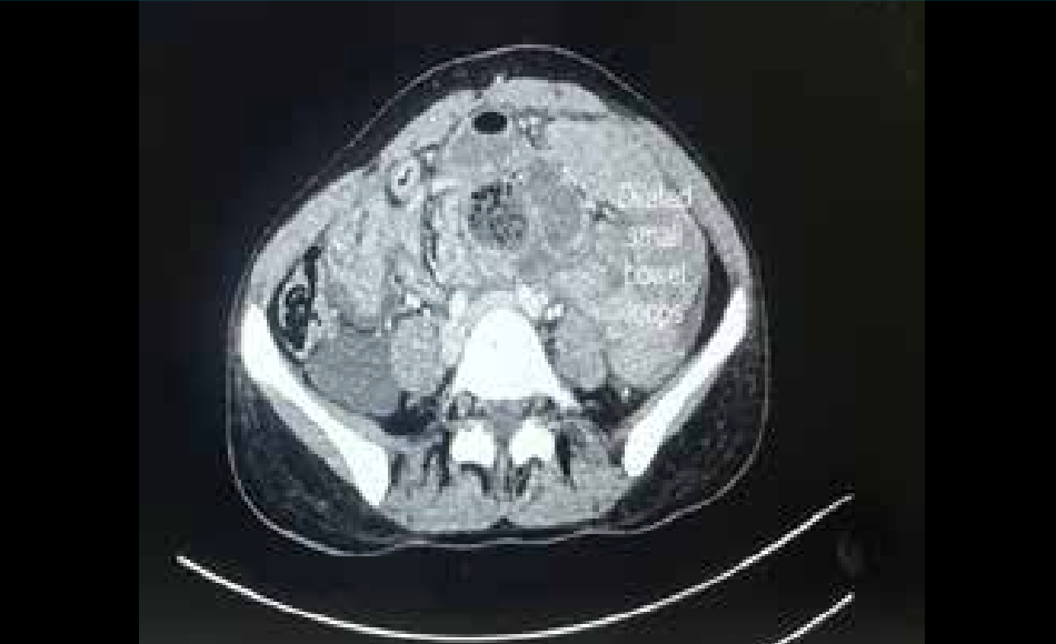
Figure 1A: Contrast-enhanced computed tomography of the abdomen showing dilated small bowel loops.
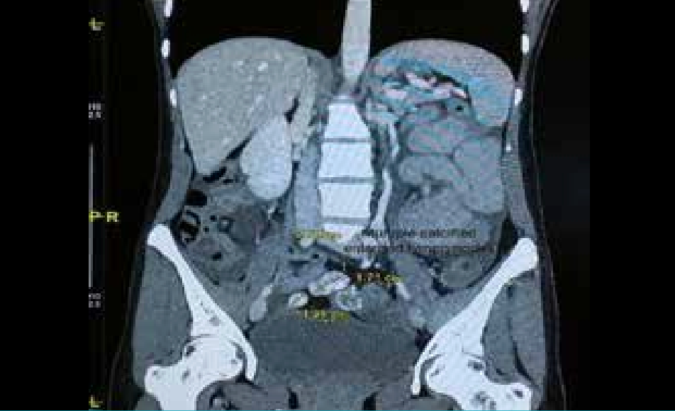
Figure 1B:Mesenteric lymphadenopathy with small bowel loops and ascites.
Given the clinical status and radiological findings, the patient was taken up for diagnostic laparoscopy, which revealed dense inter-bowel and omental adhesions forming a fibrous cocoon around the small bowel (Figure 2).
There were multiple loculated ascitic fluid collections (Figure 3), and the bowel loops were matted and adherent to the anterior abdominal wall, liver, and urinary bladder (Figure 4).
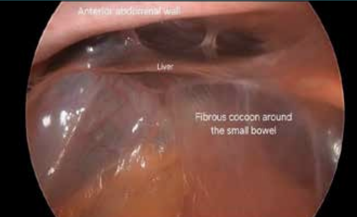
Figure 2: Diagnostic laparoscopy showing a fibrous cocoon around the intestine.
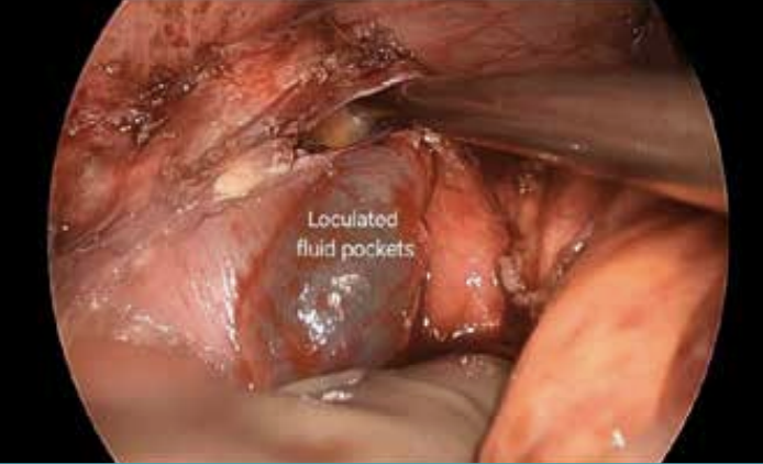
Figure 3:Multiple loculated ascitic fluid pockets.
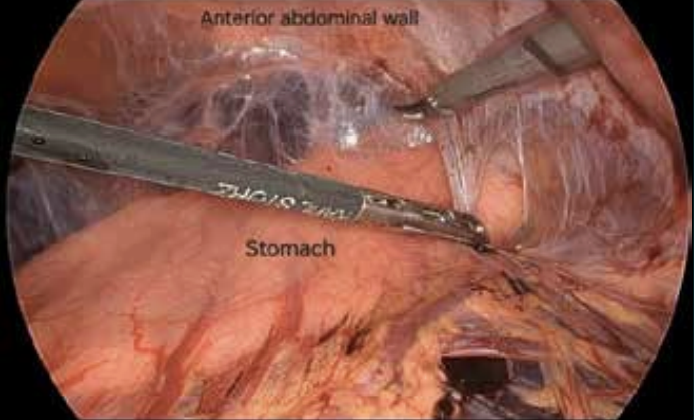
Figure 4:Dense adhesion between bowel and anterior abdominal wall.
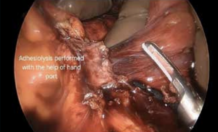
Figure 5:Adhesiolysis performed through Hand Assisted Laparoscopic Surgery (HALS)
Multiple enteric fistulous openings with dense adhesions were noted at the distal ileum. Hand Assisted Laparoscopic Surgery (HALS) was attempted to do extensive adhesiolysis through suprapubic incision (Figure 5).
An exploratory laparotomy through a midline incision was performed because of dense adhesions and multiple enteric fistulae. Thorough peritoneal lavage was carried out. Primary closure of the ileal fistulae and ileocolic anastomosis was performed.
GeneXpert testing of tissue and ascitic fluid samples confirmed Mycobacterium tuberculosis, resistant to rifampicin, isoniazid, and fluoroquinolones (levofloxacin and moxifloxacin), indicating XDR-TB.
Postoperatively, the patient was initially managed with nil per os (NPO) and was started on total parenteral nutrition (TPN) and enteral feed. She was referred to a national TB centre for the initiation of XDR-TB–specific treatment. She was started on a linezolid, clofazimine, cycloserine and bedaquiline regimen. Her postoperative recovery was further complicated by low bilious output from the anastomotic drain, which prompted a follow-up CECT that excluded major anastomotic leaks or intraabdominal collections. The pelvic drain was removed on postoperative Day 12, and the anastomotic drain was removed on postoperative Day 13. She is stable and tolerating the XDR-TB regimen at follow-up after 1 and 5 months.
Discussion
Abdominal cocoon syndrome, also known as SEP, is a rare but important cause of intestinal obstruction due to chronic granulomatous inflammation that results in fibrosis, adhesions, and encapsulation of bowel loops in TB patients.4
This patient, being non-compliant with ATT and having recurrent episodes of subacute intestinal obstruction progressing to an acute presentation, raised concern for complications of TB, including fibrotic cocoon formation, fistula, or treatment failure due to drug resistance or non-compliance.
Imaging, particularly CECT, plays an important role in raising suspicion. Surgical exploration remains the definitive method for both diagnosis and treatment.
Even on ATT, abdominal TB can cause progressive fibro-inflammatory complications. SEP in TB patients should be suspected in chronic or recurrent bowel obstruction. Drug-resistance testing, such as GeneXpert, becomes very important in the persistence or atypical presentation of TB.5 Surgical management, along with appropriate modification of ATT as necessary based on drug resistance profiling, will be key to the patient’s recovery.
Ethical approval
This case report has been reported in line with the SCARE criteria.
Sources of funding
None.
Conflicts of interest disclosure
The authors declare that they have no financial conflict of interest regarding the content of this report.
Conclusion
Secondary abdominal cocoon syndrome is an infrequent but serious complication of abdominal TB. The case also underlines the role of surgical management in an advanced disease and tissue testing in identifying drug resistance, thus allowing appropriate adjustment of ATT by a multidisciplinary team for optimal outcomes.
Vikas Panwar, D.V. Sneha, Vedant Rai. Sclerosing Encapsulating Peritonitis in an Extensively Drug
Resistant Tuberculosis Patient Presenting as Intestinal Obstruction – A Case Report.
MMJ. 2025, December. Vol 2 (4).
DOI: XXXXX
References
- Danford CJ, Lin SC, Smith MP, et al. Encapsulating peritoneal sclerosis. World J Gastroenterol. 2018;24(28):3101–11.
- Chorti A, Panidis S, Konstantinidis D, et al. Abdominal cocoon syndrome: Rare cause of intestinal obstruction—Case report and systematic review of literature. Medicine (Baltimore). 2022;101(27):e29837.
- Aziz W, Malik Y, Haseeb S, et al. Abdominal Cocoon Syndrome: A Laparoscopic Approach. Cureus. 13(7):e16787.
- Foo KT, Ng KC, Rauff A, et al. Unusual small intestinal obstruction in adolescent girls: The abdominal cocoon. Br J Surg. 1978;65(6):427–30.
- World Health Organization. WHO consolidated guidelines on tuberculosis: Module 4 — treatment: drug-resistant tuberculosis treatment. Available at: https://www.who.int/publications/i/item/9789240007048. Accessed on: 6th November 2025.