 WhatsApp Us (24/7)
 +91 926 888 0303 (24/7)


Abstract:
Homogentisic acid deposition within connective tissues causes ochronotic arthropathy, a rare musculoskeletal consequence of alkaptonuria that causes progressive degenerative changes in the joints. Significant impairment and unique intraoperative challenges during surgical management may result from advanced involvement of weight-bearing joints. We present the case of a 53-year-old adult who arrived with radiating hip and knee pain, stiffness, and limited mobility. The patient's history of dark urine prompted a biochemical analysis that showed high levels of homogentisic acid. It's interesting to note that there were no traditional outward signs of ochronosis, such as bluish-black scleral discolouration (Osler's sign) or auricular cartilage pigmentation. Both the hip and knee joints showed significant degenerative changes on radiographs. A phased surgical approach was chosen due to the advanced arthropathy, starting with bilateral total hip replacement and progressing to bilateral total knee replacement. Pathognomonic symptoms of ochronotic arthropathy, i.e. diffuse blackish-brown pigmentation of the articular cartilage and subchondral bone, were observed during surgery. Following surgery, the patient experienced a smooth recovery and reported long-lasting pain relief along with a return to his normal level of mobility and independence. The Harris Hip Score (HHS) increased from 31 to 93, and the Knee Society Score (KSS) increased from 40 to 93 at the 12-month follow-up, indicating a notable improvement in functional assessment. The current case of ochronotic arthropathy demonstrated pain relief and functional restoration after the staged bilateral total hip and knee arthroplasty. Despite the intraoperative challenges posed by pigmented and friable tissues, the use of common modern implants produced stable fixation and facilitated a smooth recovery for the patient.
Key words: Ochronotic Arthropathy, Alkaptonuria, Total Knee Arthroplasty, Osteoarthritis.
Introduction
Homogentisate 1,2-dioxygenase deficiency leads to alkaptonuria, a rare autosomal recessive condition that causes ochronosis and an accumulation of homogentisic acid.1 Connective tissues gradually deteriorate due to ochronosis, or pigment deposition in alkaptonuria. Additionally, it increases the risk of two early degenerative joint diseases: osteoarthritis and arthritis.2 Globally, the prevalence is thought to be between 1 in 250,000 and 1 in 1,000,000, with higher clusters reported in Slovakia and the Dominican Republic.3 Only isolated cases and a few small case series have been reported in the Indian population, indicating that the condition is not only rare but also probably underdiagnosed.4
Patients frequently experience early-onset osteoarthritis of the hips, knees, and spine, making musculoskeletal involvement the most incapacitating characteristic. Conservative measures such as physiotherapy, analgesics, and lifestyle modification provide only temporary relief, while medical therapies like vitamin C or nitisinone have shown limited benefit in altering disease progression.5 Once arthropathy advances, total joint arthroplasty is the definitive treatment.6 Studies have confirmed that both total hip and total knee arthroplasty restore function and relieve pain, although intraoperative findings of pigmented, brittle tissues and altered bone quality pose unique surgical challenges.7,8 Globally published literature describes single joint replacement, and reports of patients undergoing staged bilateral hip and knee arthroplasty are extremely limited.9 Furthermore, cases without classical external signs are difficult to recognise, often delaying diagnosis.10
Given the limited literature on ochronotic arthropathy without classical external signs, this report helps fill an important gap by documenting favourable short-term outcomes following staged arthroplasty. It highlights the diagnostic challenges of atypical presentations and offers clinically relevant insights to guide surgical management in comparable cases.
Case Report
A 53-year-old man arrived with a four-month history of bilateral hip and knee pain that had been getting worse over time, along with stiffness and irregularities in his gait. The pain significantly restricted patients’ daily activities, and he required the use of a cane for ambulation. The patient also described dull, nonradiating lower back pain that had recurred intermittently for the past four years but had become disabling in recent months. The present case also summarises the history of urine discolouration, characterised by progressive dark brown pigmentation on standing, with no reported family history of comparable illness
On examination, no external signs, like the scleral or auricular pigmentation, were observed. Internal and external rotation were painfully limited, and hip flexion was limited to 90°. The knees showed fixed flexion deformities of 5° on the right and 10° on the left, with further flexion possible to 120°. Lumbar spine mobility was preserved, and neurological examination was unremarkable
Urinary homogentisic acid was elevated (2637.00 mg/24 hrs), confirming alkaptonuria with routine biochemical investigations within normal limits. Radiographs revealed significant degenerative changes in the knees and hips, as well as calcification of the pelvic tendon and intervertebral disc (Figure 1). Magnetic resonance imaging (MRI) revealed multiple-level cervical and lumbar disc herniations without canal compromise. A scanogram verified the abnormal alignment and narrowing of joint spaces in both lower limbs (Figure 2).
Differential diagnoses considered included primary osteoarthritis and spondyloarthropathy; these were excluded on the basis of biochemical confirmation of homogentisic aciduria and the characteristic intraoperative pigmentation subsequently observed. The final diagnosis was confirmed as ochronotic arthropathy secondary to alkaptonuria.
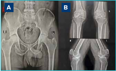
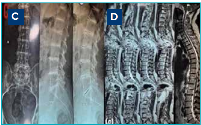
Figure 1: A. Pelvic radiograph (anteroposterior view) showing destruction of the femoral head bilaterally, B. Radiograph of the knee (anteroposterior standing and lateral view) showing severe osteoarthritis knee, C, D. Lumbar radiographs (anteroposterior and lateral views) and magnetic resonance imaging (MRI) screening of the whole spine showing multilevel narrowing of the thoracic and lumbar intervertebral spaces with osteophytosis, calcification, and subchondral osteosclerosis.
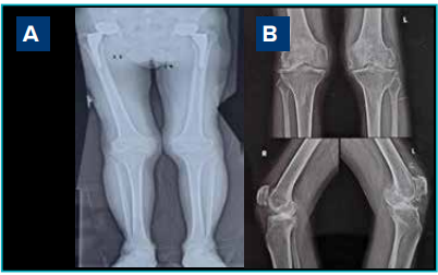
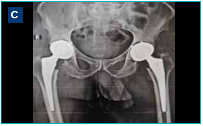
Figure 2: A. Scanogram of both lower limbs, B. Radiograph of the knee (anteroposterior standing and lateral view) showing a severe osteoarthritic knee, C. Six months follow-up of bilateral total hip replacement.
Clinical Outcomes
First surgery: bilateral total hip arthroplasty (THA)
The patient was admitted with worsening hip pain and subsequently underwent staged bilateral THA using the posterolateral (Moore’s) approach, performed 17 days apart. Intraoperatively, the femoral head, acetabulum, and articular cartilage exhibited pronounced ochronotic pigmentation varying from brown to black, whereas the subcutaneous tissue and tensor fascia lata appeared unremarkable. The femoral head was destroyed, with subchondral bone exposure in weight-bearing areas (Figure 3). Based on these findings, a macroscopic diagnosis of ochronosis was made. The pigmented cartilage, bone, and hypertrophied synovium were sent for histopathological evaluation. A hydroxyapatitecoated spiked titanium acetabular cup with a dual mobility liner and a porous-coated uncemented femoral stem was implanted for optimal osseointegration (Figure 3). The patient was mobilised on the first postoperative day with full weight-bearing and was discharged on postoperative Day 3. According to the Harris Hip Score (HHS), there was a notable improvement in joint stability, mobility, and pain relief at the six-month follow-up.
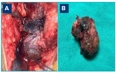
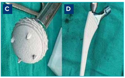
Figure 3: A, B. Intraoperative images showing severe destruction of the acetabular and femoral head cartilage with darkened subchondral bone, C, D. Porous-coated uncemented acetabular and femoral stem.
Second surgery: bilateral total knee arthroplasty (TKA)
Six months later, owing to persistent severe knee pain, the patient was readmitted and underwent bilateral TKA. The subvastus technique, spinal anaesthesia, an intraoperative ultrasound-guided adductor canal block, and no tourniquet were used during the procedure. Intraoperatively, the cartilage and synovium showed diffuse dark-brown pigmentation, similar to what was observed during THA. Closer evaluation revealed extensive destruction of the distal femoral cartilage and darkened subchondral bone in both femoral and tibial cuts, changes characteristic of ochronotic arthropathy (Figure 4). To maximise biocompatibility, longevity, and functional recovery following bony cuts, a bilateral cruciate-retaining gold knee prosthesis was implanted (Figure 5).
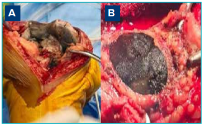
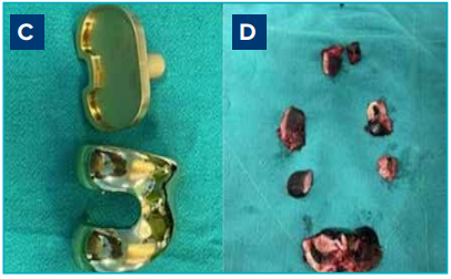
Figure 4: A, B, D. Intraoperative images showing severe destruction of the distal femoral cartilage with darkened subchondral bone in the femoral and tibial cut, C. Gold knee prosthesis.
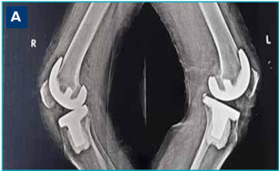
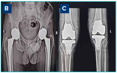
Figure 5: A, C. 6 months follow-up X-ray of total knee replacement lateral and anterolateral view, B. 12 months followup X-ray of total hip replacement anteroposterior view.
On the day of surgery, full weight-bearing mobilisation and postoperative quadriceps-strengthening exercises were initiated. By the third postoperative day, the patient was able to climb stairs and walk independently. Follow-up assessments over the next few months confirmed sustained functional recovery, which was characterised by improved mobility and a noticeable reduction in pain.
Postoperative functional outcomes after THA
Follow-up evaluations at 1, 3, 6, 9, and 12 months post-THA were conducted using the Visual Analogue Scale (VAS), Harris Hip Score (HHS), and Forgotten Joint Score-Hip (FJS-Hip). As per Nilsdotter et al. 2011, the VAS score improved from 8 preoperatively to 2 at 1 month, 1 at 3 months and remained at 0 thereafter.11 The HHS increased from 31.4 preoperatively to 62.6, 75.7, 85.0, 89.0, and 93.0 at 1, 3, 6, 9, and 12 months postoperatively, respectively
Additionally, the FJS-Hip improved from 0 preoperatively, to 29.5 at 1 month. The score continued to improve to 54.3, 79.2, 85.3, and 91.2 at 3, 6, 9, and 12 months, respectively (Figure 6).
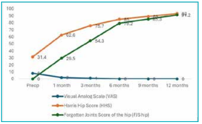
Figure 6: Trends in Visual Analogue Scale (VAS), Harris Hip Score (HHS). and Forgotten Joint Score-Hip (FJS-Hip) preoperatively and at 1, 3, 6, 9, and 12 months post-THA follow-up.
Postoperative functional outcomes after TKA
Postoperative recovery following bilateral TKA was evaluated at 1, 3, 6, and 12 months using the Visual Analogue Scale (VAS), the American Knee Society Score (AKSS), and the Forgotten Joint Score-Knee (FJS-Knee). The VAS score improved from 7 preoperatively to 2 at 1 month, 1 at 3 months, and 0 at both 6 and 12 months. After a baseline score of 40, the AKSS increased to 60 at 1 month, 82 at 3 months, 86 at 6 months, and 93 at 12 months. The FJS-Knee increased from 34.2 at 1 month to 67.5 at 3 months, 82.8 at 6 months, and 89.1 at 12 months. Given that the preoperative FJS-Knee score was 0, these postoperative values represent substantial improvement from baseline. These results show a steady and progressive functional recovery that leads to almost full joint function restoration and pain-free mobility within the first year following surgery (Figure 7).
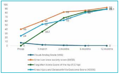
Figure 7: Trends in Visual Analogue Scale (VAS), American Knee Society Score (AKSS), Forgotten Joint Score-Hip (FJSHip), and Knee Injury and Osteoarthritis Outcome Score (KOOS) preoperatively and at 1, 3, 6, and 12-months postoperative follow-up.
Discussion
A rare musculoskeletal symptom of alkaptonuria, ochronotic arthropathy is typified by the progressive degeneration of large weight-bearing joints. Diagnosis is frequently delayed, particularly in the absence of classical external features such as scleral or auricular pigmentation, as observed in our patient.12 At one year, the HHS improved from 31.4 preoperatively to 93, the AKSS improved from 40 to 93, and both the hip and knee-FJS exceeded 90. These results demonstrate that mobility and quality of life were nearly fully restored within the first year following surgery.
When compared with larger series, our outcomes align closely. Pachore et al. 2019, reported 10 patients (12 hips) undergoing THA for ochronotic arthritis, with HHS improving from a preoperative mean of 36 to 88 at long-term follow-up (up to 24 years).13 Compared to this, our patient achieved a slightly higher HHS at 12 months. The results of Rajkumar et al. (2020), who reported improvement in the KSS from 27.2 to 89.4 and the HHS from 17.8 to 78 across 27 arthroplasties, closely resemble the results we saw (HHS 93, KSS 93).14
According to a systematic review of TKA in ochronotic patients by Lee et al. (2019), pain alleviation and functional recovery were consistent across reported cases, irrespective of implant design.15 By 6 months, our patients' knee pain VAS decreased from 7 to 0 and their KSS increased to 93. Durability was further supported by Spencer et al. (2004), who reported 11 joint replacements with outstanding function at 6–12 years.16 The extent of improvement is in line with these long-term findings, despite the case's one-year follow-up
A strength of this case is the detailed documentation of validated outcome measures over sequential followup visits, which allows direct benchmarking against existing literature. A limitation for this case study was the shorter follow-up with one patient, which restricts assessment of implant survival and generalisability.
Conclusion
This case demonstrates that staged bilateral THA and TKA can be a viable treatment option for patients with ochronotic arthropathy even in the absence of classical external features, highlighting both the diagnostic challenges and the potential for favourable early surgical outcomes.
Sujoy Bhattacharjee. Ochronotic Arthropathy Effectively Treated with Staged Bilateral Total Hip and Knee Arthroplasty: A Case Report MMJ. 2025, December. Vol 2 (4).
References
-
Sharabi AF, Goudar RB. Alkaptonuria. [Updated 2023 Aug 8]. In: StatPearls [Internet]. Treasure Island (FL): StatPearls Publishing; 2025 Jan-. Available at: https://www.ncbi.nlm.nih. gov/books/NBK560571/. Accessed on: 2nd September 2025.
-
Thapa M, Bhatia M, Maurya VK. Alkaptonuria. Med J Armed Forces India. 2018;74:394–96.
-
Ochronosis and Alkaptonuria: Practice Essentials, Pathophysiology, Epidemiology. Available at: https:// emedicine.medscape.com/article/1104184-overview. Accessed on: 2nd September 2025.
-
Sheth J, Nair A, Sheth F, et al. Burden of rare genetic disorders in India: twenty-two years’ experience of a tertiary centre. Orphanet J Rare Dis. 2024;19(1):295.
-
Wu K, Bauer E, Myung G, et al. Musculoskeletal manifestations of alkaptonuria: A case report and literature review. Eur J Rheumatol. 2019;6:98–101.
-
Alkaptonuria (Black Urine Disease): Background, Pathophysiology, Epidemiology. Available at: https:// emedicine.medscape.com/article/941530-overview?form=fpf Accessed on: 2nd September 2025.
-
Ilyas I, Kashif S, Algashiri MF, et al. Long-term follow-up of bilateral hip and knee arthroplasty secondary to ochronotic arthropathy. Arthroplast Today. 2020;6:214–19.
-
Al-Ajlouni JM, Alisi MS, Yasin MS, et al. Long-Term Outcomes of the Knee and Hip Arthroplasties in Patients with Alkaptonuria. Arthroplast Today. 2020;6:689–93.
-
Agrawal AC, Baghel A, Garg AK, et al. Total Hip Arthroplasty in Ochronotic Arthropathy with quadriceps tear. A Case Report with a Review of the Literature. J Orthop Case Rep. 2025;15(2):80.
-
Alkaptonuria - Symptoms, Causes, Treatment. NORD. Available at: https://rarediseases.org/rare- diseases/ alkaptonuria/. Accessed on: 2nd September 2025.
-
Nilsdotter A, Bremander A. Measures of hip function and symptoms: Harris hip score (HHS), hip disability and osteoarthritis outcome score (HOOS), Oxford hip score (OHS), Lequesne index of severity for osteoarthritis of the hip (LISOH), and American Academy of orthopedic surgeons (AAOS) hip and knee questionnaire. Arthritis Care Res. 2011;63:S200–7.
-
Couto A, Sá Rodrigues A, Oliveira P, et al. Ochronotic arthropathy—a rare clinical case. Oxf Med Case Reports. 2018(9):omy069.
-
Pachore JA, Shah VI, Upadhyay S, et al. Primary hip arthroplasty for the treatment of alkaptonuric hip arthritis: 3-to 24-year follow-ups. Arthroplasty. 2019;1(1):8.
-
Rajkumar N, Soundarrajan D, Dhanasekararaja P, et al. Clinical and radiological outcomes of total joint arthroplasty in patients with ochronotic arthropathy. Eur J Orthop Surg Traumatol. 2020;30(5):923–29.
-
Lee WC, Tan TL, Chan YH. Total knee arthroplasty in ochronosis arthropathy: a case report and systematic review. Case Rep Orthop. 2019;2019(1):1871856.
-
Spencer J, Gibbons CM, Sharp R, et al. Arthroplasty for ochronotic arthritis no failure of 11 replacements in 3 patients followed 6–12 years. Acta Orthop Scand. 2004;75(3):355–58.