 WhatsApp Us (24/7)
 +91 926 888 0303 (24/7)


Abstract:
Chronic total occlusion (CTO) of the superior vena cava (SVC) is a recognised cause of venous hypertension, particularly among haemodialysis patients with repeated central venous catheterisations. Although endovascular therapy is the initial treatment of choice, long-segment fibrosis frequently results in failure, necessitating surgical reconstruction. Concomitant coronary artery disease (CAD) further complicates management. We report a 53-year-old, dialysis-dependent female with symptomatic SVC CTO and triple-vessel CAD. After failed endovascular recanalisation, she underwent simultaneous off-pump coronary artery bypass grafting (OPCAB) and innominate vein–right atrial appendage (RAA) bypass. A 13-mm autologous pericardial tube graft was created intraoperatively and used as the venous conduit. OPCAB×3 and innominate–RAA bypass were performed successfully. Dense fibrosis at the SVC–right atrium (RA) junction confirmed chronicity and explained the prior endovascular failure. Postoperatively, the patient showed rapid improvement in facial and upper-limb oedema, effective venous drainage, and stable graft flows. Recovery was uneventful, and follow-up imaging demonstrated sustained patency of the coronary grafts and the pericardial venous conduit. Autologous pericardial tube grafting provides a durable, infection-resistant option for central venous reconstruction, particularly valuable in dialysis-dependent patients. Combining this approach with OPCAB minimises inflammatory and coagulopathic risk, avoids prosthetic complications, and enables effective treatment of complex chronic pathology. This case highlights the importance of individualised off-pump strategies using autologous tissue to achieve safe and durable outcomes.
Key words: Chronic SVC Occlusion, Innominate Vein Bypass, Autologous Pericardium, Complex Cardiac Surgery
Introduction
Chronic total occlusion (CTO) of the superior vena cava (SVC) is increasingly encountered, especially in haemodialysis patients with long-term central venous catheterisation. The reported prevalence of central venous stenosis ranges from 4.3% to 41% depending on catheter duration and modality of imaging.1-3 Endovascular therapy is typically the first-line treatment, but longsegment fibrotic CTOs frequently fail and require surgical bypass.4,5
Multiple graft materials have been used for central venous reconstruction (Table 1), including expanded polytetrafluoroethylene (ePTFE) prosthetic grafts, bovine pericardial tubes, spiral saphenous vein grafts, vascular allografts, and autologous pericardium.6-11 Autologous pericardium offers excellent biocompatibility and a low infection and thrombosis risk, making it especially suitable for dialysis-dependent patients.
The presence of coexistent coronary artery disease (CAD) complicates the operative strategy. Off-pump coronary artery bypass grafting (OPCAB) avoids cardiopulmonary bypass-induced inflammation, coagulopathy, and the need for additional venous cannulation — beneficial in patients with compromised venous access.12,13
This case highlights the combined use of autologous pericardial tube grafting and OPCAB to treat simultaneous SVC CTO and multivessel CAD.
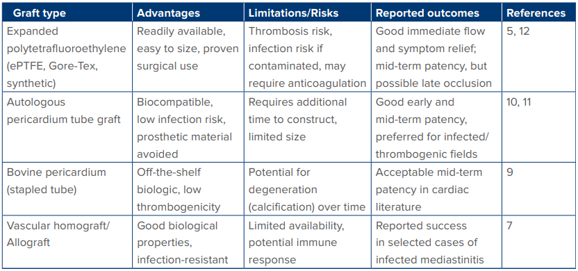
Table 1: Surgical options and outcomes with different graft materials.
Case Report
A 53-year-old female on long-term haemodialysis presented with progressive upper-body venous congestion. Repeated vascular access procedures suggested catheter-related fibrosis as the aetiology.1,2 Computed tomography (CT) venography revealed longsegment SVC occlusion with extensive azygos and hemiazygos collateralisation (Figure 1A). Endovascular attempts failed as guidewires were unable to cross the densely fibrotic obstruction, consistent with CTO.3,4 Coronary angiography confirmed triple-vessel CAD.
Surgical techniques used included OPCAB and innominate–right atrial appendage (RAA) bypass. To avoid cardiopulmonary bypass–related complications, OPCAB was performed using reverse saphenous vein grafts to the left anterior descending (LAD), obtuse marginal (OM), and posterior descending artery (PDA).12
The innominate–RAA bypass was performed using an autologous pericardial tube graft (Figures 1B and 1C). Given the thrombosis and infection risks of prosthetic grafts, and the risk of calcification in bovine pericardium,8,9 a 13-mm autologous pericardial tube graft was constructed over a Hegar dilator following established techniques.10,11 The graft was anastomosed end-to-side from the innominate vein to the RAA.
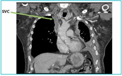
Figure 1A: Venogram showing superior vena cava (SVC) stenosis.
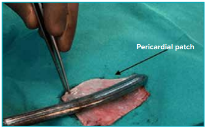
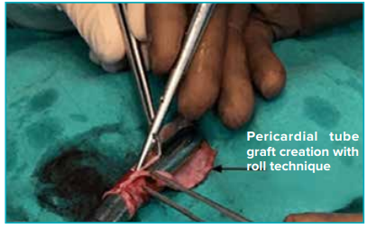
Figure 1B: Pericardium tube graft creation.
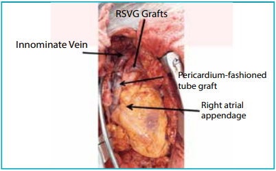
Figure 1C: Intraoperative image showing the grafts: Reverse saphenous vein graft (RSVG) and pericardium-fashioned tube graft.
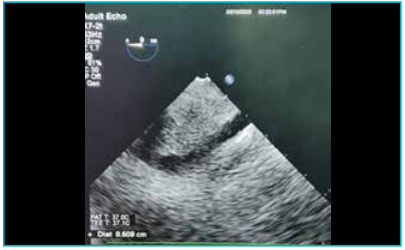
Figure 1D: Postoperative 2D echo showing patent pericardiumfashioned tube graft.
Results
Intraoperative findings: Dense fibrosis involving the innominate vein and SVC–RA junction confirmed a chronic, non-crossable obstruction, consistent with endovascular failure patterns.4 All coronary targets were adequate for grafting.
Postoperative course: Venous congestion improved significantly within 48 hours. The autologous pericardial conduit demonstrated excellent flow dynamics and handling properties, consistent with previously reported patency profiles.10,11
Follow-up: Serial imaging confirmed sustained patency of both venous (Figure 1D) grafts. The patient remained asymptomatic with resolution of SVC syndrome.
Discussion
Autologous pericardial grafts offer superior durability and resistance to infection compared with synthetic grafts, making them ideal for central venous reconstruction in dialysis-dependent patients.6,8,10,11
Prosthetic grafts such as ePTFE are convenient but associated with higher thrombosis and infection, while bovine pericardial grafts may calcify over time.9 Allografts are effective but limited by availability.7
OPCAB offers additional benefits by avoiding cardiopulmonary bypass, reducing inflammation, minimising coagulation disturbances, and eliminating the need for venous cannulation — advantages particularly significant in patients with SVC pathology.12,13 Simultaneous central venous bypass and OPCAB is feasible and effective in experienced centres.10-13
Conclusion
The simultaneous management of CAD and chronic SVC occlusion requires meticulous planning and a tailored surgical strategy. The successful use of an autologous pericardial tube graft and OPCAB in this patient highlights the versatility of biologic conduits and the safety of avoiding cardiopulmonary bypass in high-risk populations. This combined approach offers effective restoration of venous drainage and myocardial perfusion with favourable early outcomes.
Nukala Rao Mallikarjuna, Debashish Panigrahi, Shobhit Saurav, Pankaj Kumar Gupta, Rahul Dutta, Adarsh Subrahmanyam Koppula, Rajneesh Malhotra. Combined Complex Off-Pump Cardiac Surgery: Surgical Coronary Artery Revascularisation and Innominate–Right Atrial Appendage Bypass in a Patient with Chronic Total Superior Vena Cava Occlusion and Coronary Artery Disease — Our Experience. MMJ. 2025, December. Vol 2 (4).
References
-
Azizi AH, Shafi I, Shah N, et al. Superior vena cava syndrome. J Am Coll Cardiol Intv. 2020;13(24):2896–910.
-
Echefu G, Stowe I, Lukan A, et al. Central vein stenosis in hemodialysis vascular access: clinical manifestations and contemporary management strategies. Front Nephrol. 2023;3:1280666.
-
Quaretti P, Galli F, Moramarco LP, et al. Dialysis catheterrelated superior vena cava syndrome with patent vena cava: long term efficacy of unilateral Viatorr stent-graft avoiding catheter manipulation. Korean J Radiol. 2014;15(3):364–69.
-
Tzifa A, Marshall AC, McElhinney DB, et al. Endovascular treatment for superior vena cava occlusion or obstruction in a pediatric and young adult population: a 22-year experience. JACC. 2007;49(9):1003–9.
-
Kalra M, Gloviczki P, Andrews JC, et al. Open surgical and endovascular treatment of superior vena cava syndrome caused by nonmalignant disease. J Vasc Surg. 2003;38(2):215–23.
-
Doty JR, Flores JH, Doty DB. Superior vena cava obstruction: bypass using spiral vein graft. Ann Thorac Surg. 1999;67(4):1111–16.
-
Willems TP, Bogers AJ, Cromme-Dijkhuis AH, et al. Allograft reconstruction of the right ventricular outflow tract. Eur J Cardiothorac Surg. 1996;10(8):609–15.
-
Kitrou P, Katsanos K, Karnabatidis D. Management of central venous stenoses and occlusions. Cardiovasc Intervent Radiol. 2023;46(9):1182–91.
-
Hiromoto A, Sakamoto SI, Miyagi Y, et al. Bovine three-portion pericardial patch for reconstruction of the aorto-mitral curtain in infective endocarditis. Surg Case Rep. 2019;5(1):2.
-
Lapergola A, Felli E, Rebiere T, et al. Autologous peritoneal graft for venous vascular reconstruction after tumor resection in abdominal surgery: a systematic review. Updates Surg. 2020;72(3):605–15.
-
Fang C, Pan H, Li Z, et al. Autologous pericardium used for reconstruction of left innominate vein in patient with mediastinal venous aneurysm:a case report. BMC Surg. 2020;20(1):88.
-
Chen JC, Bongard F, Klein SR. A contemporary perspective on superior vena cava syndrome. Am J Surg. 1990;160(2):207–11.
-
Koga Y, Tomita M, Shibata K, et al. An experience using spiral vein graft as arterial substitute. Jpn J Surg. 1981;11(4):305–09.