 WhatsApp Us (24/7)
 +91 926 888 0303 (24/7)


Abstract:
Fungal pleural effusion (FPE) is an uncommon cause of pleural infection, accounting for only 1%–3% of all cases. It usually occurs in immunocompromised individuals, such as those with malignancy, diabetes, human immunodeficiency virus (HIV), or chronic steroid use. We report a rare case of left-sided Candida-associated pleural effusion in an 85-year-old man with no known comorbidities or immunosuppressive condition. The patient presented with left-sided chest pain and weakness for two weeks. The pleural fluid analysis showed an exudative, lymphocyte-predominant effusion with low adenosine deaminase (ADA) levels. Medical thoracoscopy with pleural biopsy revealed fungal hyphae consistent with Candida species, confirming the diagnosis. The patient was treated with oral fluconazole 200 mg twice daily for eight weeks, showing marked clinical improvement and radiological resolution without the need for surgical intervention. This case highlights that FPE can occur even in immunocompetent individuals, emphasising the need for early pleural biopsy and timely antifungal therapy in undiagnosed exudative effusions
Key words: Fungal Pleural Effusion, Candida, Immunocompetent, Pleural Biopsy, Fluconazole.
Introduction
Fungal pleural effusion (FPE) is a rare cause of pleural infection, representing only a very small proportion of pleural effusions worldwide. It usually occurs when fungal organisms invade the pleural space, either directly or through adjacent pulmonary or surgical infection. Although bacteria remain the most frequent cause of pleural infections, fungi are occasionally isolated, most often Candida species and less commonly Aspergillus. 1,2
Fungal infections of the pleura are typically seen in individuals with underlying illnesses such as diabetes mellitus, malignancy, human immunodeficiency virus (HIV) infection, organ transplantation, or prolonged use of corticosteroids.3,4 They may also develop following chest surgery, trauma, or prolonged hospitalisation.
However, pleural infections caused by fungi in immunocompetent individuals are extremely uncommon. Such presentations may delay diagnosis because they are not usually suspected in patients without risk factors. Early use of diagnostic procedures such as medical thoracoscopy and pleural biopsy can be crucial in identifying the cause.
We describe a rare case of Candida-associated pleural effusion in an elderly but otherwise healthy patient, emphasising that fungal infection should not be excluded based on only immune status. This case also focuses on the role of direct pleural sampling in the diagnosis of unusual causes of pleural effusion.
Case Report
An 85-year-old male, a non-smoker with no significant past medical history or immunosuppressive condition, presented to Max Super Speciality Hospital, Patparganj, with complaints of left-sided chest pain persisting for the past two weeks along with generalised weakness. The chest pain was significant enough to compel the patient to lie in the left lateral decubitus position for relief.
On examination, the patient had poor general condition. But he was conscious, oriented, haemodynamically stable, and maintaining oxygen saturation on room air. Respiratory examination revealed reduced air entry in the left infrascapular region.
Initial investigations included a chest X-ray (Figure 1), which showed blunting of the left costophrenic angle, suggestive of a moderate left-sided pleural effusion. Initial laboratory investigations were as follows:
-
Complete blood count (CBC):
Total leucocyte count (TLC) – 4.2 × 10⁹/L, haemoglobin – 8.3 g/dL, and platelets – 177 × 10⁹/L -
Kidney function test (KFT):
Creatinine – 1.2 mg/dL, sodium – 128.9 mmol/L, potassium – 4.31 mmol/L -
Liver function test (LFT):
Alkaline phosphatase (ALP) – 131.2 IU/L, albumin: globulin (A:G) ratio – 0.85
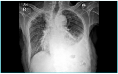
Figure 1: Chest X-ray-posteroanterior (CXR-PA) view, on presentation.
A high-resolution computed tomography (HRCT) chest (Figures 2 and 3) was done to rule out any lung parenchymal pathology and to look for any mediastinal lymph nodes. It showed a mild pleural effusion on left side with consequent atelectatic changes in left lower lobe, along with subcentimetric lymph nodes in the right paratracheal, aortopulmonary (AP) window and subcarinal regions.
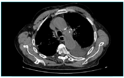
Figure 2: High-resolution computed tomography (HRCT) chest (mediastinal window), on presentation.
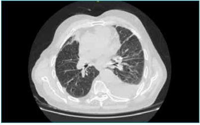
Figure 3: High-resolution computed tomography (HRCT) chest, lung window, on presentation.
A diagnostic thoracentesis was performed, revealing slightly turbid pleural fluid that was exudative, lymphocyte predominant, with low adenosine deaminase (ADA) levels. The pleural fluid analysis showed: glucose 137 mg/dL, protein 94.1 g/dL, albumin 2.29 g/dL, TLC 1250 cells/mm3 (neutrophils 10%, lymphocytes 90%, with a few macrophages and mesothelial cells), ADA 8.10 U/L, and negative malignant cytology.
In view of low ADA levels with exudative, lymphocytic effusion, malignancy was suspected, and the patient was taken up for medical thoracoscopy with pleural biopsy during which approximately 6500–7000 mL of straw-coloured pleural fluid was drained. The visceral pleura and parietal pleura were visualised. Multiple blackish to brownish discolourations were noted over the visceral pleura, from which targeted pleural biopsies were obtained. Post-thoracoscopy X-ray shows expansion of lung parenchyma (Figure 4)
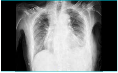
Figure 4: Chest X-ray-posteroanterior (CXR-PA) view, post medical thoracoscopy.
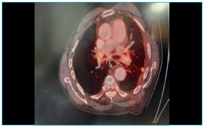
Figure 5: Positron emission tomography (PET) scan post medical thoracoscopy.
To further evaluate the cause, a whole-body ¹⁸F-fluorodeoxyglucose positron emission tomography– computed tomography (¹⁸F-FDG PET-CT) scan was conducted after thoracoscopy. Since the patient was initially unwilling to undergo PET imaging, we proceeded with medical thoracoscopy and pleural biopsy. The PET scan was performed later, once the patient subsequently consented. The scan showed (Figure 5) faint FDG-avid minimal left-sided pleural effusion with corresponding subsegmental collapse, along with necrotic mediastinal lymphadenopathy. Enlarged bilateral hilar, subcarinal, and parahilar lymph nodes were noted — the largest measuring 1.8 × 1.9 cm in the subcarinal region, 1.5 × 1.8 cm at the left hilum, and 1.9 × 1.7 cm at the right hilum. No metabolically active mass or distant organ involvement was seen.
Histopathological examination (Figure 6) of the pleural biopsy revealed pleura with mononuclear cells and fungal hyphae consistent with Candida spp., with no granulomas or malignant cells. This confirmed the diagnosis of Candida-associated FPE.
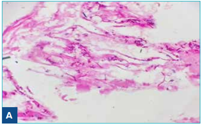
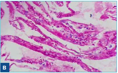
Figure 6: Pleural Biopsy showing tubular, filamentous hyphae consistent with Candida spp. under periodic acid-Schiff stain (PAS Stain) 400x magnification. A. Shows pseudohyphae and histiocytes. B. Presence of histiocytes more prominently seen.
In view of the rare findings on the pleural biopsy, the patient’s attendants requested a second opinion on the pathology slides. The samples were reviewed at Lab Core Diagnostics, which reported the following:
-
Block A: Histiocytes with yeast forms and pseudohyphae of fungus
-
Block B: Fibrocollagenous tissue showing a few reactive mesothelial cells and histiocytes
The final diagnosis was the presence of fungal organisms, with morphology favouring Candida.
The patient was initiated on oral fluconazole at a dose of 200 mg twice daily (BD) for a total duration of eight weeks, following which he showed gradual symptomatic improvement, with resolution of cough and chest discomfort.
We reviewed 7 case reports on fungal pleurisy available on PubMed, and the clinical, diagnostic along with treatment outcomes are summarised in Table 1.
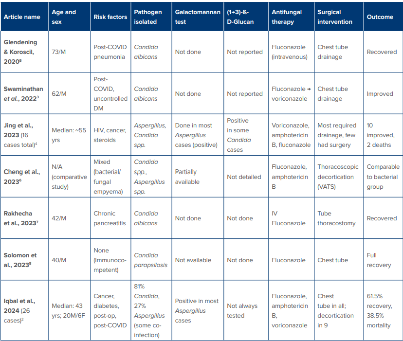
Table 1: Summary of published case reports and case series of fungal pleural effusion — diagnosis and management
Abbreviations: COVID: Corona Virus Disease 2019; DM: Diabetes Mellitus; F: Female; HIV: Human Immunodeficiency Virus; IV: Intravenous; M: Male; spp: Species; N/A: Not applicable; Post-op: Post-Operative; VATS: Video-Assisted Thoracoscopic Surgery.
Discussion
FPE is rare but important to recognise because its diagnosis can be difficult. Although the overall incidence is low, these infections often present diagnostic and treatment challenges. According to the latest European Respiratory Society/European Society of Thoracic Surgeons (ERS/ESTS) guidelines (2023), fungal infections are responsible for only about 1.75% of community-acquired and 2.68% of hospital-acquired pleural infections.
Most case series report Candida albicans as the most frequent organism, followed by non-albicans species such as C. glabrata and C. parapsilosis.2,5 Earlier studies report that affected patients usually have identifiable risk factors, such as malignancy, recent thoracic surgery, or significant immunosuppression.6,7
Our patient had none of these typical risk factors, making this presentation unusual. This suggests that fungal pleural infections can sometimes occur even in people who are otherwise immunocompetent. Advanced age or subtle mucosal injury may play a role, although such contributing factors are not always obvious.
The pleural fluid pattern in our case also differed from the patterns usually described. Fungal empyema commonly produces a neutrophilic, purulent exudate with a very low pH.2 However, our patient had an exudative, lymphocyte-predominant effusion with low ADA. This may reflect a small fungal load limited mainly to the pleural tissue, causing a slower, cell-mediated inflammatory response rather than a frank empyema.
Diagnosing FPE requires a high level of suspicion. Pleural fluid cultures may be negative, and a pleural biopsy may be necessary to confirm the diagnosis. In this case, histopathology showing fungal hyphae compatible with Candida gave a definite diagnosis.
Management includes systemic antifungal therapy, drainage of the pleural space, and surgery in more severe cases. Fluconazole is effective for most Candida species, while azoles or amphotericin B are recommended for Aspergillus infections.9,10 Our patient improved with oral fluconazole alone, without the need for surgery, highlighting the importance of early recognition and timely treatment.
Overall, this case adds to the limited body of evidence showing that FPE can occur even in patients without obvious risk factors. When pleural effusions remain unexplained and routine tests are inconclusive, fungal causes should be considered. Early thoracoscopy, pleural biopsy, and prompt antifungal therapy can significantly improve outcomes.
Conclusion
FPE remains a rare and often overlooked diagnosis, particularly in individuals without obvious risk factors. This case highlights the need for clinicians to consider less common causes of pleural effusion, especially when the pleural fluid analysis is indeterminate. In such scenarios, early use of diagnostic tools like thoracoscopy and pleural biopsy is key to uncovering unusual pathogens such as fungi. Prompt initiation of appropriate antifungal treatment can lead to good recovery — even in the absence of surgical procedures. Recognising and treating such atypical presentations early can make a significant difference in patient outcomes.
Tanya, Priyanka Aggarwal, Subhadeep Saha, Praveen K Pandey. Case of Fungal Pleural Effusion in an Immunocompetent Patient: A Case Report and Review of Literature. MMJ. 2025, December. Vol 2 (4).
References
-
Bedawi EO, Ricciardi S, Hassan M, et al. ERS/ESTS statement on the management of pleural infection in adults. Eur Respir J. 2023;61(2).
-
Iqbal N, Ali AS, Zahid A, et al. Fungal empyema thoracis, a rare but an emerging entity: a retrospective case series from Pakistan. Ther Adv Infect Dis. 2024;11:20499361231223887.
-
Swaminathan N, Anderson K, Nosanchuk JD, et al. Candida glabrata Empyema Thoracis-A Post-COVID-19 Complication. J Fungi (Basel). 2022;8(9).
-
Jing Y, Wei Q, Zeng H, et al. The clinical features and prognosis of fungal pleural infection: A case series and literature review. Medicine (Baltimore). 2023;102(48):e36411.
-
Glendening J, Koroscil M. A report of fungal empyema following recovery of severe SARS-CoV-2 infection. Chest. 2020;158(4):A566.
-
Cheng YF, Chen CM, Chen YL, et al. The outcomes of thoracoscopic decortication between fungal empyema and bacterial empyema. BMC Infect Dis. 2023;23(1):8.
-
Rakhecha D, Patil BU, Gore R, et al. Fungal empyema thoracis: A rare occurrence at the rural tertiary care. Saudi J Health Sci. 2023;12(2):167–70.
-
Solomon AE. A Rare Case of Candida Parapsilosis Empyema Thoracis. Int J Med Stud. 2023;11:S75.
-
Patterson TF, Thompson GR, Denning DW, et al. Practice Guidelines for the Diagnosis and Management of Aspergillosis: 2016 Update by the Infectious Diseases Society of America. Clin Infect Dis. 2016;63(4):e1–e60.
-
Pappas PG, Kauffman CA, Andes DR, et al. Clinical Practice Guideline for the Management of Candidiasis: 2016 Update by the Infectious Diseases Society of America. Clin Infect Dis. 2016;62(4):e1–50.