 WhatsApp Us (24/7)
 +91 926 888 0303 (24/7)


Abstract:
Perioperative management of patients undergoing resection of mediastinal masses can be challenging due to the potential for airway compromise, cardiovascular collapse, major blood loss and neurological compromise in myasthenic patients. This article aims to highlight the wide variety in pathologies, surgical and anaesthetic approaches, intraoperative and postoperative events encompassed by the term ‘surgery for mediastinal mass’. This was a retrospective, observational, descriptive study in which records of 31 patients who underwent resection of mediastinal masses were analysed. Wide variation in symptomatology, surgical approach, structures resected, intraoperative blood loss and duration of surgery was noted. Thymectomy was the most common surgery performed, accounting for a little over 50% of cases. Breathlessness was the most common presenting symptom. Additional lung resection was required in 32.25% patients, and 35.48% required major vascular or pericardial resection. Blood loss ranged from less than 100 mL to 15 L, and the average duration of surgery was 6.09 hours. All patients except one were extubated on the table. There is no single formula for providing anaesthesia to patients with mediastinal masses. Anticipation of challenges and individualised management are key to positive outcomes.
Key words:Mediastinal Mass, Thymectomy, Major Blood Loss.
Introduction
The mediastinum is a cavity within the thorax between the two pleural sacs. It lies in the midline behind the sternum and anterior to the thoracic vertebral column, and contains the heart, great vessels, hollow viscera such as the trachea and oesophagus, the splanchnic and sympathetic chains, and the thymus. In addition, it contains mediastinal lymphatics, lymph nodes and connective tissue. While any of these organs or tissues can enlarge pathologically, thymomas, lymphomas, enlarged thyroid, neurogenic tumours and germ cell tumours are the most common causes of mediastinal masses. Due to close proximity to vital vascular structures and the airway, anaesthetic and surgical management of these tumours is challenging. Vascular compression, vascular infiltration or airway compression may necessitate the use of cardiopulmonary bypass (CPB). Resection around the heart and great vessels increases the possibility of major blood loss. The asymptomatic nature of these tumours allows them to grow to a large size before causing compressive symptoms, unless they are incidentally diagnosed at an earlier stage.
Despite the multiple challenges involved, surgical treatment is worthwhile, as achieving complete surgical resection (R0) improves the prognosis of most mediastinal masses.1,2 In this case series, we retrospectively analysed 31 surgical patients in terms of the preoperative symptoms, intraoperative surgical and anaesthetic approaches, and the postoperative hospital course.
Materials and Methods (Including Statistical Analysis)
Institutional review board approval was obtained, and data were collected retrospectively from records of 31 patients who underwent surgery for excision of mediastinal masses. Notes were made of the patients’ preoperative symptoms, pre-existing comorbidities, preoperative work-up, the surgery performed, and the anaesthetic plan, including the technique of induction, intraoperative blood loss, duration of anaesthesia, whether CPB was used or kept ready, the method of postoperative analgesia and the need for postoperative ventilation. The data was tabulated in Microsoft Excel and interpreted as percentages.
Results
Out of the 31 patients with mediastinal masses, 16 (51.61%) underwent thymectomy. Of these, 11 were operated robotically, 3 via midline sternotomy, and 2 via video-assisted thoracoscopic surgery (VATS), of which 1 was converted to median sternotomy (Table 1).
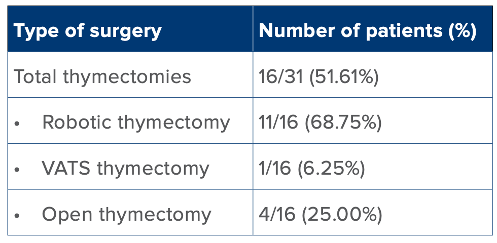
Table 1: Thymectomy approaches
Abbreviation: VATS: Video-Assisted Thoracoscopic Surgery.
Ten out of the 31 patients (32.25%) required additional lung resections in the form of wedge resection, lobectomy or segmentectomy. Eleven patients (35.48%) had pericardial, atrial or great vessel involvement requiring resection and repair (Table 2).
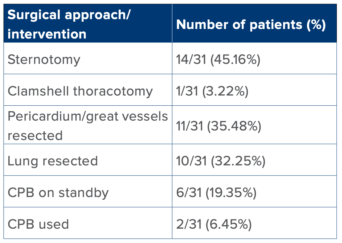
Table 2: Surgical approach and resection characteristics.
Abbreviation: CPB: Cardiopulmonary Bypass.
Twelve patients presented with breathlessness, 8 had motor weakness due to myasthenia gravis, and 6 were incidentally diagnosed (Table 3). Four of the myasthenic patients received intravenous immunoglobulin (IVIg) treatment preoperatively, and one of them also underwent plasma exchange due to myasthenic crisis.
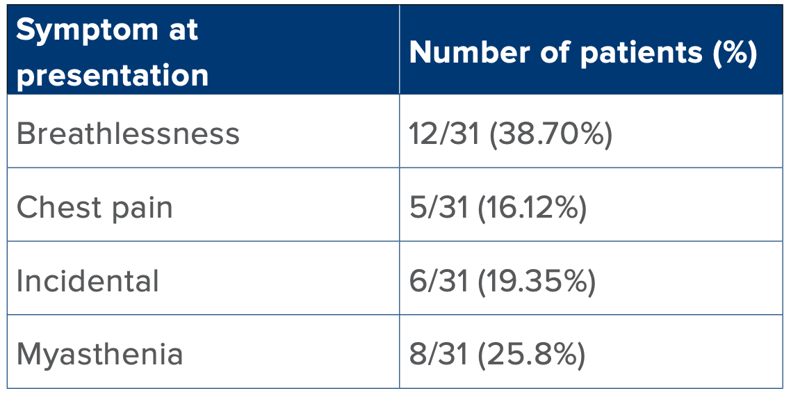
Table 3: Symptoms at presentation.
There was a wide range of intraoperative blood loss and total duration of surgery. The average blood loss was 882 mL (interquartile range [IQR]), and the average duration of surgery was 6.09 hours (IQR). The maximum blood loss encountered was 15 L in one patient, while 12 patients had less than 100 mL blood loss. In total, 8 out of 31 had ≥ 500 mL loss (Figure 1). The duration of surgery ranged from 2 to 12 hours (Figure 2). Only one patient required postoperative ventilation, while the rest of them were extubated on table.
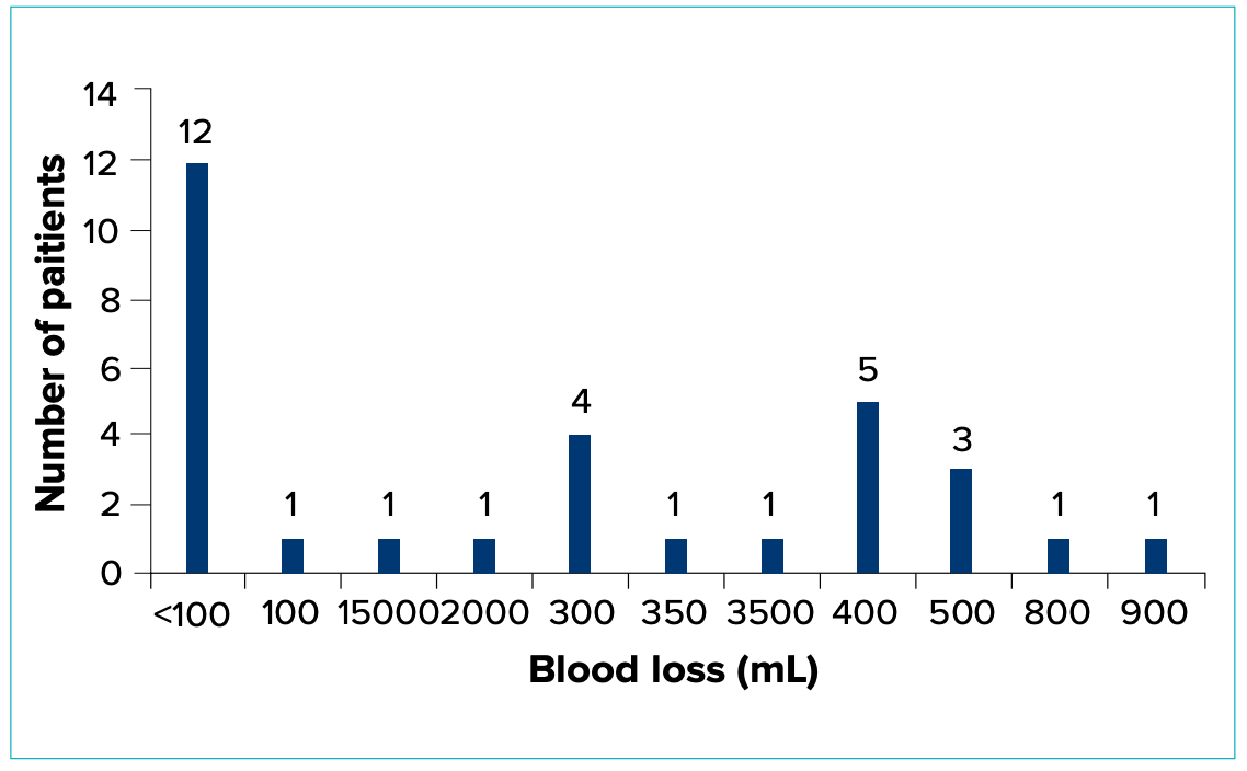
Figure 1: Patient distribution by intraoperative blood loss.
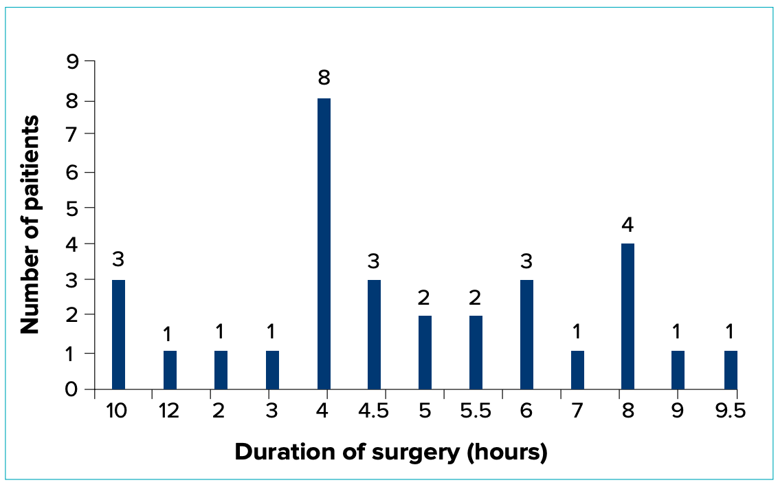
Figure 1: Patient distribution by surgery duration.
All patients received multi-modal analgesia postoperatively with intravenous (IV) paracetamol and non-steroidal anti-inflammatory drugs (NSAIDS). In addition, nearly half of the patients received IV patient-controlled analgesia (PCA) fentanyl, and 9 out of 31 (29.03%) received epidural analgesia (Table 4).
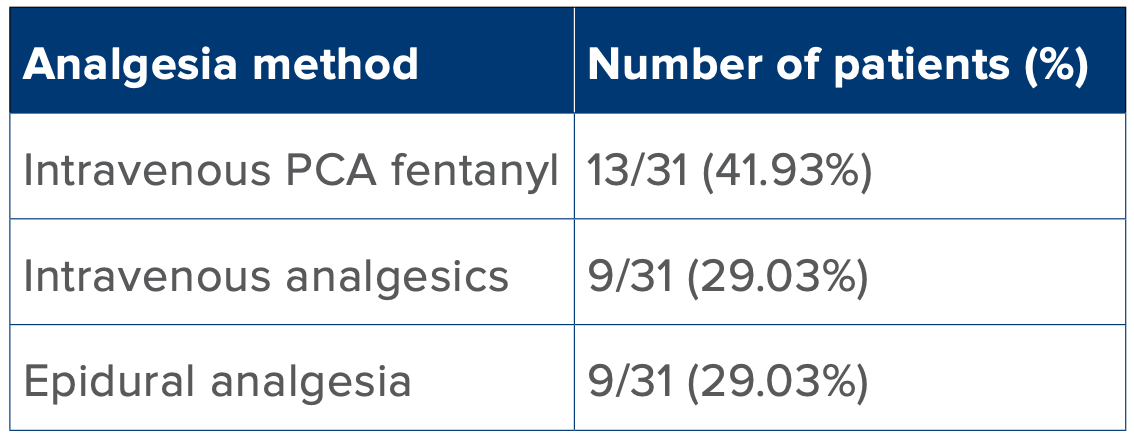
Table 4: Postoperative pain management.
Abbreviation: PCA: Patient-Controlled Analgesia
All patients were discharged home in 3–4 days after intercostal drain (ICD) removal, and there was no in-hospital mortality.
Discussion
Mediastinal masses are uncommon tumours to encounter. Just like the many organs and tissues that occupy the mediastinal cavity, mediastinal masses include a varied group of benign and malignant growths that can arise in one of the four compartments. Anterior mediastinal masses are the most common, with thymomas, lymphomas, and germ cell tumours being the most frequent pathologies in adults.3 Among the 31 patients operated on at our centre over 2 years, a large majority (29) had anterior mediastinal masses.
The anterior mediastinum allows these tumours to grow significantly before any obvious signs and symptoms appear.4 Compression of the large airways can cause breathlessness or a persistent cough. Worsening of symptoms in the supine position may be observed. This is of particular importance to the anaesthesiologist as it indicates the possibility of critical compression of the airway during induction of anaesthesia. A vague chest pain or heaviness is often reported by patients. Metabolic disturbance may occur due to thyroid or thymus involvement. Myasthenia gravis, presenting with muscular and generalised weakness, difficulty in breathing and swallowing, and dystonia, is typical of thymomas. Incidental diagnosis of the mass is possible, especially in case of previously operated patients on regular follow-up. However, about 60% patients show some clinical symptoms prompting investigation.4,5 Mediastinal syndrome refers to respiratory or haemodynamic compromise due to a mediastinal mass causing compression of surrounding structures like the trachea, heart, and great vessels. There is often a positional component, with worsening of symptoms in the supine position. Induction of general anaesthesia carries the risk of complete decompensation in these patients. While the majority of our patients presented with symptoms of breathlessness, muscular weakness or chest pain, none of them had positional symptoms of airway or haemodynamic compromise.
The preferred anaesthetic plan for surgical resection is general anaesthesia, often with invasive lines for advanced haemodynamic monitoring. Since resection occurs close to the heart and great vessels, beatto-beat blood pressure monitoring is essential. An arterial line also allows intermittent sampling to assess acid-base status, hydration and haemoglobin levels in case of massive blood loss. We secured large-bore IV access in the lower limb for selected patients. This can be lifesaving, especially when resection involves or is in the proximity of the aorta and superior vena cava (SVC). If central venous access is required, the femoral veins are cannulated in cases with cardiac or SVC involvement. Depending on the surgical approach, lung isolation using a double-lumen tube or bronchial blocker may be needed. Minimally invasive techniques like robotic or thoracoscopic surgery require the ability to selectively ventilate the lungs. In our series, 25 out of 31 patients were intubated with double-lumen tubes, allowing selective ventilation during surgery if required. This included all robotic and thoracoscopic procedures, as well as some open procedures where one-lung ventilation might have been required to facilitate easier resection.
Induction of general anaesthesia in an asymptomatic patient is generally straightforward using intravenous induction and muscle paralysis. Mask ventilation and intubation follows as per usual protocol. However, patients with respiratory symptoms in any position pose a significant challenge to the anaesthesiologist. It is important to carefully assess radiological scans to note the tumour size, extent, and involvement of surrounding structures while planning the anaesthesia management. Airway narrowing, vascular involvement and proximity can be appreciated in these. Traditional teaching advocates IV induction for the asymptomatic patients and inhalational induction while maintaining spontaneous respiration for those with respiratory symptoms.6 The rationale is that the administration of anaesthetic agents alone can cause loss of tone in the pharyngolaryngeal musculature causing the mediastinal mass to further obstruct the airway. This is expected to worsen with muscle paralysis. Inhalational induction can theoretically be more gradual and allows the patient to maintain spontaneous respiration.7 However, in practice, it is seldom possible to achieve the depth of anaesthesia required for laryngoscopy and intubation while preserving respiratory drive. In a spontaneously breathing patient with a partially obstructed airway, the depth of anaesthesia achieved with inhalational agents alone is often unpredictable. It may be insufficient to allow airway manipulation and yet still cause partial obstruction due to a large mediastinal mass. The safest approach when airway obstruction is anticipated is to perform an awake fibreoptic intubation, after topicalising the airway, and in the position the patient finds most comfortable.7,8 Once the tube is passed beyond the level of obstruction, a muscle relaxant can be given. A rigid bronchoscope and an experienced operator must be present in the theatre at the time of induction to rescue and stent the airway if needed. Haemodynamic collapse due to compressive effects of a large mass may also occur post-induction of anaesthesia. If either airway or haemodynamic collapse is expected to occur under anaesthesia, readiness for CPB by cannulating the femoral vessels is a safe option. Although rarely used, cannulation must be done pre-emptively as CPB cannot serve as a rescue technique due to the time required to cannulate the vessels and go on pump.7,8
A special work-up is required for patients with thymomas as they may have raised acetylcholine receptor antibodies (AchR Ab), with or without overt muscular weakness. Neuromuscular testing with repeated nerve stimulation and electromyography maybe needed to establish the diagnosis in patients who test negative for antibodies.9 Optimisation of respiratory reserve and muscle strength by instituting right treatment in the form of cholinergic drugs, steroids and if required immunoglobulins or plasmapheresis is essential for reducing perioperative morbidity. Anaesthesia planning must include additional monitoring like neuromuscular transmission monitor and depth of anaesthesia monitor. It is wise to avoid the use of neuromuscular blockers or at least use them sparingly while monitoring trainof-four (TOF) response intraoperatively to prevent excessive muscle weakness and postoperative respiratory compromise. The combination of rocuronium and sugammadex avoids the use of neostigmine and may provide a more reliable reversal of neuromuscular blockade when muscle relaxants cannot be avoided.10 We used TOF monitors for all thymomas and avoided infusions or timed doses of muscle relaxant, which we normally use in robotic surgeries. Instead, small boluses of atracurium were administered as and when needed, guided by TOF response. In our experience, equally important was the preoperative optimisation of muscle strength and prompt re-institution of cholinergic drugs and steroids post-surgery. Gradual tapering of treatment as tolerated by patients helps avoid precipitating myasthenic crisis. All our myasthenic patients could be extubated on table and were observed in high-dependency units for a minimum of 24 hours. Surprisingly, a previously asymptomatic patient experienced an episode of muscular weakness and respiratory distress after discharge home and required ventilatory support for five days on readmission. The patient responded well to steroids and cholinergic drugs and was discharged home after two weeks.
In mediastinal mass, whether thymoma or otherwise, involvement of cardiac chambers or aorta or SVC due to tumour infiltration might necessitate the use of CPB intraoperatively to allow tumour resection. In such cases, anticoagulation with heparin is required. This must be anticipated, and anaesthetic planning should reflect this possibility by avoiding neuraxial techniques, particularly epidural catheter placement, in patients who may require full heparinisation. These patients also tend to have higher intraoperative blood loss and may require blood transfusion due to the resection of vascular structures. Even when full-dose heparinisation is not required, such as in pericardial or SVC resection without CPB, patients often receive heparin at a dose of 100 units/kg at regular intervals to maintain an activated clotting time (ACT) of around 200 seconds.
Concomitant excision of lung tissue might be needed depending on the extent of the mass. Therefore, a preoperative assessment of the patient’s suitability through evaluation of cardiopulmonary reserve and lung function testing is essential.
Optimal postoperative analgesia is essential for early mobilisation and compliance with respiratory physiotherapy. Available options include epidural catheter, paravertebral block (with or without a catheter), erector spinae block or IV PCA with opioids. Paracetamol and NSAIDS should be used in all patients to reduce the requirement of opioids and catheterbased infusions.
In our practice, all patients received multimodal analgesia with paracetamol, NSAIDS and either local infiltration of the incision site or intercostal block administered by surgeons. We avoided using epidural analgesia in patients with a high likelihood of requiring CPB. Most of these patients received IV PCA fentanyl and remained reasonably comfortable. Epidurals were placed in open procedures without cardiac or major vascular involvement, as well as in minimally invasive surgeries with a high possibility of conversion to an open approach. IV PCA was given after minimally invasive surgeries when round-the-clock paracetamol and NSAIDS alone were insufficient to control pain and facilitate mobilisation and effective physiotherapy.
We encountered a wide spectrum of pathologies from mediastinal cysts requiring marsupialisation to infiltrative masses involving the heart and lungs. Overall, thymectomies were the most predictable surgeries in terms of minimal blood loss involved, feasibility of robotic approach, and low likelihood of requiring CPB. Two of our patients underwent redo surgeries, one for recurrence and the other for residual disease, as part of a planned staged procedure. The highest blood loss occurred in a patient with a mediastinal paratracheal mass that had expanded to fill the entire hemithorax (Figures 3 and 4).
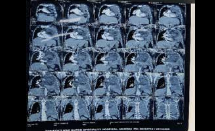
Figure 3: Imaging of a giant mediastinal mass
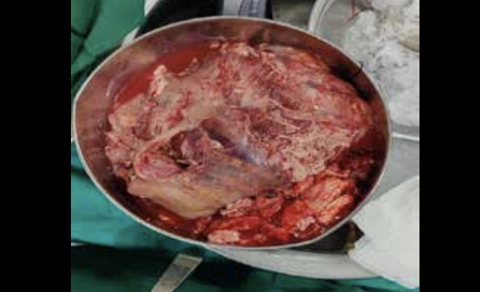
Figure 4: Excised giant mediastinal mass
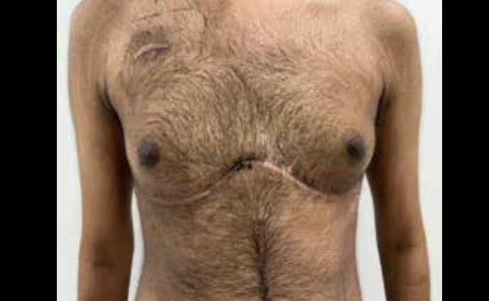
Figure 5: Clamshell thoracotomy incision.
This patient underwent a 10-hour surgery and experienced blood loss of more than 10 litres, necessitating a massive transfusion protocol that included 22 units of packed red cells, 8 units of fresh frozen plasma, 6 units of cryoprecipitate and 5 units of platelets. This was an eventful surgery that demonstrated excellent teamwork from anaesthesia, surgical, transfusion medicine and critical care teams. Another patient with a posterior mediastinal mass required a clamshell thoracotomy incision, which is unusual in adults (Figure 5). However, access to the posterior attachments of the tumour would have been impossible without this approach. There were concerns about postoperative analgesia in this young adult, but a thoracic epidural at the T7–T8 level was effective throughout the 4-day recovery period.
Multi-disciplinary meetings involving the thoracic surgeon, anaesthesia team, neurologist, cardiologist and chest physician were organised to discuss key concerns and formulate plans for preoperative optimisation, intraoperative equipment and personnel arrangements, and postoperative care. Co-ordination with the blood bank was vital in cases with potential for major blood loss. We found that pre-emptive communication with the transfusion team before induction, dedicating a single liaison person, and having designated staff and runners in place during massive transfusion cases facilitated a faster and more efficient response.
Conclusion
This article highlights the wide variety in pathologies, surgical and anaesthetic approaches, and intraoperative and postoperative events that fall under the umbrella of ‘surgery for mediastinal mass’. There is no single formula for providing anaesthesia to patients with mediastinal masses. Key considerations include signs of mediastinal syndrome, metabolic conditions such as myasthenia, involvement of major vessels, heart or large airways requiring resection, the possibility of major blood loss, the need for CPB, and prolonged operative duration. Thoughtful planning for each of these factors contributes to safe intraoperative anaesthetic management, smooth and pain-free recovery, and an uneventful discharge home.
Aditi Tilak, Shalini Saksena. Anaesthesia for Mediastinal Masses: A Retrospective Review. MMJ. 2025,
December. Vol 2 (4).
DOI: XXXXX
References
- Vaporciyan AA, Rice D, Correa AM, et al. Resection of advanced thoracic malignancies requiring cardiopulmonary bypass. Eur J Cardiothorac Surg. 2002;22(1):47–52.
- Fang Y, Qin Z. Surgical treatment of giant mediastinal tumors. Turk Gogus Kalp Damar Cerrahisi Derg. 2021;29(1):52–60.
- Dubashi B, Cyriac S, Tenali SG. Clinicopathological analysis and outcome of primary mediastinal malignancies – a report of 91 cases from a single institute. Ann Thorac Med. 2009;4:140–2.
- Napolitano MA, Werba G, Desai SA, et al. Presenting symptomatology of mediastinal masses and its effect on surgical outcomes. Am Surg. 2022;88(2):212–8.
- Azarow KS, Pearl RH, Zurcher R, et al. Primary mediastinal masses: A comparison of adult and pediatric populations. J Thorac Cardiovasc Surg. 1993;106:67–72.
- Gardner JC, Royster RL. Airway collapse with an anterior mediastinal mass despite spontaneous ventilation in an adult. Anesth Analg. 2011;113(2):239–42.
- Mackie AM, Watson CB. Anaesthesia and mediastinal masses. A case report and review of the literature. Anaesthesia. 1984;39(9):899–903.
- Shenoy A, Kamath S. Anaesthetic management of a huge mediastinal tumour with tracheobronchial compression. Ind J Resp Care. 2013;2:328–31.
- Rousseff RT. Diagnosis of Myasthenia Gravis. J Clin Med. 2021;10(8):1736.
- Daum P, Smelt J, Ibrahim IR. Perioperative management of myasthenia gravis. BJA Educ. 2021;21(11):414–19.