 WhatsApp Us (24/7)
 +91 926 888 0303 (24/7)


Abstract:
Tuberculosis (TB) has a notorious role as the “great imitator”–frequently mimicking a variety of infectious and non-infectious medical conditions- which often contributes to delays in care-seeking, diagnosis, and management. Probably, the vice versa also holds true. India, being a high endemic zone for tuberculosis, the threshold to label an unexplained illness of more than 2 weeks as TB by Indian clinicians is quite low, often leading to missed or delayed diagnosis of the underlying disease. Immunoglobulin G4-related disease (IgG4-RD) is another such “mimicker” often underdiagnosed due to its varied clinical presentation and is erroneously treated with anti-tubercular drugs. The overlapping biochemical and imaging features of IgG4-RD make prospective diagnosis difficult without a high index of suspicion. The incidence reported in literature is 0.78 to 1.39 per 100,000 person-years, which is likely an underestimation of the disease. Here, we present two cases where patients suffering from IgG4-RD were initially diagnosed and treated as tuberculosis and we review the literature associated with IgG4-RD.
Key words: Tuberculosis, IgG4-related disease, Imitators.
Introduction
Tuberculosis (TB) has a dubious distinction of frequently mimicking a variety of infectious and non-infectious medical conditions. This often contributes to delays in care-seeking, diagnosis, management, and adherence to the therapeutic regimen of tuberculosis. The vice-versa also holds true. As TB is endemic in India, the threshold to label an unexplained illness of more than 2 weeks, as tuberculosis, by Indian clinicians is quite low leading to erroneous treatment with anti-tubercular drugs. Here, we present two cases where patients actually suffering from Immunoglobulin G4-related disease (IgG4-RD) were initially diagnosed and treated as tuberculosis because of vague clinical presentations.
A time-tested medical teaching is that any patient, at high risk of TB, with an unexplained illness of >2-3 weeks of duration, should be investigated for pulmonary or extra-pulmonary tuberculosis. Patients at high risk of TB1 are:
- Resident of a region where prevalence of TB is high
- Recent exposure to a person with a case of infectious TB
- History of a positive test result for Mycobacterium tuberculosis (MTB)
- Human immunodeficiency virus (HIV) infection
- Residents and employees of high-risk congregate settings
- Medically underserved, low-income population
- Medical risk factors for TB (diabetes mellitus, conditions requiring prolonged corticosteroid and other immunosuppressive therapy, chronic kidney disease, certain haematological malignancies and carcinomas, weight >10% below ideal body weight, silicosis, gastrectomy, or jejunoileal bypass)
India accounts for highest global TB burden (28%).2 A state of persistent immune response due to stimulation by MTB antigens without any evidence of clinically manifested active TB is defined as TB infection (TBI). According to the National TB prevalence survey, 2021, the crude prevalence of TBI among individuals >15 years is 31.3%. An increasing trend of TBI is observed with increasing age and two deaths occur every three minutes from TB in India.
Establishing a definitive laboratory diagnosis of TB may not be possible in many circumstances. No specific bacteriologic confirmation is ever established in at least 15 to 20 percent of patients with a clinical diagnosis of TB. In such cases, a “presumptive clinical diagnosis" is made based on epidemiologic risks together with physical findings, radiographic findings, positive Tuberculin Skin Test (TST) or Interferon Gamma Release Assay (IGRA), analysis of sputum or bronchoscopy specimens, and/or histopathology. In high endemic zones like India, a positive result supports (but cannot be used to establish) a diagnosis of active TB disease, and a negative result does not rule out active TB disease. In the setting of high clinical suspicion for TB, initiation of empiric therapy based on these findings is considered appropriate.3
Case 1
A 66-year-old male was admitted to Max Super Speciality Hospital, Dehradun with generalised body weakness, loss of appetite, low-grade fever, weight loss, and neck swelling for the past 5-6 months. The patient was a known case of hypertension and diabetes due to chronic pancreatitis for the past 3 years. He also had a history of sclerosing cholangitis, for which stenting of the common bile duct was done twice in the last one year. On examination, the patient was emaciated with no pedal oedema. Imaging revealed multiple enlarged lymph nodes in para-aortic, aortocaval and retrocaval regions. He also presented with mediastinal and left hilar lymphadenopathy. Computed tomography (CT) findings showed bulky bilateral submandibular glands. Fine Needle Aspiration Cytology (FNAC) of these glands revealed non-specific findings because of scanty lymphoid tissue, and an ultrasound (USG) guided aspiration biopsy of the gland was suggested but was not done. Instead, considering the overall clinical manifestations, anti-tubercular treatment was initiated. A week back, the patient developed rashes all over his body and subsequent investigations revealed renal dysfunction, for which he was referred to Max Super Speciality Hospital, Dehradun. Laboratory investigations at the time of admission are detailed below. (Table 1)
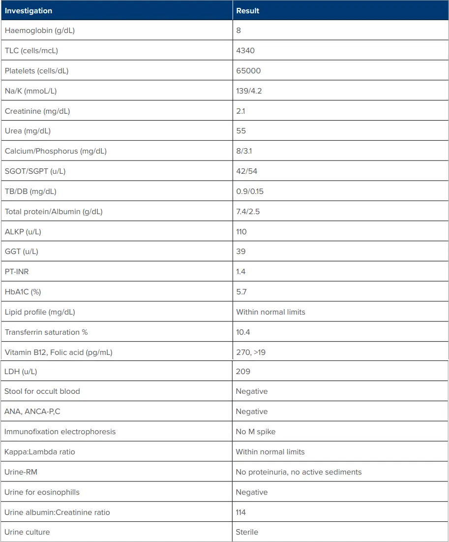
Table 1: Detailed laboratory investigations at time of admission
Abbreviations: TLC: Total Leukocyte Count; Na: Sodium; K: Potassium; SGOT: Serum Glutamic Oxaloacetic Transaminase; SGPT: Serum Glutamate Pyruvate Transaminase; TB: Total Bilirubin; DB: Direct Bilirubin; ALKP: Alkaline Phosphatase; GGT: Gamma-Glutamyl Transferase; PT-INR: Prothrombin TimeInternational Normalised Ratio; HbA1c, Glycated Haemoglobin; LDH, Lactate Dehydrogenase; ANA: Antinuclear Antibodies; ANCA-P,C: Antineutrophil Cytoplasmic Antibodies; RM, Routine Microscopy.
A provisional diagnosis of acute interstitial nephritis (drugs associated) was made, and a renal biopsy was done which revealed IgG4-RD. Prednisolone was initiated.
After 2 months of treatment with prednisolone, the fever resolved, submandibular gland swelling subsided completely, and the requirement of anti-diabetic medications decreased.
Laboratory parameters improved significantly. (Table 2)
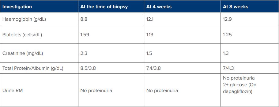
Table 2: Parameter after treatment with Prednisolone
Abbreviation: RM: Routine Microscopy.
Case 2
A 21-year-old female presented with generalised body weakness and low-grade fever from the last 2 months for which she was investigated by a general physician and found to have renal dysfunction, and hence, referred to a nephrologist for further evaluation. Laboratory values were as follows: (Table 3)
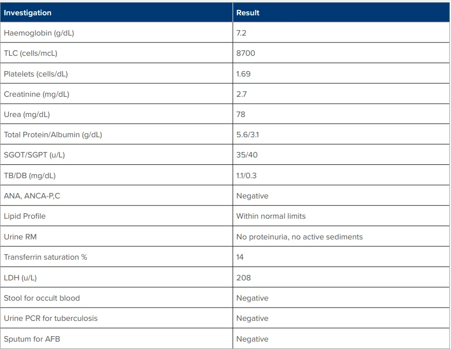
Table 3: Laboratory values
Abbreviations: TLC: Total Leukocyte Count; SGOT: Serum Glutamic Oxaloacetic Transaminase; SGPT: Serum Glutamate Pyruvate Transaminase; TB: Total Bilirubin; DB: Direct Bilirubin; ANA: Antinuclear Antibodies; ANCA-P,C: Antineutrophil Cytoplasmic Antibodies; RM: Routine Microscopy; LDH: Lactate Dehydrogenase; PCR: Polymerase chain reaction; AFB: Acid-fast bacilli.
Ultrasound was suggestive of normal-sized kidneys with altered echogenicity and maintained cortico-medullary differentiation. Renal biopsy was advised but refused by the patient and she was lost to follow-up thereafter.
Three months later, she again returned to the outpatient department (OPD) with persisting complaints, serum creatinine ~4.1, and a renal biopsy was planned for which ultrasound was done.
USG revealed normal-sized kidneys with markedly increased cortical echotexture with loss of cortico-medullary differentiation. A well-defined isoechoic mass lesion in the lower pole of the right kidney with internal vascularity was found. The mass lesion at the lower pole of the right kidney with retroperitoneal lymphadenopathy was confirmed by contrast CT. Urologist opinion was taken and considering the lesion as neoplastic, partial nephrectomy was done and tissue was sent for histopathology examination. The pathologist found no evidence of underlying malignancy in the tissue, but a granulomatous inflammation highly suggestive of mycobacterial aetiology
By this time, serum creatinine had risen to 5.5 (estimated glomerular filtration rate (eGFR) ~11). Anti-tubercular treatment (ATT) was initiated to which she had no response, rather developed hepatic dysfunction. Subsequently, serum IgG4 levels were sent and found to be high (3.28 mg/dL (0.1-1.2)). ATT was stopped, and the patient was put on prednisolone considering IgG4-RD as the primary aetiology. She responded significantly to steroids within two weeks of initiating treatment bringing her serum creatinine down to 2.5 (eGFR ~27) and her general condition improving.
Review of Literature
After its description in 2003 by Kamisawa, IgG4-RD is increasingly recognised as a serious immune-mediated condition that can affect virtually every organ in the body. It has a highly variable clinical presentation, indistinguishable from many inflammatory and neoplastic diseases. The overlapping biochemical and imaging features of IgG4-RD makes prospective diagnosis difficult without a high index of suspicion. The Incidence reported in literature is 0.78 to 1.39 per 100,000 person-years, which is perhaps gross underestimation because of underdiagnosis of the entity.4,5
Salient Features6,7
- In general, the history and physical examination are not diagnostic
- The clinical features are subacute, typically developing over several months.
- Patients often feel well at the time of diagnosis.
- Lymphadenopathy is common, but patients are generally afebrile.
- Symptoms of asthma or allergy are present in approximately 40 percent of patients.
- Patients with multiorgan disease often lose substantial amounts of weight, which may be due to IgG4-related autoimmune pancreatitis.
- Often recognised incidentally based on a radiologic finding or histopathology examination of a tissue specimen.
- Patients often present with subacute development of a mass in the affected organ or diffuse enlargement of an organ (Pseudotumours). Imaging studies may demonstrate diffuse or focal organ lesions (usually a mass or swelling).
Autoimmune Pancreatitis8 (AIP): Autoimmune etiology accounts for approximately 2% of chronic pancreatitis cases and often presents as a pancreatic mass or as painless obstructive jaundice and can be mistaken for pancreatic cancer.8 Patients with type 1 AIP may exhibit acute, recurrent, or chronic pancreatitis. Complications of AIP frequently include glucose intolerance or frank type 2 diabetes mellitus and, more frequently, exocrine pancreatic insufficiency. Most patients with type 1 AIP have another concomitant IgG4-related condition, including IgG4- related sclerosing cholangitis, lymphadenopathy, or IgG4-related tubulointerstitial nephritis.
IgG4-Related Lymphadenopathy is common (~41 to 80% of patients with AIP) and may be the initial or only manifestation of IgG4-RD in a given patient.9 It is usually non-tender, rubbery and often asymptomatic at the time of diagnosis.9 Multiple groups of lymph nodes are often involved simultaneously. Mediastinal, hilar, intrabdominal, and axillary lymph nodes are usually involved. Symptoms may occur due to impingement on other organs.
IgG4-Related Kidney Diseases: Tubulointerstitial nephritis (TIN) is the most common renal manifestation which presents with signs of acute or chronic kidney injury, mass lesion(s) in the kidney (potentially mimicking renal cell carcinoma (RCC), or both.10 TIN is more common in middle-aged or older men. Most of the cases where kidneys are involved, other organs are involved as well. Urinalysis is not a reliable tool for establishing a diagnosis of IgG4-related TIN.
Other Organ Involvement:
- Sialadenitis
- Dacryoadenitis
- Orbital pseudotumor
- Orbital myositis
- Retroperitoneal fibrosis (RPF)
- Aortitis, and periaortitis
- Thyroid involvement
- Lung involvement
High Index of Suspicion of Underlying IgG4-RD if:
- Pancreatitis of unknown origin
- Sclerosing cholangitis
- Bilateral salivary and/or lacrimal gland enlargement
- Retroperitoneal fibrosis (RPF)
- Orbital pseudotumor or proptosis
- Development of an otherwise unexplained mass lesion in any of the following organs: pancreas, biliary tree, orbits, lungs, kidneys, major salivary gland, or lacrimal gland
Nearly diagnostic of IgG4-RD:
- Combination of biliary tract and pancreatic disease
- Isolated involvement of the bilateral submandibular glands
Both features were present in one of the cases described above, still there was much delay in making the diagnosis of IgG4-RD as the eyes do not see what the mind does not know. Hence, clinicians, radiologists, and pathologists should be well aware of this entity.
Laboratory Evaluation-Serum IgG4
Measure serum IgG4 levels in all patients suspected of IgG4-RD. Serum IgG4 is above the upper limit of normal in approximately two-thirds of all patients with IgG4-RD.11 An elevated serum IgG4 levels cannot establish the diagnosis on its own. It requires clinical and histopathological evaluation as well. Very high levels of serum IgG4 do not exclude alternative diagnoses. In a study12 examining 32,206 cases, 191 patients had IgG4 serum levels >5 times the upper normal limit; 25 percent had alternative diagnoses, such as malignancies, other autoimmune diseases, and infections.
Normal IgG4 does not rule out IgG4-RD. Serum IgG4 is normal in approximately 30 percent of patients who are ultimately diagnosed with IgG4-RD. The optimal IgG4 threshold for diagnosis is unclear.11 Studies have varied from 135mg/dl to 1180mg/dL.
Imaging
Identification of a mass or organ enlargement on imaging should raise suspicion for IgG4-RD. CT is used in most cases, although it cannot distinguish active inflammation from fibrosis, even with the use of contrast. Positron emission tomography (PET) is a reasonable alternative, particularly when vascular involvement is suspected, although masses associated with IgG4-RD may not be PET-avid. No imaging findings are diagnostic of IgG4- RD, and imaging findings may not distinguish IgG4-RD from other diagnoses.
Biopsy
Confirm diagnosis of IgG4-RD by biopsy of the involved organ whenever possible. Although histopathology findings are important to the diagnosis of IgG4-RD, such findings alone are never diagnostic of IgG4-RD. Diagnosing IgG4-RD requires consideration of the clinical, serological, radiological, and pathologic data and exclusion of potential mimickers (e.g., malignancy).
Solid Organ Pathology (Characteristic Histopathology Features)
- Lymphoplasmacytic tissue infiltration of mainly IgG4-positive plasma cells and lymphocytes
- Storiform fibrosis
- Obliterative phlebitis
- Tissue eosinophilia
IgG4 lymphoplasmacytic infiltrates and even storiform fibrosis can also be observed in conditions mimicking IgG4-RD. Diagnosis cannot be predicated entirely upon pathology findings, regardless of the intensity of IgG4-positive plasma cell infiltration. Histopathologic features are more important than tissue IgG4-positive cell counts and the ratios of IgG4 to IgGpositive cells. The diagnosis of IgG4-RD cannot be predicated entirely upon the number of IgG4-positive plasma cells as many diseases are associated with large numbers of IgG4- positive plasma cells (variety of malignancies, granulomatosis with polyangiitis, Castleman disease, etc).12 Furthermore, the minimum for making the diagnosis for most tissues is from 30 to 50 IgG4-positive cells per high power field (HPF) .12 However, in some organs or tissues (e.g., kidney) only 10 IgG4-positive plasma cells per HPF may be sufficient.
Selection of Biopsy Site
In patients with multi-organ involvement, do not biopsy all involved organs. Instead, select a single organ to biopsy based on practical considerations (e.g., accessibility of organ). One should not use lip, lymph node, or gastrointestinal tract biopsies to establish the diagnosis.13 In patients with IgG4-RD with salivary gland involvement, a lip biopsy (i.e., biopsy of labial minor salivary glands) is helpful mainly to exclude other diseases such as Sjgren’s disease and lymphomas, but its sensitivity is quite low in diagnosing IgG4-RD. Biopsy of submandibular glands is more sensitive for diagnosing IgG4-RD in patients with swelling of these glands. Biopsies of lymph nodes are often problematic to interpret with regard to the diagnosis of IgG4-RD because they seldom undergo the storiform fibrosis that is so highly characteristic of IgG4-RD, and large numbers of IgG4+ plasma cells can be found in multiple diseases in which IgG4-RD is not the diagnosis, resulting in poor specificity of this finding. Biopsies of the intestinal tract often have a high concentration of IgG4+ plasma cells. Because the gastrointestinal tract is seldom affected by IgG4-RD, biopsies from the gastrointestinal tract should not be relied upon to establish the diagnosis of IgG4-RD.
Because of the poor sensitivity and specificity of available modalities, the Japanese IgG4 team has updated the 2011 comprehensive diagnostic criteria for IgG4-RD and proposed the 2020 revised comprehensive diagnostic (RCD) criteria for IgG4-RD to establish the diagnosis of IgG4-related disease (Table 4).
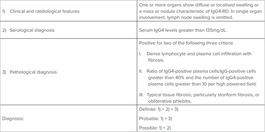
Table 4: The 2020 Revised Comprehensive Diagnostic (RCD) Criteria for IgG4-RD
Treatment
Initial therapy:
Once the diagnosis of IgG4-RD has been established, the extent of disease should be evaluated before initiating treatment. Testing should include a CT scan of the chest, abdomen, and pelvis, and may include other studies. Therapy should be initiated in all patients with symptomatic disease or having asymptomatic disease but is affecting a vital organ (e.g., aorta, retroperitoneal fibrosis causing hydronephrosis). Steroids14 (prednisolone0.6mg/kg/day), are the first line of treatment which may then be tapered, according to response, to discontinuation over a two-month period. Response to therapy can be monitored by symptomatic improvement, reductions in the size of masses or organ enlargement, improvement in organ function or a decrease in serum levels of IgG4.
Resistance to initial therapy:
- In patients who do not respond to up to 40 mg/day of prednisolone.
- In patients who cannot be tapered to <5 mg daily.
- In patients who have strong relative contraindications to glucocorticoid therapy.
Use rituximab15 (1 g intravenously every 15 days for a total of two doses). When rituximab is not feasible, azathioprine (2mg/kg/ day) or mycophenolate mofetil (up to 2.5g/day) may be used.
Subsequent therapy:
May continue treatment with a steroid-sparing agent or low-dose glucocorticoids to maintain remission; if impact on relapse rate is unclear. The use of a prolonged course of immunosuppression for remission maintenance should be considered on an individual basis.
Recurrent disease
- The ideal therapy for patients who relapse is not well established.
- Treatment options include rituximab (1g intravenously every 15 days for a total of two doses), azathioprine (2mg/kg), mycophenolate mofetil (2 to 2.5g/day), and calcineurin inhibitors.
Role for surgery
- May require surgery for relief of mechanical obstruction (e.g., hydronephrosis, obstructive jaundice, aortic aneurysms due to IgG4-related aortitis)
Treatment Response and Prognosis
The natural history and prognosis have not been defined. Spontaneous improvement can be seen, but recurrence is common. Most patients respond initially to glucocorticoids, but relapses are common following discontinuation of therapy. Significant organ dysfunction may arise from uncontrolled and progressive inflammatory and fibrotic changes in affected tissues. The possibility of increased risk of malignancy in patients with IgG4-RD has not been established and requires further studies.
The following features make IgG4-RD less likely:
- Persistent fever
- No response to glucocorticoids
- Simultaneous leukopenia and thrombocytopenia
- Hypereosinophilia
- Autoantibodies suggestive of another autoimmune diagnosis
- Radiologic features suspicious for malignancy or infection
- Rapid radiologic progression (i.e., significant worsening in four to six weeks)
- Splenomegaly
Conclusion
IgG4-RD is another great imitator, like tuberculosis, and is often underdiagnosed due to its polymorphic presentation. Any patient with an unexplained illness lasting >2-3 weeks or development of an otherwise unexplained mass lesion in any organ should also be investigated for IgG4-RD.
Puneet Arora. Are We Over-Diagnosing Tuberculosis? IgG4-Related Disease is Another “Great Imitator”:
A Review. MMJ. 2024, Dec. Vol 1 (4).
DOI: XXXX
References
- Paradkar M, Padmapriyadarsini C, Jain D, et al. Tuberculosis preventive treatment should be considered for all household contacts of pulmonary tuberculosis patients in India. PLoS ONE. 2020;15(7):e0236743.
- Chauhan A, Parmar M, Dash GC, et al. The prevalence of tuberculosis infection in India: A systematic review and metaanalysis. Indian J Med Res. 2023;157(2&3):135-151.
- Kashyap RS, Rajan AN, Ramteke SS, et al. Diagnosis of tuberculosis in an Indian population by an indirect ELISA protocol based on detection of Antigen 85 complex: a prospective cohort study. BMC Infect Dis. 2007;7:74.
- Stone JH, Zen Y, Deshpande V. IgG4-related disease. N Engl J Med. 2012;366(6):539-51.
- Kamisawa T, Zen Y, Pillai S, et al. IgG4-related disease. Lancet. 2015;85(9976):1460-71.
- Wallace ZS, Deshpande V, Mattoo H, et al. IgG4 related disease: clinical and laboratory features in one hundred twenty five patients. Arthritis Rheumatol. 2015;67(9):2466-75.
- Brito-Zerón P, Ramos-Casals M, Bosch X, et al. The clinical spectrum of IgG4-related disease. Autoimmun Rev. 2014;13(12):1203-10
- Takuma K, Kamisawa T, Igarashi Y. Autoimmune pancreatitis and IgG4-related sclerosing cholangitis. Curr Opin Rheumatol. 2011;23(1):80-7.
- Cheuk W, Yuen HK, Chu SY, et al. Lymphadenopathy of IgG4- related sclerosing disease. Am J Surg Pathol. 2008;32(5):671-81.
- Buglioni A, Jenkins SM, Nasr SH, et al. Clinicopathologic features of IgG4-related kidney disease. Kidney Int Rep. 2024;9:2462.
- Carruthers MN, Khosroshahi A, Augustin T, et al. The diagnostic utility of serum IgG4 concentrations in IgG4-related disease. Ann Rheum Dis. 2015 Jan 1;74(1):14-8.
- Baker MC, Cook C, Fu X, et al. The positive predictive value of a very high serum IgG4 concentration for the diagnosis of IgG4-related disease. J Rheumatol. 2023;50(3):408-12.
- Moriyama M, Furukawa S, Kawano S, et al. The diagnostic utility of biopsies from the submandibular and labial salivary glands in IgG4-related dacryoadenitis and sialoadenitis, so-called Mikulicz's disease. Int J Oral Maxillofac Surg. 2014;43(10):1276- 81.
- Khosroshahi A, Wallace ZS, Crowe JL, et al. International consensus guidance statement on the management and treatment of IgG4-related disease. Arthritis Rheumatol. 2015;67(7):1688-99.
- Ebbo M, Grados A, Samson M, et al. Long-term efficacy and safety of rituximab in IgG4-related disease: data from a French nationwide study of thirty-three patients. PLoS One. 2017;12(9):e0183844.