 WhatsApp Us (24/7)
 +91 926 888 0303 (24/7)


Abstract: ABinder’s facies is a rare congenital malformation that has multifactorial aetiologies. This anomaly affects the anterior part of the maxilla and the nasal cavity. A 30-year-old primigravida with a non-consanguineous marriage presented for a second opinion regarding an anomaly scan at the fetal medicine department of our institute at 21 weeks. Her scan performed elsewhere suggested a flat and short nasal bridge. A thorough 2D and 3D examination of the fetus was carried out. The midsagittal view of the face showed a flat nose with an increased fronto-nasal angle. In this study, we discuss the diagnosis of Binder syndrome during the second trimester of pregnancy.
Key words: Binder Syndrome (Flat Facies), Genetic Abnormalities, MRI-2D and 3D Imaging.
Introduction
Binder syndrome (flat facies) is a rare congenital malformation characterised by mid-facial hypoplasia involving the maxilla and nasal complex.1,2,3 It results from a growth disturbance in the prosencephalic induction centre.2,3 Along with this, mild hypertelorism and malocclusion due to overbite can be observed in some children.1,3 This is a marker for genetic abnormalities like trisomy 21 or may be seen in syndromes like chondrodysplasia punctata.
A 30-year-old primigravida presented for a second opinion regarding an anomaly scan at 21 weeks. No family history of any chromosomal abnormalities, warfarin abuse, or vitamin K deficiency was noted. The first-trimester nuchal translucency measurement was normal with a high risk for trisomy 21 of 1:105 on the double marker test. During the anomaly scan, a small, and vertically placed nasal bone was observed, and the nasal bridge was flat, with an increased frontonasal angle of 153 degrees (the normal angle is less than 140 degrees) (Figures 1 and 2). In addition, two tiny echogenic, intracardiac foci were seen in both foetal lateral ventricles of the heart (Figure 3). The biometry was normal, with normal long bones, and no evidence of any stippling was noted to suggest an association with chondrodysplasia punctata.
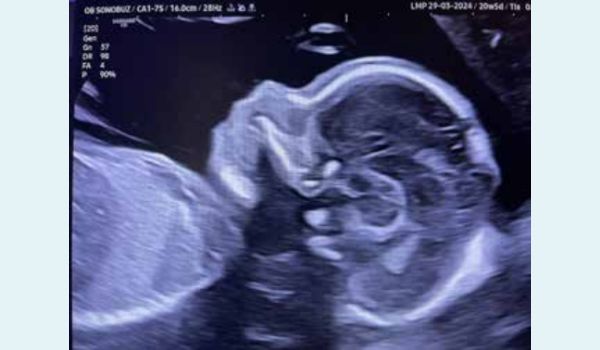
Figure 1: Normal sagittal image of the face and nasal bone.
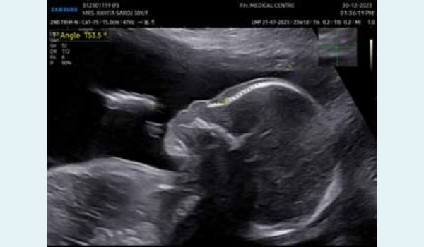
Figure 2: Abnormal flat facial profile with an increased frontonasal angle.
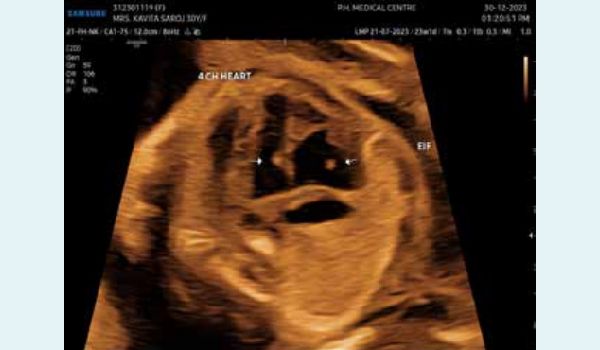
Figure 3: Echogenic intracardiac foci in both ventricles of the foetal heart.
The patient was counselled for amniocentesis and the possible association with Down’s syndrome, as indicated by the high risk on the double marker test. However, the parents declined further testing and opted for termination of pregnancy (Figure 4).
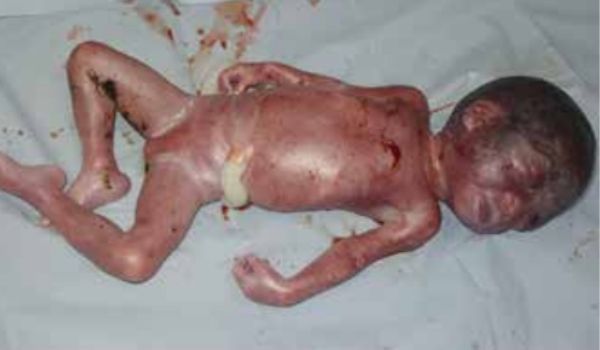
Figure 4: Post abortus image confirming dysmorphic face with mid-facial hypoplasia, short nasal bridge, and macroglossia.
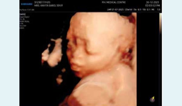
Figure 5: 3D-reconstruction of the face showing mid-facial hypoplasia and macroglossia with protuberant tongue.
Discussion
Binder’s facies is a rare congenital malformation that has multifactorial aetiologies. Common causes include isolated Binder's facies, chondrodysplasia punctata, metabolic abnormalities like Zellweger syndrome, chromosomal abnormalities like trisomy 21, autoimmune maternal illnesses like systemic lupus erythematosus (SLE), abnormal metabolism of vitamin K, hereditary factors, such as Xp22 and external influences which includes maternal chronic disorders with vitamin K deficiency, such as untreated coeliac disease, secondary short bowel syndrome, digestive malabsorption, or intractable vomiting in the early stages of pregnancy; and prenatal exposure to phenytoin, alcohol, and coumarin derivatives.3-6 The mode of inheritance is not well understood, and cases can be sporadic. There may be associated genetic abnormalities and syndromes.4,6 If isolated, Binder’s facies carry a favourable prognosis.
The diagnosis of Binder's phenotypic and other facial dysmorphisms causes considerable distress and anxiety to the parents. Hence, following the diagnosis of Binder's phenotype – in the midsagittal and coronal views of the foetal face, a thorough and careful examination of the foetus is conducted, with particular attention paid to the cardiac and skeletal systems for any association with chondrodysplasia punctata.5,6 The results of 2-dimensional imaging are enhanced by 3D imaging (Figure 5). If skeletal abnormalities are suspected, MRI imaging can offer more detailed information.6 Prenatal and preconception history should be thoroughly reviewed, including comorbidities such as hepatic disease, chronic malabsorption syndromes, connective tissue disorders, intractable vomiting, and use of teratogens like alcohol and warfarin.5 Before determining whether a trait is normal or abnormal, one should consider physiognomy, or the typical physical characteristics of a family. Isolated Binder’s to detect chromosomal abnormalities and microdeletions.1,5,6 A detailed sonogram, proper genetic testing, and counselling by a team of foetal medicine specialists, obstetricians, and geneticists will help avoid unnecessary pregnancy terminations. phenotype has an excellent outcome. It can be surgically corrected, and the bite can be adjusted with the help of an orthodontic surgeon. Amniocentesis with chromosomal microarray (CMA) is an invasive procedure recommended to detect chromosomal abnormalities and microdeletions.1,5,6 A detailed sonogram, proper genetic testing, and counselling by a team of foetal medicine specialists, obstetricians, and geneticists will help avoid unnecessary pregnancy terminations.
Alka Karnik, Arati Joshi, Rashmi Parekh. Case Report of a Second Trimester Diagnosis of
Binder Syndrome. MMJ. 2024, Dec. Vol 1 (4).
DOI: xxxxxxxxxxxxxxxxxxxx
References
- Textbook of Ultrasound of congenital fetal anomalies - Dario Paldini, Paolo Volpe.
- Cook K, Prefumo F, Presti F, et al. The prenatal diagnosis of Binder syndrome before 24 weeks of gestation: Case report. Ultrasound Obstet Gynecol. 2000;16(6):578-81.
- Kerbrat A, Ferri J. Surgical treatment for patients with Binder syndrome, clinical features, and associated symptoms: A systematic review. Journal of Craniofacial Surgery. 2022;33(2):p530-533.
- Vaid D, Vajpayee D, Rafeek S, et al. Binder's syndrome: A rare case report. Clin Rhinol An Int J. 2020;13(1):30-34.
- Nedev PK. The Binder syndrome: Review of the literature and case report. Int J Pediatr Otorhinolaryngol. 2008;72(10):1573-6.
- Veduta A, Duta S, Ciobanu AM, et al. Fetal skeletal dysplasias that involve the face: Binder syndrome and Nager Syndrome. Maedica (Bucur). 2021;16(1):140-144.